Make an appointment with a urologist by phone or by filling out the online form
| Kidney anatomy | Kidney ultrasound | Consultation with a urologist | Blood in urine |
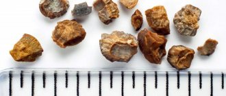
Stone in the ureter
– this phenomenon is not only unpleasant, but also dangerous. If a sufficiently large stone comes out of the kidney and gets stuck in the ureter without reaching the bladder, this in most cases manifests itself as severe renal colic.
This severe and painful condition can last for several days, but the most dangerous are its consequences: inflammation of the mucous membrane of the ureter and the kidney itself, blockage of the ureter, renal failure and even death of the kidney. Therefore, it is important to know the signs of this condition and immediately call a urologist at home at the first symptoms.
Medicinal method
The safest method to help remove kidney stones is medication. Stones can be removed by:
- Increasing diuresis with the help of special medications and increasing the amount of fluid consumed. But this method is used only if the size of the stones does not exceed 4 mm, since this ensures the possibility of their unhindered passage through the ureter and urethra.
- Dissolution of kidney stones through the use of infusions based on various types of medicinal raw materials and the use of drugs is possible only in the presence of stones that are chemically classified as organic or the class of urates and phosphates. However, the most common oxalates are practically insoluble in dissolution.
Attention! There is no guarantee that even the most complete medical treatment will help remove even tiny stones.
Removing stones at home
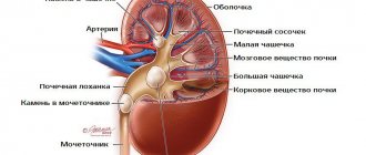
To begin with, it should be noted that the ureter is a tube narrowed at the edges that connects two organs and allows fluid to move from the kidney to the bladder.
Therefore, it is quite possible to remove a stone from the urinary duct at home. What should you do for this?
- Taking some antispasmodic drug will help you relax and increase the diameter of the ureter. You can use papaverine, drotaverine or no-shpa.
- If the pain is unbearable, you should take a pain reliever.
- In a hot bath for half an hour, you should drink a diuretic decoction (for example, from dill seeds or palya) so that the liquid helps push the pebble out of the ureter.
- Active movement, jumping rope, jogging with emphasis on the heels also helps remove the foreign body from the body.
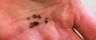
- Carefully examine the urine in a specially prepared container. If a stone comes out of the ureter, be sure to perform an ultrasound.
- Laboratory testing will determine the type of stone removed from the urinary duct.
Surgical stone removal
In cases where urolithiasis occurs with the formation of staghorn stones, severe complications, is accompanied by severe hematuria, severe pain that deprives a person of the ability to work, or leads to the development of hydronephrotic transformation and attacks of acute pyelonephritis, patients may be offered surgical treatment. But in different cases, patients are indicated for different types of surgical intervention.
The operation to remove a stone located in the kidney is called pyelolithotomy. It is performed under general anesthesia and consists of making a 10 cm incision on the patient’s side on the side of the affected kidney. Through it, the doctor can reach the kidney, cut it and remove the stone from the pelvis. Immediately after this, the wound is sutured, and the stitches are removed after a week.
Important: operations are dangerous due to the development of severe bleeding, secondary infections and other equally serious complications.
These types of surgical treatment are used exclusively in cases where all other methods aimed at removing kidney stones do not produce results. This is due to the fact that urolithiasis very often recurs, but previous surgical intervention makes such treatment impossible in the future.
Symptoms of urethrolithiasis
If some kind of calculus gets into the lumen of the ureter from the kidneys, then in this case the person almost immediately begins to be bothered by certain symptoms. The degree of their severity depends on how much the organ is blocked. With complete blockage of the ureter, they are very pronounced and appear sharply; in such patients, a clinical picture of acute renal failure is observed. With stones in the ureters, the following symptoms are noted:
- severe pain in the lower abdomen, lower back of the ribs or spine;
- increased body temperature;
- nausea, vomiting, diarrhea;
- frequent urge to urinate;
- increased blood pressure;
- headache.
Painful sensations are caused by disruption of microcirculation processes in the renal tissue and irritation of nerve endings. They can intensify with sudden movements and physical activity. When a stone enters the ureter, men and women often experience irradiation of pain to the external genitalia - the scrotum and penis or labia majora, respectively.
Important: The main reason for the occurrence of foreign stones in the ureter is a violation of phosphorus-calcium and oxalic acid metabolic processes in the body, as well as infectious diseases of the urinary system, changes in the acidity and composition of urine.
If sand gets into the ureter, the symptoms are not so pronounced and may go away if it is spontaneously removed. This is due to the fact that the grains of sand are small in size and to a small extent block the outflow of urine from the ureter.
Gentle methods for removing stones
In recent years, methods that are free of the dangers associated with abdominal surgery and do not require long-term rehabilitation have gained significant popularity. They are based on crushing the formed stones in one way or another with further removal of fragments with special tools or naturally, that is, along with urine.
Removing stones using endoscopic equipment
If doctors are faced with the question of removing a stone located in the kidney, and the patient does not have other diseases of the urinary system, with the exception of nephrolithiasis, it is best to remove it using endoscopic equipment. Removal of kidney stones is carried out by:
- Laparoscopic surgery, which involves the introduction of special surgical instruments into the renal calyces and pelvis through incisions in the lumbar region, the size of which usually does not exceed 1 cm.
Important: such operations do not require significant muscle dissection, since the incisions performed are very small, so there are no noticeable scars left after them.
- Insertion of a nephroscope equipped with video equipment into the kidney through the urinary tract.
- Insertion of a urethroscope into the lumen of the ureter to remove stones stuck in it.
If the stone is small, it is removed without preliminary destruction, otherwise it can be crushed with special tools, a laser, or by installing a directed ultrasonic wave emitter in close proximity to the stone, which is carried out to form using endoscopic equipment. If doctors are faced with the task of removing a stone located in the ureter, they can resort to pneumatic lithotripsy, which involves inserting a urethroscope through the urethra and bladder and exposing the stone to a series of shock waves. Thanks to this, the stone is destroyed in a few seconds, and the resulting fragments are removed from the patient’s body using special loops or forceps.
External lithotripsy
The method involves the destruction of stones due to wave action on them without making any cuts or punctures. The fragments formed during the procedure are subsequently excreted from the body along with urine. As a rule, ultrasound is used as shock waves. More information about the method of ultrasonic lithotripsy can be found in the article: Features of ultrasonic crushing of kidney stones.
Remote lithotripsy is effective in the presence of small stones, the size of which does not exceed 2 cm. Doctors usually talk about its painlessness, but patients often complain of quite severe discomfort and even pain experienced during manipulation.
Laser lithotripsy
Laser crushing of stones in the kidneys and ureters is the most modern and safe method for removing stones of any size. The procedure is carried out through a urethroscope or nephroscope inserted through the urethra, using special equipment in which a laser beam is created using holmium. Under its influence, even large stones quickly break down into dust, and thanks to the monitor, to which the image from the inserted equipment is transmitted, the doctor can completely control the course of the procedure and, if necessary, make changes to its course.
Crushing kidney stones with a laser is an absolutely painless, bloodless procedure in which the risk of damage from fragments to the vulnerable mucous membranes of the urinary tract, and therefore the development of bleeding, is minimal. Thus, today laser lithotripsy is the safest and most effective method for removing kidney stones, including coral stones. Therefore, it is a worthy alternative to abdominal surgery, helping to cope with staghorn stones, which are common today. The only disadvantage of laser crushing is the high cost of the procedure.
Attention! To destroy stones of any size, just 1 session of laser lithotripsy is enough, unlike other methods.
But perhaps it would be more correct to treat not the effect, but the cause?
We recommend reading the story of Olga Kirovtseva, how she cured her stomach. Read the article >>
Kidney stones are not immediately life-threatening, but they are one of the most unpleasant illnesses a person can experience. Unfortunately, this is a fairly common disease. However, there are several treatments available to remove kidney stones and make them pass quickly and easily.
Some substances contained in urine can form crystals that settle in the kidneys and ureters. Gradually they accumulate and form stones, which then bring numerous troubles to a person.
Removing kidney stones: conservative treatment
One way is to cleanse the kidneys. Drinking plenty of water (2 to 3 liters per day) helps produce enough urine to push the stone through the urinary tract and out of the body. At the same time, you can take painkillers to make the process somewhat less painful. Physical activity along with drinking plenty of water will also help remove the stone, although pain may ruin all efforts.
Lithotripsy
Ultrasound crushing of kidney stones (lithotripsy) is the most common procedure for removing stones that are too large to pass out of the body on their own. It does not require surgery: instead, a special device sends ultrasound waves into the abdominal cavity, where they break the kidney stones into pieces small enough to be passed through urine. However, this method also has side effects, such as abdominal pain and blood in the urine. Lithotripsy is performed on an outpatient basis. It often takes more than one session to achieve a positive effect and remove kidney stones successfully.
Urethroscopic stone removal
Sometimes kidney stones become lodged in the ureter. In this case, urethroscopic removal of kidney stones is necessary. The process does not require surgery. Instead, the doctor uses a urethroscope to remove the stones through the urethra.
Fortunately, most stones can be treated without surgery, which may be recommended if the stones are too large, cause persistent pain, block the flow of urine, or damage kidney tissue. In addition, surgery is usually indicated in cases where treatment with other methods is impossible.
Nephrolithotomy
If the kidney stones are too large or the sound waves cannot reach them, the surgeon may perform a nephrolithotomy to remove the stone. The surgeon makes a small incision in the back and, using an instrument called a nephroscope, enters the kidney and removes the stone. Before surgery, stones may need to be crushed using ultrasound. After nephrolithotomy, patients must remain in the hospital for several days, but the procedure has one advantage: the patient does not experience pain, because everything is done under anesthesia.
Pyelolithotomy
The surgery involves opening the affected area and removing the stone(s). It is performed under general anesthesia. The incision is made parallel to the costal arch. The doctor removes the stone with forceps, after which the kidney or ureter is sutured with self-absorbing material.
Urolithiasis or urolithiasis is a widespread disease. It occurs in 1-3% of the working population. Urolithiasis is a multi-causal disease. Kidney stones are salt deposits that can form due to poor diet, metabolic disorders, too hot climate, vitamin deficiency or hypervitaminosis D. Certain compounds contribute to the development of the disease, including medications - glucocorticoids, tetracyclines, etc.
Kidney stones can be extremely painful, cause problems with urination, and provoke inflammation. To prevent unwanted complications, timely diagnosis and rapid adequate treatment are necessary. Most urologists are in favor of surgery, since it allows you to quickly and reliably solve the problem. Removing kidney stones using modern methods allows the use of minimally invasive methods, which significantly reduces the risk of complications and relapses.
Folk remedies for getting rid of kidney stones
Treatment with traditional methods for kidney problems is very common.
Urologists quite often themselves prescribe herbal remedies to patients, as well as home procedures as auxiliary measures in the conservative treatment of nephrolithiasis.
Traditional medicine experts say that many natural medicines help get rid of kidney stones, and it’s not for nothing that almost all pharmaceutical preparations for treating kidneys are made on the basis of herbs. Of course, the fact that natural ingredients can completely dissolve and remove stones is an exaggeration.
However, the diuretic, anti-inflammatory, antiseptic, and antispasmodic properties of some plants may well be useful in the treatment of urolithiasis. In addition, many infusions and decoctions can affect the acid-base balance of urine, helping to soften salt formations.
Here are some popular folk recipes:
- Drink a glass of compote made from dogwood berries and wild pear 4 times a day.
- Prepare an infusion of parsley and take it throughout the day: pour a teaspoon of chopped root and leaves into a glass of boiling water and leave for 3 hours.
- Drink a decoction of rose hips as tea for 3 months, then take a break.
- Take the roots of ripened sunflowers and chop them finely. Boil a glass of crushed root in 3 liters of water for about 5 minutes, cool. Drink the resulting medicine within 3 days.
It is worth remembering that traditional medicine for urolithiasis is effective only in the initial stage of the disease, as well as for the prevention of urolithiasis and the prevention of its relapses. In cases where neither decoctions nor pharmaceutical preparations can help, surgery is not necessary. You can try to crush the stones.
Indications for surgery
Surgery may be performed in the following cases:
Depending on the degree of damage, the methods of surgical intervention may vary:
- Unilateral urolithiasis. Localization of stones in one kidney makes it possible to preserve the functions of the genitourinary system in case of unsuccessful surgery.
- Bilateral urolithiasis. If the location of the stones is established, the operation can be performed on two kidneys simultaneously. Otherwise, it is carried out in two stages, with a break between them of 1-3 months.
The best treatment is prevention
In order to prevent the onset of the disease, it is necessary to maintain proper nutrition and drinking regime. To quickly remove mucus, microorganisms, sand, and leukocytes, you need to drink about two liters of natural water per day.
A decoction of rose hips, dried fruits, carrot juices - all this will be a good prevention of stone formation. It is also recommended to consume vegetables and fruits in their pure form.
At the first symptoms that indicate the presence of pathology in the urinary system, it is necessary not to delay treatment. Chronic foci of infection should also be sanitized. For example, inflammatory processes in the organs of the urinary system can provoke the formation of sand, so they need to be treated in a timely manner.
It is also recommended to do gymnastic exercises at the stage of stone release:
- jumps;
- run;
- jumping.
I would also like to mention diet. Experts recommend excluding from your diet foods that have a high content of oxalic acid, namely:
- legumes;
- cabbage;
- sorrel;
- nuts;
- spinach, etc.
Types of surgery
The following methods for removing stones are distinguished:
- Lithotripsy. The stone is crushed by exposure to ultrasound through the skin, after which it is removed through the ureter or catheter to the outside.
- Endoscopic operations. A special instrument, an endoscope, is inserted through the ureter or urethra and approaches the location of the stone. Removal is carried out through it.
- Open surgery. Involves direct incision of the kidney and surgical removal of salt deposits.
- Resection. The operation is a type of open, but involves partial removal of the kidney.
Lithotripsy
The essence of the procedure
Since its discovery and introduction into practice (in Russia - in the late 90s of the last century), lithotripsy has earned recognition and taken a leading place in urolithiasis surgery. It eliminates the trauma of surgical intervention and the risk of infection, since the effect is performed percutaneously without any incision.
The essence of the method is based on the effect of ultrasound on various environments of the body. It spreads freely in the soft tissues of the body without causing any harm. When ultrasound encounters a dense salt deposit, it creates cavities and microcracks in it, which leads to a violation of the integrity of the stone.
Modern lithotriptors are generators of ultrasonic shock waves; depending on the country of origin, they can be driven by an electromagnetic, electrohydraulic, piezoelectric element, or even a laser. However, there are no significant differences between them. Visual control of the location and condition of the stone can be carried out using x-rays or ultrasound.
Indications and contraindications
Lithotripsy is performed to remove small stones (up to 2 cm), the localization of which can be unambiguously determined by one of the indicated methods, from living kidneys. In the fifth and final stage of urolithiasis, using this removal method can be dangerous. Note. Some authors (O.L. Tiktinsky) believe that even with large coral deposits it is possible to use ultrasound. But in this case, constant monitoring of the location of all their fragments and readiness for additional endoscopic surgery is necessary.
Surgery is not performed in the following patient conditions:
- Pregnancy.
- Injuries to the musculoskeletal system that do not allow you to take the correct position on the couch.
- The patient's body weight is above 130 kg, height is more than 2 m or less than 1 m.
- Blood clotting disorder.
Progress of the operation
At the very beginning of the use of the technique, general anesthesia was widely used, but today it is obvious that in most cases it is not necessary and doctors limit themselves to epidural anesthesia. Analgesics are injected into the lumbar spine. They begin to act within 10 minutes, and the duration does not exceed 1 hour. In emergency cases and when epidural anesthesia is contraindicated, they are administered through a vein.
The operation is performed in the prone position on the stomach or back, depending on the location of the stone. In the second case, the patients legs will be raised and secured. After the onset of anesthesia, a catheter is inserted into the ureter, through which a contrast agent enters the kidney for better visualization. The patient will not feel any discomfort.
If the stone is larger than 10 mm, a needle is inserted into the renal pelvis. Through the puncture, the formed channel expands to the required diameter, which allows you to place a tube with a tool in it to extract sediment fragments. This lithotripsy is called percutaneous or percutaneous. Smaller stones, after crushing, are excreted in the urine - a remote version of the technique.
A catheter inserted into the ureter is filled with saline solution . It is designed to facilitate the passage of the ultrasonic wave and protect neighboring tissues from unwanted effects. The device is located at the exact location of the stone projection. When it acts, the patient feels soft, painless tremors. Sometimes several approaches are required to break the stone.
Important! In rare cases, the patient may experience pain of varying degrees of intensity during the procedure. You must remain calm and not move. You should report pain to your doctor.
With non-invasive lithotripsy, the patient moves to the ward after the operation and the end of anesthesia. There he is asked to urinate into a jar in order to control the removal of stone fragments. Unpleasant sensations are possible. There may be blood in the urine - this is normal; it is formed as a result of sand scratching the epithelium of the ureter. The release of salt residue may take up to several days after surgery. With percutaneous lithotripsy, the stone will be removed through a tube, but parts of it may be passed out in the urine.
After 2 days, the doctor performs an ultrasound of the kidneys to study their condition. If the operation is successful and there are no complications, the patient is sent home.
Endoscopic operations
Depending on the location of the stone, the endoscope can be inserted into the urethra (urethra) or higher into the bladder, ureter, or directly into the kidney. The lower the deposits are located, the easier the operation is. It is performed under general anesthesia or intravenous anesthesia to remove stones up to 2 cm. Indications are:
- Ineffectiveness of lithotripsy;
- Long-term presence of a stone in the path of the ureter;
- “Stone paths” (residual formations) after exposure to ultrasound.
The operation, despite its apparent simplicity, requires a highly qualified surgeon and high-quality modern equipment. A urethroscope is inserted into the patient's urinary canal. This device consists of a tube with a mirror that allows the surgeon to directly detect stones. Once the tube reaches them, they will be removed. The most modern technique is laser removal of kidney stones. The action of the beam is transmitted through a special fiber, which is inserted into the urethroscope.
In some cases, the installation of a stent is required - this is a catheter that prevents compression of the ureter (obstruction). It is placed for up to several weeks. Removal also occurs without incisions using an endoscope.
Stones by localization
There are these types of stones depending on their location:
- in the lower third of the ureter. In this case, the patient experiences pain, if he is a man, then in the scrotum area, if a woman, in the vulva area. Besides this, I am bothered by a frequent and painful urge to urinate. Blood may appear in the urine. The insidiousness of the disease is due to the fact that its symptoms may be similar to other pathological processes. So, for example, if a stone is at the mouth of the ureter, that is, it is located near the entrance to the bladder, but has not yet penetrated into it, then the clinical symptoms are difficult to distinguish from manifestations of prostatitis, urethritis or acute cystitis;
- in the middle and upper parts. In this situation, the intensity of the pain is so pronounced that the patient simply cannot tolerate it. Painful sensations spread in the lumbar region. The stones move, therefore blocking the passages, which is why the unpleasant sensations either subside or worsen again. If the stone does not move, the pain may be mild or completely absent.
Open surgery
In recent years, this type of intervention has been performed extremely rarely. Indications for it are:
- Constant relapses;
- Large stones that cannot be removed in any other way;
- Purulent inflammation.
Open surgery is performed under general anesthesia and is abdominal . This means that it affects the body cavity. Excision occurs through all layers of tissue. The presence of a stone in the renal pelvis is considered favorable. This reduces the invasiveness of the operation. It is also possible to open the ureter and remove the stone from there.
The modern option for performing the operation is laparoscopy. Removing the stone through a small incision. A camera is inserted into it to transmit images to a large screen. Laparoscopic stone removal is carried out only for special indications and is often replaced by endoscopic operations.
How to remove stones from the ureter at home
Unfortunately, urolithiasis today is a fairly common and very unpleasant phenomenon. This is how the disease urolithiasis manifests itself, in which a stone formed in the renal pelvis migrates into the urinary duct. Much less often, stone formation is observed directly in the ureter itself. In the altered tissues of the ureter, infection quickly spreads, ascending pyelonephritis, cystitis, and periureteritis develop. In places where the stone is located, bedsores or perforations form. The disease is complex and painful, so it is important to prevent its development.
Removal of part of the kidney
Indications and contraindications
This operation allows you to save the organ, which is especially important if you have only one working kidney. Resection is performed in the following cases:
- Multiple (multi-locular) stones located at one pole of the organ.
- Constant relapses of the disease.
- Necrotic lesions.
- The last stages of urolithiasis.
Important! A contraindication is the patient's serious condition, if doctors assume that the operation may aggravate it.
Progress of the operation
Resection is performed under general anesthesia. The patient is placed on his healthy side, under which a cushion is placed. The surgeon makes an incision. After this, he pushes the underlying layers of tissue apart. A clamp is applied to the part of the kidney with the ureter to prevent bleeding, since this is where the maximum concentration of blood vessels is located.
After this, the affected area is excised. The edges are stitched. A drainage tube is removed from the kidney. After this, the wound is sutured. The drainage tube remains in the kidney for 7-10 days after surgery; after this period, provided there is no separation of pathological contents, it is removed.
Complications
Each of the described types of operations may have a different probability of undesirable consequences, but in general they can be presented in the form of the following list:
- Relapses. Recurrent stone formation is not uncommon with urothiasis. The operation only combats the consequences, but does not eliminate the cause. That is why it is important in each specific case to find out why urolithiasis developed, to give the patient recommendations for lifestyle changes, diet and, possibly, taking medications.
- False relapses. This is the name given to the remaining unremoved stone fragments. This outcome of the operation is becoming less and less common due to the improvement of methods of its implementation and constant monitoring of its progress.
- Infection. Even with minimally invasive operations such as endoscopic ones, there is a risk of pathogens entering internal organs. To prevent infection, a course of antibiotics is prescribed even if the patient is in good condition.
- Acute pyelonephritis is inflammation of the renal pelvis. It occurs due to the displacement of stones, prolonged residence of their fragments in the kidney and the accumulation of infiltrate (fluid) around them.
- Bleeding. Most often occur during open operations. To prevent them, irrigation of the kidney with a solution containing antibiotics is used.
- Progression, exacerbation of renal failure. For prevention, hemodialysis (connection to an artificial kidney machine) is used before and after surgery.
- Heart rhythm disturbances, hypertension (high blood pressure). The complication occurs more often after ultrasonic destruction of stones due to an incorrect assessment of the patient’s condition.
Diet therapy
Complex therapy of urolithiasis involves dietary nutrition. Thanks to a properly selected diet, it becomes possible to dissolve urate stones at the initial stage of their formation.
In addition, the diet helps prevent the appearance of new oxalate and phosphate stones. In addition to a properly selected diet, it is necessary to follow a drinking regime. You should drink 2.5 liters of liquid per day. In this case, the amount of fluid is increased if a person has a predisposition to the formation of kidney stones.
The diet is compiled based on the type of stones identified. If calcium deposits have been identified, the patient is advised to reduce the consumption of dairy products. There are cases when they are strictly prohibited, since new stones quickly form and increase in size due to the individual characteristics of the body.
- Oxalate stones require exclusion from the diet of foods that contain oxalic acid. These include citrus fruits, sorrel and lettuce leaves, and spinach. Magnesium preparations accelerate the process of acid binding. They are recommended to be taken at a dose of 2 g per day.
- Urates appear in the kidneys due to an increase in the level of uric acid in the body. To prevent their occurrence, the patient is advised to avoid foods that provoke increased production. These include meat by-products, concentrated broths, and vegetable fats. Urine alkalizers will help neutralize the acid. Prescribed medications containing sodium citrate (Blemaren, Kataria), as well as citrus fruits.
- Phosphates, on the contrary, cause urine to become alkaline and require acidification. Meat and fish dishes, as well as baked goods and vegetable oil can help with this. The volume of fluid consumed in case of phosphate stones should be reduced.
Cost of surgery for urolithiasis, carried out under compulsory medical insurance
The most common type of intervention is lithotripsy. It is carried out in most clinics and medical centers dealing with urological diseases. The average price is 20,000 rubles. The operation is free of charge only for persons under 18 years of age in public medical institutions.
According to the compulsory health insurance policy, endoscopic, open surgeries and kidney resections are usually performed in hospitals. The first type of procedure in private clinics costs from 30,000 rubles. The price does not include medications necessary for rehabilitation and a place in a hospital. Open abdominal surgeries are rarely performed in private clinics; the price must be determined privately. The cost of partial kidney removal starts from 17,000 – 18,000 rubles and can reach 100,000 rubles. The price is shown only for the procedure.
Patient reviews about the operation
The largest number of reviews on the Internet are devoted to lithotripsy. Many patients were satisfied with the result. As a rule, the following negative aspects are noted:
- High price. Often the decision to undergo surgery must be made suddenly and as soon as possible. Not every patient has a reserve of several tens of thousands of rubles.
- Painful sensations during surgery. This happens quite rarely, and patients note that the discomfort cannot be compared with the agony of renal colic.
- Risk of relapse and lack of guarantees.
With other operations to remove kidney stones, especially those performed free of charge, patients are concerned about the tactics of the chosen treatment. Not every doctor explains to the patient the essence of his actions and prescriptions, especially when it comes to elderly patients or their relatives. The wrong choice of the type of surgical intervention and the lack of improvement are usually difficult for people entering a medical facility.
Urolithiasis is a common disease that develops as a result of the combined action of many factors. And although modern methods of surgical treatment can successfully solve this problem, the latest developments in the field of ultrasonic crushing are not available to everyone. The outcome of treatment cannot always be predicted, and with any type of therapy the risk of relapse remains. Therefore, if there is a predisposition to the disease or its presence in relatives, all measures must be taken to prevent urolithiasis.
Causes of the disease
Ureteral calculi are formed from various substances:
- uric acid;
- cystine;
- calcium phosphate;
- struvite.
Most often, the process of stone formation is influenced by the following factors:
- Genetic predisposition. Doctors say that the disease is more often diagnosed in patients who have a family history of urolithiasis.
- Impaired outflow, stagnation of urine. The development of the disease may be based on congenital pathologies. Most often, the disease is provoked by narrowed ureters in women, their underdevelopment, kinks or abnormalities of the bladder.
- Chronic urinary tract diseases. Infectious diseases can lead to the development of pathology. For example, pyelonephritis.
- Disturbed exchange. Acquired or congenital ailments may be accompanied by the penetration of lithogenic substances into the urine - calcium (if hyperparathyroidism is diagnosed), urate (in the case of gout).
- Diseases of the digestive system. If the absorption function is impaired, stones may form.
- Use of medications. Some medications can lead to the development of the disease. For example, such consequences are provoked by uroseptics from the nitrofuran category.
Doctors say that uroliths often form in women living in hot and dry climates. High-calorie foods rich in animal proteins can trigger the development of the disease.