Excess amounts of these amino acids are excreted in the urine. However, the solubility of cystine (cystine is a dimeric form of cysteine) in an aqueous environment is low, which causes the development of clinically significant stone formation. Cystine stones are detected in approximately 1% of patients with kidney stones. However, in children, kidney stones of this type are found in 5-8% of cases. Cystinuria affects 1 in every 15,000 people (US statistics). Pure cystine stones are formed only in homozygous carriers of the anomaly. Normally, an adult excretes in urine no more than 19 mg of cystine for every gram of creatinine per day. Due to the presence of sulfhydryl residues in cysteine molecules, cystine stones are radiopaque.
Causes of cystine kidney stones
The solubility of cystine in water is approximately 250 mg/l. This figure increases as pH increases. The pKa value of cystine is 6.5, so the increase in its solubility is especially noticeable with increasing pH in the range from 6.5 to 7.5. If the concentration of cystine in urine is above 250 mg/l, the urine is in a state of supersaturation with this compound. When the concentration of cystine in urine is kept at a level of less than 200 mg/l, cystine stones do not form. In homozygous carriers of the above anomaly, from 800 to 1000 mg of cystine are excreted in the urine per day. To prevent the formation of stones, such patients must excrete at least 4 liters of urine per day with a normal pH.
content .. 11 12 13 ..CYSTINE STONES (URINAL STONES)
General information Examination Treatment Metaphylaxis
General information
The etiological factor in the formation of cystine stones is a hereditary (autosomal recessive) disorder of tubular reabsorption of the four main amino acids - cystine, ornithine, lysine and arginine (COLA) - Abderhalden-Lignac syndrome. Ornithine, lysine and arginine have good solubility, but cystine is poorly soluble, which in the presence of cystinuria more than 200 mg per day is the main cause of the formation of cystine stones. The disease is inherited in an autosomal recessive manner and is more common in men (70% of patients). In healthy individuals, approximately 40-80 mg of cystine is excreted in urine per day. Cystinuria is a condition characterized by a defect in the transepithelial transport of cystine in the intestine and kidneys. A decrease in the level of magnesium in the urine is also a risk factor for the development of cystine urolithiasis. The model of risk factors for cystine urolithiasis is presented in Figure 36
Figure 36. Risk factors for stone formation from cystine.
The formation of cystine stones can even occur in the fetus, although the first manifestation most often occurs in the second decade of life. Individual dietary habits may contribute to cystine stone formation. High urine density, high consumption of animal protein and sodium chloride increase urinary excretion of cystine.
Survey
Laboratory program
Anamnesis
Familial cystinuria, the presence of cystine stones in close relatives. The first manifestation of the disease is in young children or at 15-20 years of age.
Visualization
Poorly radiopaque stones are visualized as shadows similar to frosted glass.
Ultrasound examination shows high stone density with typical echo shadowing.
Laboratory program
Presence of characteristic hexoganal crystals in urine sediment.
Urine pH is normal.
Positive cystine test [17]. Quantitative determination of cystine in urine is necessary only in cases of cystinuria to select appropriate treatment. This is only possible with a special analysis to identify this amino acid.
Urinary cystine levels:
normal level: 0.17 -0.33 mmol/day (40 -80 mg/day);
in patients: more than 4.16 mmol/day (1000 mg/day);
start of treatment: "= 0.8 mmol/day (192 mg/day);
solubility limit: 1.33 mmol/day (320 mg/day).
In addition to cystinuria, additional metabolic abnormalities such as hypercalciuria, hyperoxaluria, and hyperuricuria may occur. Therefore, it is necessary to follow the examination standard and know the levels of calcium, oxalate and uric acid. To exclude a urinary infection, it is necessary to perform a nitrite test and bacteriological examination.
Serum examination
When examining blood serum, the levels of biochemical parameters are normal.
Stone analysis
When analyzing the stone, cystine is determined.
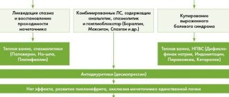
Treatment
The increased risk of stone formation with this type of urolithiasis is expressed through the activity of the products (AR) and is calculated using the formula:
Despite the fact that the solubility of cystine increases with increasing urine pH (more than 7.0), drug-induced litholysis through oral administration of drugs is rarely achieved. Cystine stones are sometimes very difficult to treat with radiotherapy. Contact crushing methods are very effective, but it should be borne in mind that stents and catheters installed after this are very quickly encrusted with cystine.
Metaphylaxis
Without appropriate metaphylaxis, it is impossible to avoid recurrence of cystine stones.
Cystinuria is an inherited metabolic defect, so adequate therapy is necessary throughout life. The following therapeutic measures should be applied:
- Reducing the concentration of cystine by intensive dilution of urine. To obtain a soluble cystine level of 1.33 mmol/day (320 mg/day) with a cystine excretion of more than 4.16 mmol/day (1000 mg/day), it is necessary to achieve a urine volume of at least 3.5 l/day. Depending on the activity of physical activity and ambient temperature, the amount of fluid consumed should be 3.5 -4.0 l/day. This volume of liquid should be evenly distributed over 24 hours. It is recommended to develop the habit of drinking liquid before each urination and before going to bed at night. Preferred are mineral waters with a high content of bicarbonate and sodium (HCO3 - maximum 1500 mg/l and 500 mg/l). The daily volume can be regulated with kidney tea, fruit tea, apple juice, and citrus juice. You should limit coffee and tea (no more than two cups per day). You should not drink juices and lemonades containing sugar, or any alcoholic beverages.
- Reducing cystine excretion by eating a low protein and sodium diet. Considering that cystine is formed during the metabolism of methionine, a diet low in this amino acid will be effective. Unfortunately, such a diet very significantly reduces the quality of life due to a poor diet of food. Patients suffering from cystinuria are better off eating a well-balanced diet with low protein content, not exceeding 0.8 g of protein per 1 kg of body weight per day. You should limit your consumption of protein-rich foods: meat, fish, sausages, eggs, cream, hard cheeses. A vegetarian diet meets these requirements. Vegetables, greens, fruits and grains should be the main components of the diet. In children, iron and iodine intake should be monitored. Children should be given one or two meat and one fish dishes per week. Cystine excretion increases with high salt intake, so salt intake should be limited. Instant foods, canned foods, and processed foods contain large amounts of salt and should be avoided.
- Increasing the solubility of cystine by alkalization of urine. The solubility of cystine increases at a urine pH of 7.5 (Fig. 37). This is a successful therapeutic moment.
Rice. 37. Cystine solubility depending on urine pH
Alkalization of urine is achieved by regulating acid-base metabolism. Contraindications to this measure are persistent urinary tract infection, arterial hypertension, renal failure, acute metabolic alkalosis. Side effects include gastrointestinal disorders and the risk of phosphate stones. Citrate mixtures are prescribed under urine pH control. Of the medications, Blemaren and Uralit-U deserve attention. Sodium bicarbonate can also be prescribed, however, sodium increases hypercalciuria. Therefore, the use of bicarbonate is possible only in patients who are tolerant to the prescription of citrate mixtures.
- A decrease in urinary cystine concentration is also achieved with the help of sulfite-containing components. The drug mercaptopropionylglycine (Tiopronin, Thiola) is prescribed when cystine excretion is more than 3.0 -3.5 mmol/l (720 -840 mg/day). This drug forms a complex compound with cystine. The dose should be increased slowly, starting from 250 mg/day under the control of cystine excretion, but a maximum of 2.0 g/day. The daily dose should not be exceeded. About 20% of patients have complications such as gastritis, dermatosis, and nephrotic syndrome.
- If the daily excretion of cystine is less than 3.5 mmol, ascorbic acid is prescribed at a dose of 3-5 g/day; if the excretion of cystine is more than 3.5 mmol, Captopril is prescribed at a daily dose of 75-150 mg (depending on the degree of cystinuria). These drugs form chelate complexes with cystine in urine, converting cystine into a soluble disulfide form. As-corbic acid is prescribed when cystine excretion is less than 3.0 -3.5 mmol/l (720 -840 mg/day). Ascorbic acid promotes the conversion of cystine to cysteine, which is a highly soluble substance. However, a side effect of using ascorbic acid in the required therapeutic dose (3-5 g/day) may be a risk factor for hyperoxaluria. The effectiveness of ascorbic acid for the prevention of cystine stone formation has been studied in several studies.
Serum and urinary parameters should be carefully monitored throughout the life of these patients. Serum: creatinine and uric acid. Urinary: volume and density of urine, cystine, pH, nitrite test, calcium, phosphorus, oxalate, uric acid.
The general principles of metaphylaxis of cystine stones are presented below:
- Change in urine pH to 7.5 -8.0;
- Reducing urinary cystine concentrations with
o increasing the daily volume of urine” 3.0 l, o reducing sodium intake from food;
- Conversion of cystine into more soluble forms: o alpha-mercaptopropionylglycine (10 -30 mg/kg/day), o D-penicillamine (15 -40 mg/kg/day),
o ascorbic acid (3 -5 g/day).
content .. 11 12 13 ..
Symptoms and signs of cystine kidney stones
Cystine stones usually form before the age of 40. Patients usually have staghorn stones in both kidneys, which often lead to urinary tract obstruction. Urine, especially early morning urine (which usually has a low pH), may contain characteristic hexagonal crystals. Heterozygous carriers of the mutation may have urolithiasis with stones that do not contain cystine at all or contain it in minimal quantities. In such cases, cystine crystals serve as initiating centers that trigger the crystallization of other substances (for example, calcium oxalate or calcium phosphate).
Uric acid stones
Uric acid stones account for 3-10% of kidney stones. Uric acid stones usually look like pebbles. Some of the stones may be hard on the outside but soft on the inside because they are composed of different types of uric acid and calcium oxalate monohydrate.
Struvite stones
Struvite stones are the next most common type of kidney stones (7-8% of cases) and are larger than the others.
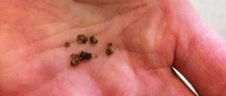
Cystine stones
Cystine stones are the result of cystinuria
, which can be inherited. The kidneys produce cystine, a type of amino acid.
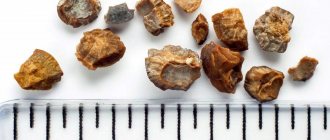
Cystine stones are compact, partially opaque, and amber in color.
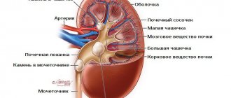
Diagnosis of cystine kidney stones
A sign of cystinuria is characteristic hexagonal crystals detected in the first morning urine sample. However, such crystals are rare. The direct primary test for the presence of cystine in urine is the sodium nitroprusside test, which detects cystine in amounts of 75 mg/g creatinine or higher. It should be noted that nitroprusside forms complexes with any sulfhydryl groups, so the test may give a false-positive result in patients taking medications containing such groups. As an alternative, the phosphotungstic acid test can be used. If the results of the initial testing are positive, a daily urine sample should be obtained from the patient and the amount of cystine excreted in urine per day should be determined. Cystine stones are more radiolucent than calcium-containing and struvite-carbonate stones. They are usually homogeneous in structure and have no layering.
Urats
These are smooth, hard stones that are yellow-orange or brick-colored. They are not visible during X-ray examination, but are visible on ultrasound; urate salts are often detected in a general urine test.
- Formed when there is an excess of uric acid in the urine (gout, psoriasis, some blood diseases, a diet with excess purines - animal proteins, especially in combination with alcohol).
- The formation of urates is promoted by the acidic reaction of urine - this can occur in diseases of the digestive system, accompanied by diarrhea, and tubular disorders.
They respond well to conservative treatment; a combination of diet, abundant alkaline drinking and citrates often allows you to do without surgery.
Treatment of cystine kidney stones
Water is considered the best cure for cystinuria. Its required amount is determined based on an assessment of the daily excretion of cystine in urine.
The goal of increasing water intake should be to reduce urinary cystine concentrations below 250 mg/L. This often requires that the patient excrete at least 4 liters of urine per day. To do this, he must drink 250 ml of water every 4 hours. Since the patient does not drink water at night, he should drink 500 ml of water at once before going to bed. To assess the effectiveness of therapy, the patient's urine should be periodically examined for the presence of cystine crystals.
Alkalinization of urine gives some effect in the treatment of cystine stone formation. The dissociation constant of cystine is 6.5. This means that at pH 7.5, about 90% of this substance is in ionized form. However, at a pH of 7.5, calcium phosphate stones are more likely to form. Therefore, alkalinization of urine in cystine nephrolithiasis should be considered only as an adjunct to therapy. Potassium citrate is used as an alkalizing agent.
The use of sodium-containing alkalis should be avoided: the increase in the volume of the intravenous fluid caused by them leads to the activation of the excretion of cystine in the urine.
If all of the above measures are ineffective, drugs such as penicillamine, α-mercaptopropionylglycine and captopril are prescribed. All of these compounds are thiols and selectively bind to cysteine, forming complexes that are much more soluble in water than cysteine dimers (cystine). α-mercaptopropionylglycine has fewer side effects than penicillamine. Penicillamine can also bind pyridoxine. Therefore, when using it to prevent the development of pyridoxine deficiency, it is prescribed in an amount of 50 mg/day. Sometimes penicillamine and α-mercaptopropionylglycine cause loss of smell and taste disturbances. To prevent these side effects, it is necessary to add zinc to the diet in the form of dietary supplements. Although captopril has the fewest side effects, it is the drug least effective at reducing urinary cystine concentrations.
How do kidney stones pass?
Kidney stones can pass without medical intervention. Smaller stones are more likely to pass, but larger ones may block the urinary tract. The stones can take up to 6 weeks to pass; the patient should drink plenty of water during this time.
People often experience pain when a kidney stone passes through the ureter. If the pain is unbearable, you should consult a doctor. The doctor may prescribe strong pain medications, anti-nausea medications, and a drug called tamsulosin, which relaxes the ureter, making it easier for the kidney stone to pass.
Passage of stone depending on size
The average size of the ureter is 3-4 millimeters wide. The chances of passing stones depend on their size:
| Stone size(mm) | Probable percentage of passage through the ureter (%) |
| Less than 4 | 80 |
| 4–6 | 60 |
| More than 6 | 20 |
Reasons for development
Experts identify the following reasons for the formation of sand and stones in human kidneys:
- A small volume of fluid consumed by a person leads to an imbalance in the water balance in the body and its oversaturation with salt crystals.
- Imbalance of ions in the human body - low parameters in the urine of elements that help retain salts in a dissolved state.
- The presence of infectious pathologies in the urinary tract.
Due to the small volume of excreted urine or excessive intake of salts from food, the amount of ions in the excreted urine increases. The deficiency of natural crystallization inhibitors causes the salts to become insoluble, i.e. crystallize. They become the core of the future calculus, which can reach gigantic sizes.
Types of existing kidney stones
Kidney stones are:
- A single stone or multiple clusters of stones.
- Single-sided or with double-sided formation.
- Shape: flat or round, with spikes or with various edges.
- The size parameters are also varied: from microscopic, invisible to the naked eye, to gigantic ones - occupying the entire renal pelvis.
What types of kidney stones are there:
- Urates - are formed as a result of the concentration of urate salts and high acidity of urine.
- Salt compounds of natural oxalic acid are oxalates.
- Phosphates – contain calcium compounds of phosphoric acid.
- Calcium salts of natural carbonic acid, carbonic apatite: calcifications.
- Struvite - formed under the influence of infectious agents that promote changes in the composition of urine (magnesium ammonium phosphate).
- Cystine, as well as xanthine, are amino acid stones in nature.
- Protein - clots of settled fibrin interspersed with bacteria, as well as salts.
- Cholesterol - black, very soft, almost invisible during radiography.
Only a doctor can determine what type of kidney stones he has through diagnostic procedures. To do this, you will need to take blood and urine tests, undergo an ultrasound or x-ray of the kidneys.
In most cases of urolithiasis (urolithiasis), it is revealed that the stones have a mixed composition. The number of stone-forming elements is no more than three, and all others are diagnosed as separate impurities.
Oxalates
The most common and dangerous types of kidney stones are oxalates, which are formed from calcium varieties of oxalic acid salts. It is distinguished by significant density parameters, as well as black-gray coloring and a prickly structure. They injure the ureters, as a result of which they acquire a blacker color. Oxalates can reach several centimeters in diameter.
It is quite difficult to get rid of this type of stones. The pain as they pass through the urinary tract is significant and can radiate to the groin, thigh, and stomach. The person does not know where to escape from the pain, takes a forced position, and may lose consciousness.
Types of crushing kidney stones:
- Open surgical option
- Lithotripsy - wave action on a stone to crush it, with further removal of small fragments
Diet therapy comes down to excluding from the individual diet foods with a significant amount of oxalic acid: beets and cocoa, chocolate and sorrel, lettuce or spinach.
Phosphates
For the most part, they have a very diverse shape, with a smooth surface, and a high content of calcium salts of phosphate acid. They have a light gray color, as well as a rapid rate of increase in volume and ease of crushing at the time of treatment. It is recommended to take decoctions of barberry, as well as rose hips and grape rhizomes, followed by removal using wave techniques.
Diet therapy is based on an optimal reduction in vegetables and fruits in the diet, the most complete exclusion of fermented milk products, as well as an increase in the proportion of fish and meat dishes.
Urats
The stones are yellow-brown in color and are smooth and have a hard structure. They consist, in most cases, of uric acid and its salts.
These types of kidney stones are quite soluble with the help of a variety of medications. The specialist prescribes a course of treatment, followed by a follow-up examination. Drinking a significant amount of liquids will also benefit.
Struvite
A fast-growing type of stones, characterized by a light gray color, as well as a soft individual structure and a rough surface. Thanks to these features, coral-shaped varieties of kidney stones can form.
They are formed due to frequent stagnation of urine with the addition of a bacterial infection. They are practically insoluble, and therefore, when conceived, lithotripsy is prescribed to extract them.
Cystine and xanthine stones
Such stones are typical for persons with a hereditary negative predisposition. If the absorption of natural cystine, as well as diaminomonocarboxylic acids, is significantly impaired, a cystine version of stones is formed. Xanthine stones are formed when there is a deficiency of the molybdenum-containing natural enzyme xanthine oxidase.