A parenchymal cyst is a special formation ranging in size from 3 to 10 cm with liquid contents. The formation usually occurs on one kidney: left or right, and has a hereditary predisposition. The shape of the cyst is usually round or oval. The formation contains pus or blood inside and occurs mainly after physical injury. In 50% of cases, several cysts are diagnosed on one kidney at once.
The danger of the pathology lies in the fact that it practically does not bother patients until a certain point. When the formation reaches a large size, it begins to put pressure on neighboring organs. In this case, the patient feels severe pain and has difficulty urinating.
The disease can develop into dangerous complications. The cyst may rupture or compress the kidney vessels, resulting in increased pressure.
Causes of parenchymal cyst formation
Several factors take part in the development of pathology. The process of the appearance of a parenchymal cyst can be described as follows:
- formation of free space between tissues;
- filling this space with liquid;
- production of collagen by tissues around the cavity, which becomes insoluble, as a result of which the tissue is torn away from the space and takes the form of a capsule.
More than half of cases of the disease are congenital. It is very difficult to determine the factor in the appearance of parenchymal cysts. This is due to the fact that the manifestations of a cyst in the right or left kidney are often masked under the underlying disease. If the pathology is congenital, the symptoms of the disease may not manifest themselves at all. They are diagnosed randomly and in this case it is impossible to identify the cause of their formation.
Acquired parenchymal cysts are much less common. Here are the main reasons:
- kidney diseases, among which a special place is occupied by urolithiasis and pyelonephritis;
- physical damage, injury, radiation exposure is also dangerous.
The prognosis of the pathology depends on the cause of its development, the presence of concomitant diseases, location and size. The larger and closer the formation is to the kidney, the higher the risk of complications. Some of the most dangerous are bleeding and purulent discharge.
You'll like this:
Renal cysts are cavity tumors filled with fluid. Less common are dermoid cysts filled with other tissues. Kidney cysts often form in the upper layer of the organ. This is usually a benign neoplasm, but there is a risk of its malignant degeneration. This disease affects men and women equally and is more common after 40-50 years of age. There are several types of cystic renal formations. The course of the disease at the initial stage is hidden. Since there is a risk of malignant degeneration of the cyst, its growth and development must be constantly monitored, which is not always possible due to the absence of symptoms.
Characteristic symptoms of the pathology
Symptoms of the presence of a parenchymal cyst are very mild. The patient may not feel anything and live a normal life. This asymptomatic course of pathology has been observed for a very long time. Cysts are often diagnosed randomly during an ultrasound for another reason.
When the formation begins to grow rapidly and put pressure on accompanying organs, the patient begins to feel unpleasant symptoms. The following signs are characteristic of a renal parenchyma cyst:
- pain in the lumbar region, increasing after physical exertion and sudden movements;
- lower pressure indicators increase;
- there are traces of blood in the urine;
- depending on the side of the lesion, right or left kidney, circulatory disturbance occurs;
- problems with urination;
- aching pain in the lower abdomen;
- increase in kidney size.
With a weak immune system, an infection may occur. In this situation, the patient will feel manifestations of pyelonephritis: loss of strength, frequent urge to go to the toilet, dull regular pain, increased body temperature.
Classification of cystic kidney formations
The following types of renal parenchyma cysts are distinguished:
- Solitary - occurs in 70-80% of cases, is a single-chamber thin-walled cavity filled with serous fluid, dimensions can vary from a few millimeters to 10-12 centimeters;
- Multilocular - the chamber of the neoplasm is divided by partitions into separate sections, develops due to burdened heredity, and has a tendency to become malignant;
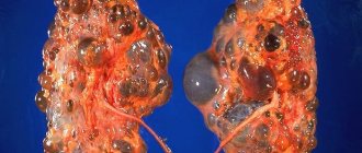
- Polycystic disease - many cysts of various shapes and sizes, which often affect the parenchyma of the right and left kidneys, are the result of congenital abnormalities of the urinary system.
The cyst can be located under the capsule of the organ (subcapsular), in the thickness of its tissues (intraparenchymal), in the area of the hilum or renal pelvis.
Methods for diagnosing parenchymal cysts
Typically, the study begins with x-rays and screening. CT (computed tomography) and puncture cystography are also performed. Ultrasound is used to make a diagnosis and prescribe effective treatment for the patient. In this case, be sure to indicate the location of the parenchymal cyst: left or right kidney. The stage of the pathological process is determined using ultrasound and MRI to identify pathologies such as a malignant parenchymal cyst. If diffuse changes were detected, this indicates the presence of various concomitant pathologies.
Diagnostics
Using hardware diagnostics, a specialist can obtain all the necessary information about the cyst: location, type and size. When a patient complains of pain in the kidneys, the diagnosis will be “parenchymal cyst”. To make a diagnosis, the following methods are used:
- analysis . Thanks to it, it is possible to establish inflammation. Biochemical analysis helps to detect symptoms of a pathological process even before adverse consequences occur.
- Ultrasound. Ultrasound examination makes it possible to identify the condition of any parenchymal organ, its size, and see the general picture of the formation.
- CT. Computed tomography helps visualize the organ, including pathological neoplasms inside it.
- MRI. For diagnostic purposes, magnetic resonance imaging is performed on soft tissues. Based on the emitted waves, it is possible to accurately determine the location and specificity of the cystic formation.
- Urography. CV is administered intravenously, which after a certain period of time appears in the circulatory system of the organ being diagnosed. In this way, it is possible to identify the patency of hollow organs and learn about the extent of their damage.
A comprehensive examination allows you to choose the most effective treatment.
Treatment methods
Timely treatment and removal of parenchyma cysts leads to rapid recovery and preservation of the organ due to the restorative ability of the parenchyma. If the size of the cyst is no more than 5 cm and it is benign, no intervention is performed. To regularly monitor the patient's condition, periodic examination is indicated. Today, surgery is the most optimal solution for treating parenchymal cysts of both the right and left kidneys. It is best to perform surgery on patients at a young age.
Ultrasound-guided puncture is the most popular treatment method. In this case, the contents of the formation are removed using a needle, into which a special substance is introduced to glue the surfaces. This is an invasive technique that is accompanied by local anesthesia.
Laparoscopic surgery is a non-invasive innovative method that allows you to completely remove a parenchymal cyst. The surgical technique consists of introducing a special substance to expand the operated field, after which a laparoscope is inserted into it. Laparoscopic resection can be performed, that is, removal of the parenchymal cyst along with part of the kidney tissue. After the operation, the patient is prescribed antibiotics and painkillers. If necessary, treatment with anti-inflammatory drugs may be prescribed. Sutures are usually removed one week after surgery. To prevent complications, the patient is advised to undergo breathing exercises.
In very severe cases, when tissue necrosis develops, kidney removal or nephrectomy is performed.
Treatment
The tactics for treating a simple cyst depend on several factors: size, location, presence or absence of clinical manifestations, presence or absence of complications.
If the size is small and there are no obvious signs, dynamic ultrasound observation is demonstrated.
Indications for invasive treatment methods are the enormous size of the cyst and/or the presence of signs associated with renal tissue atrophy or complications.
Invasive treatment methods:
Percutaneous puncture followed by the introduction of a sclerosing (gluing) solution into its cavity. Laparoscopic excision. Timely opening and excision (for huge and multi-chamber cysts, for suppuration, and for suspected malignant degeneration).
Nutritional Features
Patients with parenchymal cysts need proper nutrition. Diet treatment is based on the following principles:
- Exclusion from the diet of salt and salt-containing products.
- Limiting the amount of fluid you drink, especially with high blood pressure and cardiovascular pathologies.
- Exclusion of spicy foods, spices and herbs, fried foods and alcohol, especially beer.
- You should forget about foods such as chocolate, coffee, and seafood during the treatment period.
- Minimum protein intake. This reduces the elimination of toxic substances.
Compliance with the principles of dietary nutrition for kidney disease is an effective treatment. Of course, it is impossible to cure a parenchymal cyst with diet alone, but if all medical recommendations are followed, recovery will not be long in coming.
The paired bean-shaped organ that produces urine is a protective filter for the body suffering from toxins and processed compounds. If the kidneys malfunction, toxic substances negatively affect the body. The human body is indivisible: if the activity of one anatomical part of the body is unstable, another organ loses its properties. A dangerous pathology is the kidney parenchyma.
What is a benign formation?
Parenchyma is formed from:
- Cortical cells
- Brain substance
Epithelial cells of a healthy person are 15-24 millimeters. Past diseases of the kidneys, ureters, bladder, and urethra negatively affect thinning. The renal epithelium is characterized by acute and chronic diseases.
A kidney parenchyma cyst is a thin-walled round, oval formation filled with liquid hemorrhagic, serous exudate with impurities of pus and blood. The parameters of the disease are 3-10 centimeters. The tumor appears on the left and right sides and is inherited. Several cysts are simultaneously found on the left and right kidneys.
Neoplasm is not the least important among urological problems. 30% of men and women over 50 years old equally suffer from the disease. The male sex is more susceptible to pathology. Having noted the anatomical features of the structure of the body, a cyst of the parenchyma of the left kidney is observed less frequently than the parenchymal condition of the right-sided bean-shaped organ.
Small cysts are not dangerous; the functioning of the organ is not disrupted. Benign tumors rarely turn into cancer. Timely diagnosis and therapeutic observations are necessary in order to eliminate the serious risk of deterioration.
For some time, the pathology does not bother the person. As the disease increases, it puts pressure on the liver, intestines, stomach, pancreas, and small intestine. Feeling discomfort, the patient finds it difficult to walk “small”. Education happens to fester, to burst. By squeezing the kidney vessels, arterial hypertension occurs. Progressive urolithiasis affects the appearance of neoplasms. An examination of the body will reveal “problems” with the organ.
Possible complications
Parenchymal cysts are sometimes associated with infection, which provokes the formation of pyelonephritis.
In such a situation, an increase in temperature is observed; urine tests can show an increased content of white blood cells.
With prolonged absence of therapy, the patient develops chronic renal failure.
Large dimensions of the growth can provoke kidney atrophy due to compression of important vessels.
Reasons for the appearance of the disease
It is difficult for doctors to identify the cause of the disease. The sequence of cystic manifestations of parenchyma:
- Free space appears in tissues
- The free zone fills with yellow liquid exudate
- Tissues produce insoluble fibrillar protein, epithelial tissue is rejected, and a capsule appears
Signs of a parenchymal cyst:
- acute, chronic infectious process in the urinary organ;
- nonspecific inflammatory process of damage to the renal tubular system;
- urolithiasis;
- increased blood pressure;
- BPH;
- hereditary genetic disorders;
- injury;
- urolithiasis disease
To determine the factor causing the disease, you need to go to a urologist. The doctor examines. Based on the study, the factor of kidney dysfunction will be announced.
The underlying disease does not show hints of a cyst. Hereditary pathology, diagnosed by chance during a medical examination, does not manifest itself at all. Acquired formations occur when:
- urolithiasis, pyelonephritis;
- physical damage, trauma, radiation
Particularly dangerous: internal bleeding, purulent exudate. A growing cyst close to the kidney has the highest probability of complications.
Causes
Depending on the cause of the occurrence, pathologies are divided into two large groups:
Congenital
Cystic formation develops from germinal tubules that have lost connection with the urinary tract. Such cysts are completely aseptic (the cavity is filled with serous contents) and extremely rarely cause clinical manifestations.
It can be detected in the prenatal state using ultrasound; it has no effect on the body of the mother or child. Surgery is rarely performed.
Purchased
- A consequence of inflammatory processes in the kidney (glomerulonephritis, pyelonephritis). As a rule, such neoplasms are a complication of the normal course of the disease and arise as a result of prolonged lack of treatment or treatment with incorrectly prescribed drugs (self-medication).
- Urolithiasis disease . It has two specific manifestations that lead to the formation of cysts: mechanical damage to the collecting system and obstruction of the lumen of the urinary tract. Both of these phenomena, with prolonged exposure, have a predisposing effect on the formation of cysts.
- Malignant formations of the retroperitoneum . In this case, there is an indirect effect on the functioning of the kidney (mechanical pressure from the outside as the tumor grows), which leads to disruption of the outflow of urine. As the disease progresses, the pyelocaliceal system expands and a prerequisite for the formation of cysts is formed.
- Kidney tuberculosis . This is always a secondary phenomenon that occurs against the background of the underlying disease (pulmonary tuberculosis). Mycobacteria can affect any part of the kidney (bark, parenchyma, pyelocaliceal system). At the site of implantation, multiple dense formations appear, which, as they progress, lead to compression of healthy areas of the organ. Neoplasms in this case arise due to the indirect influence of tuberculosis foci.
- Kidney infarction . A rare disease that is associated with a sharp disruption of blood flow in the renal artery or smaller vessels and the development of ischemia and necrosis in the kidney area. It is extremely rare that the affected area can be replaced by a cystic cavity some time after a heart attack.
- Traumatic injuries . In this case, the cystic formations will be serous-hemorrhagic in nature due to hemorrhage into the cavity. It is necessary to distinguish between post-traumatic neoplasms (acute pathology, there is no evidence of the presence of a cystic cavity in the anamnesis before the incident) and parenchymal cysts complicated by trauma (the presence of a confirmed cyst in the anamnesis before the injury).
- Overweight . Obesity of the III-IV degree leads to a pronounced load on the kidneys and serves as a prerequisite for the formation of cysts.
In the pathogenesis, regardless of the cause, there are two main points: occlusion of the renal tubules and subsequent ischemic changes.
Symptoms
Signs of the disease are not shown. The person does not feel anything, does not assume that he is sick. The pathology is asymptomatic for a long time. Ultrasound examination performed for another reason helps to identify the disease.
Inconveniences appear with rapid growth and pressure on nearby components of the body. Becoming more than 1 cm, the neoplasm pinches the thin connective membrane. Kidney activity is impaired.
Symptoms of parenchymal condition:
- lumbar pain that becomes stronger with physical activity;
- diastolic pressure numbers increase;
- bloody discharge in the urine;
- development of a pathological condition due to changes in blood volumes;
- difficulty urinating, frequent trips to the toilet;
- discomfort above the groin area;
- disorders of the urinary system;
- swelling of the face, legs;
- weakness, fatigue, insomnia;
- desire to drink
Weak immunity affects urinary function. With a weak immune system, pyelonephritis develops with fever, soreness, and loss of strength.
Symptoms
There are several options for the clinical course of parenchymal cysts (the division is conditional):
Throughout life, it does not manifest itself and does not cause disturbances in kidney function (general urine analysis and general blood test without changes). They are discovered accidentally by ultrasound. In this case, it is recommended to be examined once a year from the moment of detection (dispensary registration).
Type II (subtype B)
· constant dull pain in the lower back;
· feeling of fullness or heaviness in the lumbar region;
· slight increase in blood pressure;
· violation of the excretory function of the kidneys (frequent urge, incomplete emptying of the bladder);
· the occurrence of swelling of the limbs and face.
This conditional group includes complicated cystic formations (suppuration, pyelonephritis), so the clinic may include:
· intoxication syndrome (increase in temperature to subfebrile levels, weakness);
Dyspeptic disorders (nausea, vomiting);
· the pain becomes sharp;
· blood pressure surges over a wide range.
In case of peritonitis or retroperitoneal hematomas, the phenomenon of acute abdomen occurs. Most often, the only treatment option is surgery.
Type III (subtype B)
All congenital pathologies belong to this type. In this case, the cyst is an independent disease (incidence 1:10,000). In children, in some cases it is possible to palpate an enlarged kidney. Often, manifestations begin with changes in laboratory parameters (general urine analysis). As the tumor grows, pain occurs. Observation and planned surgical intervention are indicated if necessary.
In accordance with the clinical picture, laboratory and instrumental diagnostics, the doctor (urologist) determines the tactics for further management of the patient.
Complications, consequences
A nephrologist surgeon diagnoses “parenchyma cyst of the right, left kidney.” After registration, annual examinations are necessary to prevent the danger of starting treatment on time.
If a man or woman does not begin effective treatment, the formation festers, nearby organs become infected, and deterioration occurs:
- necrosis of the renal parenchyma;
- disturbance of urine outflow;
- acute, chronic renal failure;
- transformation of a cyst into oncology;
- infection
Forecast
If you ignore the manifestations of pathology, parenchymal kidney cysts can cause unpleasant consequences:
- infection of the tumor;
- necrosis of the kidney parenchyma (tissue);
- suppuration;
- rupture of the membrane of the cystic formation;
- development of acute renal failure.
Contrary to popular belief, experts believe that a simple parenchymal cyst does not degenerate into a malignant tumor. With timely treatment, diet and doctor's prescriptions, the prognosis is very favorable.
Renal parenchymal cysts appear at birth or during life. To prevent pathology, it is necessary to promptly eliminate kidney problems, follow the principles of proper nutrition, and give up bad habits.
Diagnosis
The examination is carried out, starting with an x-ray with a contrast agent, screening, and using computed tomography. Ultrasound diagnostics is used to make a diagnosis and prescribe effective treatment. The localization of the formation plays a role: on the right, on the left there is a parenchymal cyst. The degree of the problem—malignant formation—is determined by ultrasound and magnetic resonance imaging. When tissue structure changes, concomitant diseases are looked for.
- History - complaints, chronic diseases of the patient, taking medications
- Feeling the patient's body, tapping the kidneys - noticeable enlargements, dislocation of the bean-shaped organs,
Research in the laboratory:
- OAM - protein, leukocytes, bacteria appear
- OAC - propulsion of erythrocyte sedimentation rate, changes in the cellular composition of the blood
- Biochemistry - the main indicators of kidney function increase - creatinine, urea
When diagnosing a parenchymal cyst, the doctor prescribes a special diet that helps reduce kidney loads. Diet principles:
- salt is reduced to 2.5 grams of daily dose;
- fatty, fried, protein foods are excluded - toxic substances are eliminated faster;
- Smoked, spicy, spicy foods are dangerous;
- You cannot drink alcohol, smoke, drink strong tea, coffee, eat chocolate, or eat seafood;
- body fluid control
Proper balanced nutrition - plant foods, natural fresh products - is effective against illness. The disease is not treated solely by diet, but affects the course of the process.
Before surgery, the urologist uses drug anti-inflammatory, antibacterial, and uroseptic therapy. Medication treatment is also used if the size of the benign cyst is less than 5 centimeters. Waiting and prevention influence the course of further surgery. The presence of arterial hypertension and pain are relieved with the necessary medications that eliminate the symptoms.
A growing cyst requires surgical help - the most effective way to eliminate the disease. Individual neoplasms are punctured, extracting the contents. Puncture is a safe method with minimal risks of complications. Under ultrasound control, the skin and soft tissues are punctured. A needle is inserted into the organ and pierces the capsule. After removing the liquid exudate, the wound is filled with a medicine that kills “harmful” cells.
Cysts with complications - abscess, pus, difficult to reach - are operated on under general anesthesia. The rehabilitation period lasts up to one year. The patient is placed on his side, an incision is made, opening the abdominal cavity. The kidney is removed by hand and the cyst is removed. Next, the nephrologist regularly monitors the patient, paying attention to chronic diseases of the bean-shaped organs.
It is important to treat diseases associated with urology in a timely manner. Following the instructions for chronic kidney disease leads to remission. Bad habits and poor nutrition affect the urinary system. Following the doctor's recommendations will help you recover.
The human body is not a set of separate organs, but a single, coherently working system. If one part of it fails, this necessarily affects the full functioning of all systems. This is especially true for the excretory system.
If kidney function is impaired, the entire body suffers, since processed products can poison all tissues and organs. However, there are diseases that can “lurk” in the kidneys for years and are detected by chance, during a routine examination or during examination of other organs. Such diseases include cysts.
Parenchymal cyst of the kidney: left and right, what it is, causes and treatment
Parenchymal cyst of the left and right kidney is a benign neoplasm that occurs in 30% of the population.
The disease, which causes diffuse changes in the parenchyma of the paired bean-shaped organ, affects predominantly the male population of pre-retirement and retirement age, but cases of the disease have also been recorded in women.
The tumor is localized in one kidney and in rare cases is diagnosed simultaneously on the right and left sides. In the article we will analyze in more detail the classification of cysts in the kidney parenchyma, factors contributing to their occurrence, symptoms, diagnosis and treatment and preventive measures.
Location of the renal parenchyma cyst.
Description and types
Often patients, upon hearing the diagnosis, wonder what it is - a parenchymal cyst of the left or right kidney. Inside the thin-walled hollow formation is a capsule filled with liquid or semi-liquid exudate. Usually the exudative fluid is yellowish in color and resembles plasma, but sometimes the sac is filled with serous fluid, blood or purulent impurities.
Often therapy does not lead to cystic resorption, but only has a symptomatic effect, which is also important: it reduces pain, lowers blood pressure, and prevents the occurrence of urolithiasis.
Dietary requirements
Parenchymal cyst requires diet. Dietary measures help reduce the load on the kidneys. The diet is based on foods of plant origin.
Main dietary directions:
- Reduce salt intake to 0.5 teaspoon per day.
- Reducing daily fluid intake - no more than 1.5 liters.
- A minimum of protein products - you need to remove meat, fish, seafood, dairy, and beans from your diet.
- Do not irritate the gastrointestinal tract with smoked, fried, spicy foods.
Puncturing
Puncture is a minimally invasive method of getting rid of a cyst. Under ultrasound control, the skin and soft tissues are pierced, then a needle is inserted into the tumor and the liquid is drawn out. Immediately after extracting the exudate, a special solution is introduced that kills the cells that secreted the liquid contents. Relapses after puncture are unlikely.
Surgical intervention
For small cystic formations, laparoscopy is used. Small incisions are made on the skin, a laparoscope is inserted, and the surgeon’s actions are reflected on the monitor screen. The doctor cuts the kidney, draws out the liquid contents, and then sutures the cystic walls to the parenchyma. After surgery, only a small scar remains on the patient's skin.
Surgery to remove a renal parenchyma cyst.
If the tumor is actively progressing, complications may develop, or negative consequences have already appeared, doctors resort to a radical method - abdominal surgery, characterized by complex postoperative rehabilitation. The lateral wall of the patient's abdominal cavity is incised, the abdominal cavity is opened, and the tumor is excised.
When it reaches a large size and threatens the life and health of the patient, the cyst is removed along with the kidney.
Preventive actions
Unlike congenital parenchymal anomalies, acquired ones are easy to prevent. For the purpose of prevention, it is necessary to undergo timely examinations and, in case of chronic diseases, to follow all medical prescriptions.
Parenchymal tumor is an insidious disease that is difficult to diagnose at an early stage. If you discover an illness, you should not panic - trust the specialists, because it is easier to cure the pathology at an early stage than to develop life-threatening complications.
Source: https://kistateka.ru/pochki/parenhimatoznaya
| The term solitary indicates that the cystic formation has a solitary temperament. Uncomplicated (solitary) renal cyst in most cases it has a round or oval shape, often has a superficial temperament and can be localized in different parts of the organ. A simple kidney cyst may be congenital (developmental anomaly) or acquired. The mechanism of development in both cases is the same: the basis is blockage of the tubule with subsequent retention (stretching) of the latter, due to the continued formation of primary urine in the glomerulus. In the case of a congenital cyst, there is a disruption of the connection between the developing tubules during intrauterine development, while a purchased simple kidney cyst is formed due to a violation of the outflow of urine through the tubule as a result of various pathological processes (pyelonephritis. kidney tuberculosis. kidney tumors...). Classification of simple kidney cysts: Intraparenchymal - located in the thickness of the renal tissue. Cortical - located in the cortical layer of the kidney. Peripelvic (parapelvic or sinus) - located near the pelvis, but does not communicate with the latter. Multilocular – multi-chamber. Rarely seen. Subcapsular - located under the kidney capsule. SymptomsThe clinical picture of the disease strongly depends on the degree of disturbance of the trophism of the renal tissue near the cyst, which in turn depends on the size and location of the formation. In many cases, a solitary kidney cyst does not manifest itself in any way and is detected by chance during ultrasound examination (ultrasound). If the location of the formation and its size lead to disruption of the trophism of neighboring tissues and their atrophy, the following complaints can develop:
The following methods are used for diagnostic purposes: Ultrasound diagnostics (ultrasound). X-ray study. To exclude the presence of a tumor in the cyst, the following can be used: computed tomography and percutaneous cystography (x-ray examination after the introduction of a radiopaque substance into the cavity of the cystic formation). TreatmentThe tactics for treating a simple cyst depend on several factors: size, location, presence or absence of clinical manifestations, presence or absence of complications. If the size is small and there are no obvious signs, dynamic ultrasound observation is demonstrated. Indications for invasive treatment methods are the enormous size of the cyst and/or the presence of signs associated with renal tissue atrophy or complications.
Percutaneous puncture followed by the introduction of a sclerosing (gluing) solution into its cavity. Laparoscopic excision. Timely opening and excision (for huge and multi-chamber cysts, for suppuration, and for suspected malignant degeneration). You'll like this:Renal cysts are cavity tumors filled with fluid. Less common are dermoid cysts filled with other tissues. Kidney cysts often form in the upper layer of the organ. This is usually a benign neoplasm, but there is a risk of its malignant degeneration. This disease affects men and women equally and is more common after 40-50 years of age. There are several types of cystic renal formations. The course of the disease at the initial stage is hidden. Since there is a risk of malignant degeneration of the cyst, its growth and development must be constantly monitored, which is not always possible due to the absence of symptoms. Features of the diseaseCystic formations in the kidneys are cavity capsules with serous contents Cystic formations in the kidneys are cavitary capsules with serous contents. The form of this neoplasm can be simple or complex (multi-chamber). Small cystic capsules are more common, but sometimes they can reach 100 mm in diameter. Renal cysts account for about 70% of tumors of this organ. Despite the frequency of diagnosis of this disease, the mechanism and reasons for its development are still not fully understood. The main reason for the formation of such capsules is considered to be pathologies of the kidney tubules, through which primary urine is excreted from the organ. If urine begins to stagnate in such a tubule, this leads to protrusion of the wall. Gradually it begins to transform into a cystic cavity.
Inside the cystic cavity there is often serous fluid, but sometimes it may contain other tissue, an admixture of blood, pus and kidney fluid. Some cysts develop simultaneously with the tumor process in the walls of the organ. By origin, renal cysts are divided into acquired and congenital. In addition, polycystic kidney disease also occurs, when not a single cavity is formed on the parenchyma of the organ, but many small cystic formations. ClassificationThe following types of kidney cysts are distinguished: The following types of kidney cysts are distinguished: A solitary cyst is an oval or round benign neoplasm that is not connected to the ducts and does not have constrictions. The cavity is filled with serous fluid, sometimes mixed with pus and blood. More often, this form of the disease affects one kidney after its injury. In 50% of cases, this type of cyst is found on one kidney in several places. A characteristic feature of this form is that damage to the left kidney is more common and in most cases is diagnosed in men. Multicystic disease is a congenital lesion of one organ, which is diagnosed very rarely. During the development of this disease, the kidney transforms into one continuous cystic formation, losing its functions. But if even the smallest area of kidney tissue remains healthy, it produces urine, which accumulates in the cavities of the cyst. Polycystic disease. This form of the disease affects two kidneys at once.
This is a congenital disease that is characterized by dilation of the kidney tubules and the formation of multiple small cystic cavities. Dermoid cyst. These are cavity formations, inside which it is not liquid that accumulates, but other tissues - hair, epidermis, teeth, adipose tissue, bone inclusions, etc. There are also congenital cysts, the formation of which is caused by concomitant hereditary pathologies (Zellweger syndrome, tuberculous sclerosis, Meckel syndrome, etc.). The kidney cysts listed above are most often congenital. As for acquired formations, the provoking factors are often various infections, inflammatory processes, injuries and other renal pathologies. In this case, organ damage can be one- or two-sided. Depending on the location, a renal cyst can be classified as follows:
If the cyst grows beyond 5 cm, it can only be treated surgically. The cortical (sinus) renal formation is located in the sinus of the organ. The capsule cavity in no way communicates with the pelvis and urinary tract. To treat this type of disease, puncture is usually used. The parapelvic cyst is localized in the renal pelvis and sinuses. This type of cystic formation most often affects the right organ and is a rather rare disease that is diagnosed after 50 years. A solitary neoplasm or simple cyst is often represented by a single cavity, which is localized on one organ (usually the left). The disease is asymptomatic for a long time. With a significant increase in the cyst, complications such as renal failure, hydronephrosis and secondary infection may develop. A complex tumor - a capsule of connective tissue is localized in the upper layer of the kidney and consists of several compartments (multi-chamber cyst). The cavity can be filled not only with liquid, but also with other tissues. Puncture treatment is not suitable in this case. Surgery is required to remove the cyst. Depending on the content, all cystic renal formations can be divided into the following types: Serous serous fluid is a clear yellowish liquid medium. It penetrates into the cavity through the walls of the capillaries; hemorrhagic contents are liquid mixed with blood. Typically, such cyst contents appear due to injury or a heart attack; purulent contents are an admixture of pus in the liquid, which is the result of an infection; calcifications are stones in a cyst. CategoriesDepending on the characteristics of cystic formations, several categories are distinguished Depending on the characteristics of cystic formations, the following categories are distinguished: The first category includes the most common form of benign cystic neoplasms, which can be easily diagnosed by ultrasound. The second category is benign kidney formations that have septations or characteristic changes. These include overdense, infected and calcified cysts. They usually reach 30 mm in size.
This type of cyst is immediately removed during surgery to eliminate the possibility of degeneration into a cancerous tumor. ComplicationsThe most dangerous complication that occurs against the background of a cystic formation is its rupture. Even minor exposure can cause it. As a result of rupture, the contents of the cyst spill into the abdominal cavity, which leads to peritonitis (inflammation). This condition requires immediate surgical intervention. In addition, cystic neoplasms can be complicated by suppuration. In this case, the patient feels general weakness, his temperature rises, and his lower back hurts. Suppuration occurs due to infection, therefore it is treated with antibiotics. Additionally, a puncture or surgery is required. As the connective tissue capsule increases in size, it begins to put pressure on surrounding tissues, organs and important renal vessels. In the latter case, the functioning of the organ is disrupted, uremia and renal failure develop. This complication occurs when there is a bilateral pathological process in the organs. In case of pathology of one kidney, the functions of the affected organ are taken over by the healthy kidney.
|
Characteristics of education
Renal parenchymal cyst is a benign neoplasm
A cyst is a cavity filled with fluid, which can be single or multiple. Cysts form in various organs, including the kidneys. The most common is a parenchymal cyst, that is, a benign neoplasm located in the parenchymal tissues of the kidneys. In most cases, this formation occurs in the right kidney. People over the age of 50 are most susceptible to such damage, especially those who have numerous acute and chronic diseases, especially those related to kidney function, as well as various infections.
Parenchymal cyst of the right kidney is extremely rare as a congenital disease. This is mainly due to various “failures” during intrauterine development of the fetus or a number of hereditary lesions. Also, an important reason for the appearance and development of kidney cysts is exposure to various toxic substances, including alcohol, tobacco, radiation, chemical compounds, drugs and other harmful drugs, including some medications.
If the presence of several neoplasms is diagnosed at once, this condition is called polycystic disease.
Cysts are mostly harmless if they are located in places that do not interfere with life, do not increase in size and do not fester. The assumption that a cyst can degenerate into a malignant tumor is incorrect and is not confirmed by modern scientists. However, polycystic disease, inflammation and suppuration of the cyst, its twisting, necrosis, proliferation and compression of adjacent tissues and blood vessels, nerves can pose a serious danger and even threaten the patient’s life.
Causes
Cyst formation can be caused by various infections and diseases
In cases where a parenchymal cyst of the right kidney is detected, it can be argued that the cause of its occurrence was one of the following diseases or conditions:
- Hereditary disorders of the structure and functioning of the kidneys associated with genetics or external influences.
- Acute and especially chronic infection affecting the kidneys.
- Inflammatory kidney diseases.
- The presence of calculi (stones) in the kidneys of various origins.
- Chronic form of arterial hypertension.
- Prostate adenoma (mainly in older men).
In some cases, the cause of a cyst may be several diseases at once. The doctor will try to accurately determine the cause of the disease by conducting various studies and tests.
Symptoms
Lower back pain, frequent urination and changes in the smell of urine are possible signs of pathology
While the neoplasm is small in size, it does not manifest its presence in the body in any way. Such a cyst is sometimes discovered by chance, during examination for other diseases. Symptoms appear when the parenchymal cyst of the right kidney begins to grow and puts pressure on neighboring tissues and organs.
The most common and obvious symptoms of a kidney cyst are the following:
- Constant and frequent urge to urinate.
- Prolonged aching and nagging pain in the lower back.
- The presence of edema, especially of the lower extremities or face, with a normal volume of fluid intake.
- Change in color and odor of urine (indicates infection).
- Traces of blood in the urine.
- Weakness and lethargy, constant malaise.
- Sleep disturbance.
- Increased thirst.
Such symptoms do not clearly indicate a kidney cyst; they are characteristic of many other diseases, and therefore are the basis for visiting a doctor and conducting a full examination. The specialist will prescribe a series of tests and hardware studies that can identify the true cause of the symptoms that appear.
Characteristic symptoms of renal parenchymal cyst: methods of diagnosis and prevention
A parenchymal cyst is a benign neoplasm that forms in the kidney and other organs.
Pathology can be congenital or acquired. The symptoms and treatment of the disease are closely related.
The development of renal parenchymal cyst is most often observed in men. People over 50 years of age are at risk.
Classification and varieties
Renal parenchymal cysts are classified according to several criteria. Depending on the developmental characteristics, the disease can be:
- Innate. The cause of the disease is the fusion of the renal tubules. A parenchymal cyst develops in the fetus if a woman smokes, drinks drugs or drinks alcohol during pregnancy. Against this background, disruptions in the intrauterine development of the child are observed.
- Acquired. The appearance of a cyst is observed after injury to the kidneys, as well as after diseases in which the tubules become clogged. The pathology may be a consequence of inflammation in the prostate gland, hypertension, or kidney stones.
Depending on the size of the formation, it can be small or large. Small neoplasms are characterized by the absence of manifestations or mild symptoms. They appear predominantly in the left organ. A small parenchymal cyst of the right kidney develops asymptomatically in most cases.
If the formation reaches a large size, it ruptures, which leads to internal hemorrhage. Parenchymal cyst in advanced cases leads to the development of hydronephrosis.
Against the background of a cyst, kidney function may be impaired. This is explained by the fact that the neoplasm compresses the tissues of the organ, which leads to impaired blood circulation and trophism. The healthy part of the kidney cannot withstand excessive stress. An infection may develop in the area of the parenchymal cyst.
A parenchymal cyst leads to serious complications, therefore, if the disease is suspected, it is recommended to diagnose and treat the neoplasm.
Diagnostics
A parenchymal cyst is a neoplasm that requires consultation with a doctor. He will listen to complaints and study the medical history. During the examination of the patient, the kidneys are palpated and percussed.
This allows you to assess the size and location of the organs of the urinary system. If a patient develops a cyst, this leads to an enlargement of the affected kidney.
After examining the patient, the doctor can make a preliminary diagnosis. To confirm this, instrumental and laboratory studies are recommended.
The patient is sent for a general urinalysis. The presence of a parenchymal cyst is indicated by protein in the urine. If an infectious process occurs, bacteria and an increased content of leukocytes are observed in the urine.
Diagnosis of pathology includes a general blood test to detect leukocytosis. A biochemical blood test allows you to determine the level of urea and creatinine, which are the main indicators of kidney function.
Patients are recommended to undergo an ultrasound examination of the kidneys, which will determine the location, parameters and structure of the tumor. Patients are prescribed computed tomography and magnetic resonance imaging. With the help of CT and MRI, the information obtained from ultrasound examination is clarified.
If a pathological process is suspected, excretory urography is prescribed, which is an X-ray method for examining the urinary system. Before the examination, a contrast agent must be injected into the human body.
Treatment of renal parenchymal cyst
There are various methods for treating renal parenchymal cysts, the choice of which depends on the characteristics of the pathological process.
First of all, the doctor prescribes a diet that reduces the load on the kidneys. The diet includes limiting salt intake. Her patient should eat no more than 2.5 grams per day.
The patient is not recommended to eat fatty, fried and protein-rich foods.
Dietary nutrition requires the exclusion of sour, salty foods that have an irritating effect. Smoked meats, spicy and hot dishes are not recommended. Patients are strictly prohibited from drinking alcoholic beverages, strong tea and coffee. The patient must comply with the drinking regime.
The basis of dietary nutrition is plant foods. Vegetables and fruits that cause fermentation in the intestines are excluded from the diet. Patients are recommended to consume fermented milk products.
If the parenchymal cyst is small, a wait-and-see approach is used and medications are used to relieve symptoms.
Drug treatment
If signs of the disease appear, the following is recommended:
- antibiotics, the action of which is aimed at eliminating the inflammatory process;
- antispasmodics, which help relax the smooth muscles of the ureters and restore their patency;
- if the patient has a kidney parenchyma cyst, it is recommended to restore the water-salt balance in the body with the help of medications;
- antihypertensive drugs. Their action is aimed at lowering blood pressure. With the help of medications, the elasticity of blood vessels is restored.
When using drug therapy, do not self-medicate, as this may worsen the condition.
Surgery
If renal parenchymal cysts are large in size, then treatment is performed surgically. Cyst removal is carried out using three methods - abdominal surgery, puncture and laparoscopy.
Abdominal surgery is performed when the tumor is suppurated. It is also recommended for capsule rupture. If the formation reaches a large size and threatens a person’s life, it is removed along with the kidney.
Laparoscopic surgery is performed through three small incisions. Control over the operation is carried out using a miniature camera, which is inserted through a puncture. The use of this type of surgical intervention is recommended for simple single-chamber cysts.
To ensure optimal visibility during surgery, a special gas is injected into the abdominal cavity. This causes the abdomen to bloat. The surgeon opens the organ and removes fluid from the cyst. At the next stage, the walls of the capsule are sutured to the parenchyma. A scar will form in this area over time.
The least traumatic surgical method for removing a cyst is puncture, which causes complications in extremely rare cases. Under the control of an ultrasound machine, a puncture is made above the location of the tumor. A special needle is inserted into the kidney. It is used to puncture the cystic formation and remove the contents.
If you suspect the presence of a cystic formation, it is recommended to immediately begin treatment, as it can cause a variety of complications. If the tumor reaches a large size, it becomes inflamed, suppurates or ruptures.
A frequent complication of the pathology is necrosis of the kidney tissue, which makes it impossible for the organ to perform its functions. Against the background of a parenchymal cyst, patients develop renal failure. If this complication is not treated, it will cause death.
Prevention
To avoid the development of kidney parenchyma cysts, it is recommended to carry out timely prevention, which consists of following certain rules. It requires the patient to exclude factors that provoke the disease.
In order not to provoke the development of a cyst in the unborn child, a woman is recommended to stop smoking during pregnancy. She is strictly prohibited from consuming alcoholic beverages and drugs.
Prevention of acquired cysts requires regular examinations by specialists. If kidney diseases are detected, it is recommended to treat them promptly and correctly.
The patient should adhere to the rules of a balanced diet. The consumption of spices, fatty, fried and protein foods is strictly prohibited. Pickled vegetables, strong tea and coffee are recommended to be consumed in minimal quantities.
A parenchymal cyst is a dangerous pathology that remains asymptomatic for a long time.
The use of traditional medicine for this disease is useless. Therefore, treatment is carried out medically or surgically. The choice of treatment method for the disease depends on the characteristics of the disease.
Source: https://propochki.info/bolezni-pochek/harakternye-simptomy-parenhimatoznoj-kisty-pochki-sposoby-diagnostiki-i-profilaktiki
Diagnosis and treatment
Kidney ultrasound is an effective diagnostic method
To identify a cyst, the doctor prescribes blood and urine tests, as well as ultrasound, computed tomography, MRI, radioisotope radiography, urography, and, if necessary, some specific examination techniques. A very large cyst or cluster of cysts (polycystic) larger than 6-8 cm can be felt manually through the abdominal wall.
Medicines that can directly combat the formation and development of cysts have not yet been invented. But it is quite possible to cope with inflammation and suppuration. For this, the doctor will specifically select special treatment methods and medications for this. Mostly antibiotics, anti-inflammatory and painkillers, and drugs to normalize salt balance will be used.
Modern examination methods will demonstrate the shape, size, location and condition of the cyst and will become the basis for further treatment.
In addition to specific treatment, weakened patients may be prescribed restorative and supportive agents, vitamin preparations and other medications at the discretion of the attending physician.
Traditional treatment
Traditional medicine tips
Impact on the cyst with a variety of traditional medicine can only be carried out with the permission of the attending physician and exclusively as an auxiliary effect.
A parenchymal cyst of the right kidney can remain in the body for years without affecting its normal functioning. But if it begins to grow, becomes inflamed, becomes deformed, or negatively affects tissues and organs, adequate measures must be taken. Incorrect or excessive use of medicinal herbs can cause increased growth of the formation, leading to inflammation or other health problems.
- In folk medicine, it is recommended to take burdock juice, which should continue for two weeks. Then a mandatory break of 7 days is required, followed by resumption of use. Drink a tablespoon of juice before meals, unless the doctor recommends otherwise.
- Also considered a good remedy for kidney cysts is an infusion of burdock leaves, as well as medicinal plants such as elecampane, golden yam, knotweed and aspen bark.
Herbal treatment is usually accompanied by a special diet that excludes substances, foods and dishes that are considered to contribute to the occurrence and development of cysts in various organs, including the kidneys.
In very rare cases, the cyst disappears on its own.
This mainly happens when you change your diet, change your lifestyle, stop using toxic and harmful foods and substances, as well as certain medications. Sometimes the cyst disappears when the underlying disease is treated, for example, when kidney stones are removed or high blood pressure is treated. But, since this phenomenon is extremely rare, you should not fully rely on it, and follow the instructions and prescriptions of your doctor.
Proper nutrition for kidney disease
All kidney diseases require a special diet. If a parenchymal cyst of the right kidney is diagnosed, a special diet may be recommended to help reduce its size or try to make the tumor disappear.
In any case, special dietary nutrition helps reduce pain and reduce the risk of inflammation and cyst growth.
The first thing to do is:
- Significantly reduce the use of cooking water. This applies not only to home cooking, but also to purchased products. Canned food, some cheeses, and sausages contain a lot of salt.
- All hot, spicy, sour and smoked dishes have an irritating effect, so they are also excluded from the menu.
- Fried and fatty foods, as well as an abundance of protein foods, are prohibited.
- Alcohol, especially beer, as well as cigarettes, coffee, and chocolate turn into poison for the kidneys.
- The patient must strictly control the volume of fluid absorbed. Too much water increases blood pressure, which can negatively affect kidney function and cause cyst growth.
- The food should be dominated by vegetables, fruits, cereals with a minimum amount of fats, proteins and sugars.
- It is worth giving up store-bought products, ready-made sauces, various instant products, thick decoctions, broths and soups, sorrel, spinach and other products containing oxalic acid (it is very irritating to the kidneys and can cause the formation of sand and stones).
- Fruits and vegetables that enhance fermentation are used to a limited extent.
- Fried, smoking, and salting should be completely excluded from food processing methods. Preference is given to boiled and baked food, steamed or in the oven.
You need to be especially careful with your diet if you have other diseases besides the cyst. These can be diseases not only of the kidneys, but also any acute and chronic processes, especially those related to metabolic processes in the body.
Renal parenchymal cyst
A parenchymal kidney cyst is a benign urological pathology characterized by the formation of a cavity in the tissues of a paired organ, which is covered with a biological capsule and contains a light-colored liquid. The cystic formation is round or spherical in shape and usually appears on only one of the kidneys. The disease is equally common among both sexes.
Parenchymal cysts of the left kidney, as well as the right, for the most part do not pose a serious danger if they are small in volume, a single formation and there are no disturbances in the functioning of the urinary system.
Despite the fact that experts deny the likelihood of malignancy (malignancy) of these neoplasms, their timely detection and subsequent treatment are extremely important.
This is due to the high risk of various complications.
Classification of renal parenchymal cyst
Kidney tissue is represented by a complex structure. Under the influence of certain factors, it can grow, fence off from the renal tubules and transform into a cyst. Parenchymal formations are divided into:
- Intraparenchymal renal cyst - localized in the thickness of the renal structure.
- Peripelvic (sinus) - located in close proximity to the pelvis, but does not communicate with it.
- Cortical - located in the parenchyma layer of the same name.
- Subcapsular - localized under the renal capsule.
- Multilocular – multi-chambered and rare.
Depending on the type of formation in the kidney parenchyma, a cyst can be:
- Congenital. It occurs against the background of mutations as a result of disturbances in intrauterine development during the formation of internal organs. The tendency to form cystic cavities is a genetic predisposition and passes from parents to children.
- Acquired. It develops as a consequence of diseases that have a damaging effect on the renal canals, which inevitably leads to their blockage. Hypertension, urolithiasis, and prostate adenoma act as catalyst pathologies. Formations of this type can develop throughout a person’s life, but most often appear in elderly patients.
According to the number of parenchymal cysts on the kidney, there are:
- Single – represented by a single capsule, most often affecting the kidney on the left. The development of pathology occurs without obvious symptoms, however, having reached a certain diameter, it provokes the occurrence of a number of disorders.
- Multiple – when it appears, a person experiences constant aching pain.
Parenchymal cysts of both kidneys are rarely encountered in urological practice. They belong to a number of superabnormal pathologies.
Causes of formation of renal parenchymal cyst
The key reasons for the formation of intraparenchymal renal cysts are:
- Failure of intrauterine development (with congenital pathology);
- Diseases of the urinary system, including infections and urinary tract infections;
- Kidney injuries;
- Tuberculosis;
- History of surgical intervention on a paired organ.
The mechanism of formation of this cyst is due to the expansion of nephrons (structural renal elements). The dimensions of the cavity can vary from 2 mm to 9 - 12 cm. It is noteworthy that congenital cysts have the ability to resolve on their own.
A parenchymal cyst of the right or left kidney is formed as a result of changes in the tubular apparatus, which leads to disruption of the outflow of urine. With such a development of events, the urine stagnates, provoking protrusion of the parenchyma wall and then a cystic capsule is formed in this area.
Symptoms of parenchymal cyst on the kidneys
As often happens, a person may not realize that a cystic neoplasm is forming in his body until the need arises to undergo an ultrasound examination of the genitourinary system.
If the capsule has a diameter of 1 - 1.5 cm, then specific symptoms in this case are practically absent or appear in a mild form. If the parenchymal formation is more than one and a half centimeters in size, then the patient may be concerned about the following negative manifestations:
- Frequent urge to urinate;
- Pulling and aching pain in the lumbar region;
- The appearance of swelling on the legs, arms and face in the morning after waking up;
- A sharp change in the color of urine;
- Restless sleep;
- An oppressive feeling of fatigue and malaise.
When such symptoms appear, it is important to understand that they may not always indicate the presence of a parenchymal cavity. Often, such ailment signals other urological problems. Therefore, it is extremely important to conduct regular examinations of the whole body.
Diagnosis of parenchymal cyst in the kidneys
Diagnostic measures aimed at examining the renal parenchyma consist of a set of mandatory studies, which include:
- A general clinical examination, which includes the collection of existing complaints and patient history, palpation and percussion of the paired organ, allowing one to assess the approximate volume and location of the capsule.
- Laboratory testing, consisting of a blood test (ROE, leukocyte formula), general urine analysis (protein, bacteria, leukocytes), biochemical blood test (urea and creatinine level).
Instrumental examinations are presented:
- Ultrasound examination of the kidneys, which allows one to determine the size, location and nature of the contents in the tumor capsule;
- Magnetic resonance and computed tomography, used to create a more complete picture and clarify the results obtained through ultrasound;
- Excretory urography is a technique for visualizing the kidneys using x-rays and introducing a contrast component.
In addition to the above methods, angio- and cystography of the kidneys, retrograde pyelography can be prescribed for diagnostic purposes.
Cyst removal
Surgical intervention is performed in the following cases:
- Lack of positive reaction when using conservative drug treatment.
- Growth of formation that threatens the normal functioning of an organ or the entire organism.
- Inflammatory process.
- The occurrence of suppuration of the contents of the cyst.
- Twisting of the cyst stalk with threatened or already developed necrosis.
- Rupture of the cyst capsule with spillage of contents.
- Cyst size exceeding 5 cm with a tendency to further growth.
There are several methods of surgical intervention. For small single cysts, a puncture (puncture) through the surrounding tissue can be performed without complications. In this case, the contents of the cyst are removed, and a special sclerosing substance is pumped into its place, eliminating the formation. The procedure is usually performed under ultrasound guidance.
Laparoscopic surgery is minimally invasive, as it is performed using special equipment through small punctures or incisions.
However, such intervention is only possible if the cyst is single, small, non-inflamed and not purulent. It will also not be possible to remove a cyst laparoscopically if it is in a hard-to-reach place or is combined with other kidney diseases, for example, stones.
More information about kidney cysts can be found in the video.
Full abdominal intervention is indicated in cases where all of the above methods are powerless, and the cyst is large, located in a difficult place to treat and/or is combined with other health problems, for example, adjacent to another tumor, which also requires immediate removal.
In this case, surgery is performed under general anesthesia followed by a postoperative recovery process. If it turns out that the kidney is very badly affected by cysts or there are other diseases besides the cyst, it may be necessary to completely remove the entire affected organ. To prevent this, it is necessary to undergo all examinations and undergo the necessary treatment in a timely manner in order to recover and maintain a normal, healthy and fully functioning organ.
Parenchyma is the main tissue of the kidneys. Together with the urinary system, it forms a capsule that performs the function of maintaining the body’s environment and removing harmful metabolic products. Depending on age, the parenchyma of a healthy kidney can be from 15 to 23 mm, in older people - from 10 to 11 mm. Thickness is affected not only by age, but also by health status. As a result of diseases of the urinary system, it becomes thinner. The kidney parenchyma has a high ability to recover.
Description and types
Kidney parenchyma is a tissue with a complex structure. When exposed to certain factors, the tissue particle grows, gradually separates from the tubules that purify the blood, and forms into a cystic formation. A cyst is a fluid inclusion surrounded by a capsule of connective tissue. If the cyst is the result of a kidney injury, its contents include blood and pus. Often a cyst of the left or right kidney is diagnosed separately. Involvement of both kidneys is rare.
Renal parenchyma cysts are:
- Congenital. Develops due to mutations that fuse the renal tubules. A possible cause is also considered to be the use of drugs or alcohol by a pregnant woman, or smoking, which negatively affects the development of the fetus. The influence of toxins and infections on the fetus during intrauterine development also contributes to the formation of parenchymal cysts. There are dermoid cysts containing teeth, hair, adipose tissue and epidermis.
- Acquired. It develops as a result of diseases that affect the tubules, leading to their blockage. Such diseases include: kidney stones;
- inflammation of the prostate gland;
- hypertension;
- chronic infectious kidney diseases;
- renal tuberculosis.
Depending on the number, parenchymal cysts are:
- Single. A single cystic formation, often affecting the left kidney. It develops asymptomatically, but when it reaches a significant size, it leads to serious disorders.
- Multiple. Even small, numerous cysts provoke pain.
The concept and consequences of this disease
As the main tissue of the kidney, the parenchyma is sensitive to acute and chronic diseases, as well as to various types of neoplasms. In our case - cysts. This is a formation ranging in size from 3 to 10 cm, filled with liquid. When fluid is retained in the nephron, a simple kidney cyst is formed. It is usually a solitary, thin-walled neoplasm that develops from parenchyma.
The cyst always forms on one side only. Often this disease can be a genetic predisposition. It is oval or round in shape, which includes: serous and hemorrhagic substance. Sometimes the serous fluid contains pus or blood. About half of the cases are characterized by the detection of several cysts on the kidney at once. Solitary cyst usually affects men.
This disease is serious, because it goes away with virtually no symptoms after a certain period. The parenchymal cyst of the right kidney is a hollow single neoplasm with thin walls. But when it grows, it begins to put pressure on neighboring organs. During this period, pain occurs when urinating.
It is important! The right kidney cyst may fester and burst. It happens that it compresses the vessels of the kidneys and because of this the pressure increases.
The diffuse nature of the parenchymal cyst of the right kidney can change for a reason. This is either the first stage of urolithiasis or a disorder. In any case, only a comprehensive study can fully determine the dysfunction of the organ.
Main symptoms
Pathology develops in a latent (hidden) form. Often, a cyst is discovered by chance during a routine examination or diagnosis of another urological disease. Rarely, a cyst manifests itself as pain that increases during physical activity. The pain is concentrated directly in the area where the tumor is located.
As the cystic formation increases, it compresses the kidney parenchyma, ureter and ureter. Because of this, pain and urination problems appear. If you have these symptoms, you should definitely consult a doctor and undergo an examination that will indicate the type, size and number of tumors. Only after this the doctor will prescribe the necessary treatment. Independent attempts to eliminate the cyst lead to serious complications, including its rupture.
Symptoms of the disease and its causes
This serious disease has many symptoms divided into types:
- Presence of blood in the urine (hematuria).
- High blood pressure.
- Pain in the lumbar region when palpated.
- Neoplasms (you can feel them with your fingers, but it’s better to go to a specialist).
As for the etiology of this disease, the causes of cyst formation are usually the following:
- genetic disposition;
- injuries;
- congenital anomaly;
- kidney disease.
Features of the disease
Cystic formations in the kidneys are cavity capsules with serous contents
Cystic formations in the kidneys are cavitary capsules with serous contents. The form of this neoplasm can be simple or complex (multi-chamber). Small cystic capsules are more common, but sometimes they can reach 100 mm in diameter.
Renal cysts account for about 70% of tumors of this organ. Despite the frequency of diagnosis of this disease, the mechanism and reasons for its development are still not fully understood. The main reason for the formation of such capsules is considered to be pathologies of the kidney tubules, through which primary urine is excreted from the organ. If urine begins to stagnate in such a tubule, this leads to protrusion of the wall. Gradually it begins to transform into a cystic cavity.
As for the reasons for stagnation of urine in the tubules, there can be many of them, because various organ pathologies and kidney dysfunction lead to impaired urine excretion. So, the causes of the disease can be ICD, kidney tuberculosis, oncology, pyelonephritis and even ordinary trauma.
Inside the cystic cavity there is often serous fluid, but sometimes it may contain other tissue, an admixture of blood, pus and kidney fluid. Some cysts develop simultaneously with the tumor process in the walls of the organ.
By origin, renal cysts are divided into acquired and congenital. In addition, polycystic kidney disease also occurs, when not a single cavity is formed on the parenchyma of the organ, but many small cystic formations.
How is this disease treated and diagnosed?
Each time, diagnosis must begin with screening, x-rays and other methods. This is followed by tomography, and later by percutaneous puncture of the cyst. To determine the condition of the parenchyma, CT and ultrasound examinations are used.
In order to accurately determine the parenchyma (its current state), as a rule, specialists perform ultrasound and the so-called CT scan. During diagnosis, it is necessary to indicate which kidney is diseased.
It is necessary to treat the disease of the parenchyma cyst of the right kidney not only with medication, but also with surgery. Most likely, the tendency to cysts is inherited. The condition of the parenchyma is determined by magnetic resonance imaging and ultrasound. If the examination reveals diffuse changes, this indicates diseases (urolithiasis or vascular diseases).
Timely removal of the cyst usually leads to a quick recovery. When the tumor is no more than 5 cm, it is considered benign. And if the patient has no complaints, then the operation is not performed, but therapeutic prophylaxis and systematic examination are carried out for some time.
Categories
Depending on the characteristics of cystic formations, several categories are distinguished
Depending on the characteristics of cystic formations, the following categories are distinguished:
The first category includes the most common form of benign cystic neoplasms, which can be easily diagnosed by ultrasound. The second category is benign kidney formations that have septations or characteristic changes. These include overdense, infected and calcified cysts. They usually reach 30 mm in size. The third category is cystic formations that are predisposed to malignancy - thickening of the septa and membranes, which can lead to malignant degeneration. Such pathologies are very difficult to detect during x-ray examination. This type of cyst is immediately removed during surgery to eliminate the possibility of degeneration into a cancerous tumor.