From the moment the fact of pregnancy is confirmed, every expectant mother is advised not to neglect routine ultrasound examination procedures, which make it possible to assess the progress of pregnancy and timely identify certain pathologies of intrauterine development in the fetus. One of these pathologies are single or multiple cysts, the location of which is the kidneys.
Despite the fact that cysts are benign in nature, their occurrence is always accompanied by disruption of the paired organ, with the subsequent development of its functional failure. Timely diagnosis of a kidney cyst in the fetus allows a number of measures to be taken that will reduce the rate of tumor development and also prevent complications.
Characteristics
Benign cystic formations can form on almost any organ, and the kidneys are no exception. The mechanism of cyst formation, regardless of their location, is standard. Under the influence of external or internal negative factors, a cavity gradually forms in the intercellular space, which is subsequently filled with interstitial fluid. As it develops, the cavity forms boundaries, which are represented by a connective tissue capsule consisting of collagen fibers.
Depending on the cause of the cyst, the internal contents of the tumor may be serous fluid, pus or blood.
The formation of kidneys in the fetus begins at 10-11 weeks of gestation. The formation of renal cysts at the stage of intrauterine development can occur during the formation of renal tubules or blood vessels. As a rule, until the renal tubules begin to function, cystic neoplasms are very difficult to identify, since their size remains constant.
There are also clinical situations in which polycystic kidney disease is diagnosed in the fetus, characterized by the formation of multiple cysts located over the entire surface of the paired organ. This serious condition can cause serious complications that require qualified assistance in a short period of time.
Symptoms
The process of development of a cystic neoplasm in childhood is accompanied by almost the same symptoms as in adults. To be more precise, the cyst is generally asymptomatic for a long time.
Regular increase in body temperature
Against the background of the formation of a foreign body in the child’s body, as well as inflammatory processes, the immune system responds in the form of an elevated temperature, which often reaches 38-39 degrees. In this case, fever and general weakness do not appear.
Hematuria
This is a pathological process, as a result of which blood or purulent impurities are observed in the urine. In this condition, the urine will be orange or red in color, which is the first sign of kidney disease.
Increased blood pressure
However, such symptoms make themselves felt only in situations where there is an intensive growth of a cystic formation, which negatively affects the functioning of nearby abdominal organs.
Anemia
Most often it manifests itself with a congenital type of pathology. It is characterized by a decrease in the concentration of red blood cells in the blood fluid, as a result of which dark circles form under the eyes and the skin turns pale.
Rarely, there are cases when the cyst is a pseudo-formation, that is, it is not attached to the kidney by channels, and it does not have an epithelial base. This type of cyst in most cases has a predisposition to spontaneous resorption during the first year of a child’s life.
Causes
Single renal cysts or cystic renal dysplasia are congenital conditions that were formed at the stage of the formation of a paired organ. With cystic kidney dysplasia, a characteristic change in the size and shape of the organ is observed, which is deformed due to the formation of multiple tumors.
In 70% of cases, the true cause of the development of this pathology is gene mutations that occurred at one of the stages of the child’s intrauterine development. Other, no less probable reasons for the development of this kidney pathology in the fetus include:
- Heredity factors. If a similar condition was previously diagnosed in close relatives, the child, even at the stage of intrauterine development, is at significant risk of developing single or multiple renal cysts.
- The impact of external adverse factors on the body of a pregnant woman and the developing fetus. A common cause of this pathology is the use of certain medications that have a teratogenic effect, unfavorable environmental conditions in the area of residence, exposure to ionizing radiation on the body of a pregnant woman, drinking alcohol and smoking during pregnancy, as well as frequent use of household chemicals, which includes its composition of acid and chlorine.
- Infectious and inflammatory diseases previously suffered by a pregnant woman, including sexually transmitted infections.
In addition, there are so-called idiopathic cysts, the true reason for the formation of which remains poorly understood.
Why and under what conditions does a cyst form in the fetal kidney?
A fetal kidney cyst is a benign tumor on the kidney or in the renal pelvis, which includes walls and a hollow space containing serous fluid. This tumor is located in the upper or cortical layer of the kidney. The shape of a cyst can be spherical, ellipsoidal, and cysts are classified into single-chamber, simple and complex.
Causes of cystic tumor formation
When a cyst forms in the fetus, the following etiological factors are mainly identified:
- Pathologies during intrauterine development due to exposure to environmental factors - irradiation of the mother's body, poor ecology, drug abuse at the stage of formation of the organs of the fetal urinary system, harmful addictions of the mother and exposure to household chemicals on the mother's body.
- Hereditary factors. When pathology is detected in relatives, the fetus develops the disease very often. Based on the manifestation of the disease in family members, fairly accurate predictions can be made.
- Pathologies of the body at the cellular level that occur during fertilization of the egg. This becomes possible due to non-compliance with contraceptive rules or during an unplanned pregnancy.
- Infectious pathologies or inflammations suffered by the mother during the formation of excretory organs in the fetus.
Sometimes the reasons for the formation of a cyst in the fetus cannot be determined.
How is kidney damage in the fetus diagnosed?
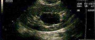
Modern medical equipment makes it possible to detect a cystic tumor as early as the fifteenth week of fetal formation. The main diagnostic method is ultrasound examination. The doctor determines the number of cysts, their location, size, and is also able to analyze the safety of kidney function.
Signs of disease development in the fetus also include:
- Compensatory kidney hypertrophy.
- Violation of the homogeneity of the contours of the organ.
- Hyperechoic tissue between cysts.
- Low water.
After the baby is born, he immediately undergoes an ultrasound to clarify the diagnosis or analyze kidney function. A month later, another ultrasound is performed.
Basic methods of prevention
- Pre-pregnancy planning. The entire female body must be prepared for bearing a child, and acute and chronic diseases must be treated.
- Timely registration of a pregnant woman - this should be done at 12 weeks of pregnancy.
It also requires constant visits to the gynecologist, and at least three ultrasound scans during the entire period of gestation, usually at the following stages:- 10 - 14 weeks;
20 - 24 weeks;
- 32 - 34 weeks.
- Refusal of a woman from harmful factors.
Such as drinking alcohol, smoking, taking medications, the influence of household chemicals, stressful situations. - Doctors recommend avoiding abortions and promptly treating inflammation of the genital organs.
- Also, during pregnancy, it is necessary to prevent any infectious damage to the body.
It is important! According to statistical data, at present, cystic tumors in the kidneys of the fetus are rare, and multiple cysts are generally considered isolated unique cases. If such a diagnosis has been made, it is important to follow the doctor’s recommendations.
Modern medical science makes it possible to effectively resist this pathology, and when diagnosing the disease in the early stages, the doctor can monitor the progress of the process and intervene when the need arises.
Prognosis and complications of renal cyst
When single small formations form in one of the kidneys, the prognosis remains favorable. Doctors monitor that the cyst does not grow and does not affect the flow of nutrients to the kidneys.
Children can be safely born with this disease and most often do not require additional treatment. All that is needed is regular diagnostics, prevention of child injuries and infections in his body.
In case of polycystic disease, experts advise the woman to terminate the pregnancy. This kidney damage can cause kidney failure, especially if both kidneys are diseased.
It becomes possible to maintain pregnancies provided that treatment is immediately organized immediately after birth. An additional negative side of polycystic disease is the tendency to affect several organs simultaneously.
When a diagnosis is made, cysts can also be diagnosed in the liver and gallbladder.
It is important! The most unfavorable prognosis is established when the fetal kidneys are affected by multicystic disease. The bilateral form is incompatible with life and the pregnancy is terminated even at long terms.
Complications can develop immediately after the birth of the child or in adulthood. The most common complications are the growth of the cyst, pressure from the neoplasm on the surrounding organs, hemorrhage into the cyst cavity, and degeneration into a malignant tumor.
(Not yet )
Source: https://TvoeLechenie.ru/urologiya/pochemu-i-pri-kakix-usloviyax-obrazuetsya-kista-v-pochke-u-ploda.html
Diagnostics
If a pregnant woman does not neglect a routine medical examination, including ultrasound diagnostic techniques, then the timely detection of single or multiple cysts in the fetus will not cause difficulties. Ultrasound diagnostic method is the main way to assess fetal development at the intrauterine stage.
From a statistical point of view, multicystic dysplasia of the left fetal kidney is detected more often than a similar right-sided lesion (as of 2020). Using the ultrasound method, it is possible to detect cysts, accurately count them, determine their location and size. In addition, this method allows you to assess the degree of decrease in the functional ability of a paired organ in the fetus. The main ultrasound signs of the development of this pathology include:
- Decreased amniotic fluid volume (oligohydramnios).
- Increased echogenicity of the renal parenchyma located between the cysts.
- Change in the homogeneity of the renal contours.
- Compensatory hypertrophic changes in the paired organ.
In addition, every newborn is shown an ultrasound examination of the kidneys immediately after birth. A repeat study is performed 1 month after birth.
Features and classification
The main characteristic feature of neoplasms of this kind is that the presence of small cavities with fluid does not manifest itself in any way, while the genitourinary system functions absolutely normally. Pathology is often detected by chance, for example, during an ultrasound examination, which, naturally, is a big surprise and shock for parents, however, not everything is as scary as it might seem at first glance.
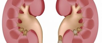
The photo shows what polycystic kidney disease looks like
Cysts up to 3-5 cm, as a rule, do not cause inconvenience and do not pose a particular danger to the health and life of the child. Some people live long and fulfilling lives with them, without any discomfort or discomfort. More significant formations or rapidly growing ones require radical treatment.
The following types of cystic neoplasia may occur in children:
- Multiband is a certain number of single-chamber neoplasms, between which the renal parenchyma has a normal unchanged structure. A characteristic feature is the significant magnitude of the pathology.
- A pyelogenous cyst is distinguished by the fact that its contours consist of transitional type epithelial cells. It has a small hole through which it communicates with the calyx of the kidney.
- Perirenal - formed directly under the kidney capsule. There are congenital and acquired neoplasias, in the latter case after rupture of the organ on which it was formed.
- Polycystic disease is a situation when quite a few cysts form on one kidney, so the organ becomes several times larger. The pathological process is congenital.
The note. A common practice is radical treatment of a kidney cyst immediately after the birth of a child, if it was identified during embryonic development. This measure helps prevent malignancy and exclude the development of cancer.
Treatment
The tactics of medical specialists when diagnosing this pathology in a developing fetus directly depends on factors such as the number and size of cysts, as well as the degree of functional failure of the damaged organ.
If the fetus has been diagnosed with a single cyst on one of the kidneys, then doctors prefer to conduct observational tactics, assessing the dynamics of the pathology using ultrasound examination.
If the fetus has been diagnosed with multicystic dysplasia of both kidneys, the woman is advised to terminate the pregnancy, since this anomaly can be fatal in a short period of time.
After the child is born, if characteristic clinical symptoms are present, the child can undergo a procedure for draining the cyst, followed by the introduction of a sclerosant. The degree of renal impairment in this pathology is assessed through laboratory techniques such as measuring the level of potassium, urea and creatinine in the body.
If a newborn develops chronic renal failure against the background of a cystic lesion, he is prescribed a hemodialysis procedure or transplantation of a damaged organ.
If the size of the neoplasm exceeds 50 mm in diameter, the child may be prescribed radical removal of the damaged organ (nephrectomy). As a rule, this intervention is not performed in the neonatal period. The optimal age for nephrectomy is from 1 year to 1.5 years. Children with a similar diagnosis undergo removal of the damaged kidney through endoscopic retroperitoneal access, which does not involve dissection of the muscle layer.
Methods of therapy
Conservative medicine is not effective in the treatment of multicystic disease, for this reason it is not carried out. Taking medications will not help get rid of cysts or reduce their size.
Surgery is the most effective; it is performed using several methods. The operation is performed on both children and adults.
Surgery
There are 2 methods that will help get rid of cysts:
- laparoscopic nephrectomy;
- open nephrectomy.
Laparoscopy is characterized by low trauma; it allows you to get rid of cysts and does not involve removing the kidney (only a certain part of it).
It is performed under general anesthesia and has a short recovery period.
Open nephrectomy is a complex abdominal operation and is performed under general anesthesia. The kidney is completely removed. This method of intervention can hardly be called low-traumatic.
But if it is not possible to remove part of the organ using laparoscopy, then open nephrectomy is used. If the tissues are not affected, the disease is just beginning to develop, but cysts have already appeared on the surface, then you can resort to puncture.
Using a puncture, the formation is opened and the fluid contained in it is taken for a biopsy.
Treatment of children
If multicystic disease has been diagnosed in a child, then before the age of 5, surgery is performed only if there is a serious threat to the baby’s life.
Preference is given to puncture, with opening the cyst and collecting its contents. If the baby has reached the age of 5 years, then surgical interventions can begin.
Indications for surgical procedures are:
- presence of a pathological process;
- damage to only 1 kidney;
- no complications.
Multicystic disease is treated surgically only if it affects 1 kidney, progresses rapidly and threatens severe complications.
Otherwise, therapy is not required; doctors simply monitor the development of the disease and, if necessary, intervene in the process.
ethnoscience
Due to ineffectiveness, it is not carried out, as is drug therapy. Taking herbal decoctions and plant extracts can negatively affect the patient's health.
Prevention
Subject to certain recommendations, every woman of reproductive age has the opportunity to protect the body of the unborn child from the occurrence of not only renal cysts, but also other anomalies of intrauterine development. Such recommendations include:
- Complete cessation of bad habits such as smoking, drinking alcohol and using drugs.
- Timely treatment of infectious and inflammatory diseases of the urinary and reproductive systems, as well as prevention of abortions.
- Timely registration with a doctor in case of pregnancy. The recommended period for registration at the antenatal clinic is 12 weeks of gestation. In addition, pregnant women are recommended to consult with a gynecologist, as well as three ultrasound examinations during the entire period of pregnancy.
- Prevention of hypothermia, as well as minimizing contact with household chemicals throughout the entire period of bearing a child.
Timely diagnosis of this pathological condition makes it possible to dynamically monitor the growth of a benign tumor.
Forecast
The most unfavorable prognosis concerns multicystic lesions of both kidneys in the fetus. The bilateral form of this disease is incompatible with life, so the woman is recommended to terminate the pregnancy regardless of the gestational age.
If a child was born with a single renal cyst, then as he grows up he may encounter a list of complications such as a rapid increase in the volume of the tumor, mechanical compression of the kidney parenchyma, malignant degeneration of the cyst, as well as rupture of the tumor capsule and hemorrhage into the tumor cavity.
Share:
How does kidney dysplasia manifest?
Symptoms and their manifestations largely depend on whether both kidneys are involved in the pathological process or only one. In addition, the severity of clinical manifestations may depend on whether there is complete tissue degeneration or whether renal function is preserved to a certain extent.
If one kidney is affected, the child has a good chance of survival, and puncture or removal of the affected organ eliminates the threat to life. Often, with a small number of cysts, symptoms may not appear for a long time, and the child will feel absolutely healthy. However, in the future, an increase in the size of the kidney and the addition of stagnant processes can provoke the following manifestations of the disease:
For many parents, a diagnosis such as renal dysplasia is a real blow, since with bilateral damage the child’s life lasts no more than a few days.