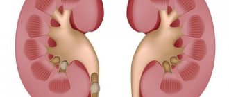
What is acute pyelonephritis? This is an inflammation of the kidneys that is caused by bacterial agents. The process does not extend to the glomerular structures, but to the pyelocaliceal system. This is a short definition of the term. If the process occurs chronically, the tubulointerstitial zone of the kidneys is affected, with subsequent dysfunction of the organs. In acute pyelonephritis, symptoms and treatment depend on what infectious factors cause the inflammation.
The problem of pyelonephritis in children
Acute pyelonephritis in children under one year of age is more often registered in boys, since they have a high frequency of detection of defects of the urinary system; after one year, cases in girls are several times higher than in boys. Acute pyelonephritis often debuts at an early age, but only the correct approach to the treatment of this disease determines the outcome: recovery, remission, chronicity of the process.
| Pyelonephritis can lead to chronic renal failure. |
The relevance of pyelonephritis lies in the fact that the disease is widespread. And also the chronic form of the disease often causes chronic renal failure, second only to malformations of the urinary system and glomerulonephritis.
How to treat pyelonephritis
The first treatment measure is to eliminate the causes that led to the improper outflow of urine. This is often done surgically - removal of stones, adenomas, urethral plastic surgery or other necessary operations. Antibacterial therapy is then carried out. Drugs are prescribed taking into account the sensitivity of the microorganisms that cause the disease to them. In general, the methods of treating kidney pyelonephritis depend on the form of the disease, age and gender of the patient.
Treatment regimen
The main drugs in the treatment of kidney inflammation are antibiotic therapy, which are prescribed on the basis of an antibiogram. Before receiving its results, the patient is prescribed broad-spectrum antibiotics for an initial course of 6-8 weeks. This may be Ceftriaxone, Nolitsin or Ampicillin, which can also be prescribed in the form of injections. In addition to antibiotics, the patient is prescribed other medications:
- analgesics to relieve pain;
- Diclofenac or Metamizole to reduce kidney inflammation;
- Furadonin, which normalizes kidney function;
- Phytolysin to restore immunity during remission.
Treatment of the chronic form
Therapy against the chronic form can be carried out at home. The basis is also antibacterial drugs. Along with them, non-steroidal anti-inflammatory drugs are prescribed. They help antibiotics reach the site of kidney damage. Pyelonephritis - it is already known that this disease can be treated with physiotherapy and symptomatic drugs such as Adelphan, Reserpine and Cristepine. They normalize blood pressure during exacerbation. These are the main ways to treat the chronic form.
Acute form
If the diagnosis is confirmed, treatment of acute pyelonephritis in children and adults is carried out in a hospital. Complex therapy immediately includes:
- Bed rest. Its timing is determined depending on the course of the disease.
- Diet. The patient is prescribed a balanced diet with sufficient vitamins and fluids.
- Antibacterial therapy. Includes broad-spectrum antibiotics from the group of cephalosporins or fluoroquinols. The course of treatment should be less than 2 weeks long.
- Antifungal drugs. They are prescribed for prolonged antibacterial therapy. It could be Levorin or Nystatin.
- Antihistamines. Also prescribed for long-term use of antibiotics. Suprastin, Diphenhydramine, Tavegil are most often used.
Treatment in children
The most difficult thing is the treatment of childhood pyelonephritis. The baby will have to take several medications at once – the doctor will tell you what these medications are. Antibiotics, homeopathic medicines, and antihistamines will be prescribed. How long does it take to treat pyelonephritis? For complete recovery in different cases it takes from 2 to 8 months. At the end of treatment, the child will also be prescribed probiotics to restore normal intestinal microflora.
Among women
The methods for treating pyelonephritis in women are not particularly different. They are also prescribed antibacterial drugs, bed rest in case of acute forms, plenty of fluids and diet. Ways to treat pyelonephritis in women include anti-inflammatory and restorative agents, multivitamin complexes and herbal medicines. Among the latter, medicines based on ginseng and eleutherococcus are particularly successful.
Treatment at home
Chronic inflammation can be cured not in the clinic, but at home. Taking antibiotics remains mandatory. Using herbal infusions based on oats, chamomile, plantain, nettle or rosehip will help. The same effect will be obtained from taking herbal medicines Canephron, Fitolysin. Additionally, you need to monitor your fluid intake - at least 1.5-2 liters per day. Under no circumstances should the kidneys be heated. This is the basic advice on how to treat pyelonephritis at home.
Causes of the disease
Pyelonephritis is caused by E. coli in more than 90% of cases.
The causative agent of pyelonephritis in children
Pyelonephritis in a child is usually caused by one type of pathogen. Up to 90% of cases of the disease are caused by E. coli infection. Of lesser importance are: Klebsiella, Proteus, enterococci, enterobacteria and staphylococci. In premature infants and children with immunodeficiency, an association of pathogens - bacteria and fungi - is more often detected. Of course, one pathogen is not enough in the development of the disease.
Predisposing factors
There are a number of predisposing factors to the appearance of pyelonephritis:
- hereditary burden;
- immaturity of the organ and metabolic disorders in the renal tissue;
- congenital malformations of the urinary system;
- birth injuries, hypoxia and asphyxia during childbirth;
- neurogenic bladder dysfunction, which is characterized by normal, regular emptying of the organ;
- children's age (up to 2 years);
- constipation;
- infectious and inflammatory diseases of the external genitalia;
- bladder catheterization;
- hypothermia, which results in muscle spasms and hemodynamic disturbances.
Mechanism of development of pyelonephritis
After the child’s body is infected with pathogenic microorganisms, bacteria penetrate into the organs of the urinary system. This can occur in 3 ways: ascending, hematogenous, lymphogenous. Ascending is the most common path of disease development. Most common in children. The source of infection is most often the rectum. After overcoming the vesicoureteral barrier, the bacteria quickly multiply and release toxins that act on the kidney tissue. The hematogenous route is observed in children with immunodeficiency with the development of sepsis. Bacteria are carried through the bloodstream throughout the body, also reaching the kidneys. The lymphogenic pathway has not been studied enough. With this type of infection, migration of bacteria from the intestine is possible.
Classification of pyelonephritis
By form:
- primary (more often the cause of development cannot be determined, that is, an infection of an absolutely healthy organ occurs);
- secondary (develops against the background of certain disorders and diseases).
Secondary can be divided into:
- obstructive (impaired uprodynamics);
- non-obstructive (develops against the background of tubolopathies, neurogenic dysfunction, metabolic and metabolic disorders).
With the flow:
- acute (pathological process lasting less than 6 months);
- chronic (inflammatory changes persist for more than 6 months).
Chronic can be:
- recurrent (frequent exacerbations of the disease);
- latent (sluggish inflammatory process, manifested only by urinary syndrome).
According to disease activity:
- peak period;
- subside;
- remission (partial or complete clinical and laboratory remission).
According to the state of kidney function:
- saved;
- impaired (tubular, glomerular changes, acute or chronic renal failure).
Clinical picture of pyelonephritis in children
Symptoms of pyelonephritis in children can be divided into general intoxication, dysuria, as well as manifestations included in the urinary and pain syndrome.
General symptoms of acute pyelonephritis
- Intoxication (headache, weakness, lethargy, loss of appetite, sleep disturbance, nausea, vomiting, high temperature);
- pale skin;
- increased blood pressure, anemia due to impaired renal function;
- changes in the blood (increased number of leukocytes, neutrophils, C-reactive protein, procalcitonin, increased erythrocyte sedimentation rate).
Dysuric symptoms
- Nocturnal or daytime enuresis (urinary incontinence);
- in case of impaired renal function, nocturia (increased urine output at night), polyuria (increased urine output during the day) or, conversely, oliguria (decreased urine output during the day).
Pain syndrome
- Stomach ache;
- pain in the lumbar region or a positive symptom of tapping (the appearance of pain when tapping the lumbar region in the projection of the kidneys).
Urinary syndrome
- Increased number of leukocytes, protein in a general urine test;
- the appearance of bacteria in the urine.
Clinical manifestations of pathology
The main complaint that patients come with is a violation of the urination process. Adult patients experience pain, pain and other types of discomfort when emptying the bladder. At night they have to wake up and go to the toilet more often to urinate.
The clinical picture of acute pyelonephritis depends on the localization of the process. With right-sided lesions, the manifestations are similar to those of appendicitis and cholecystitis. It is extremely important to carry out adequate differential diagnosis.
Left-sided pyelonephritis is accompanied by nagging pain on the affected side. They can mimic diseases of the large intestine, as well as damage to the spleen.
Bilateral localization of the inflammatory process in the kidneys is common. Isolated damage to one organ is rare in modern conditions. Patients complain of pain in the lumbar region. With bilateral pyelonephritis, these sensations are constant, aching. Patients define them as stupid. At home, patients tend to put shawls and scarves on their belts. Sometimes heating pads and other thermal effects are used.
In a horizontal position, the symptoms of the disease become less pronounced. This is a clear differential sign that distinguishes acute pyelonephritis or exacerbation of chronic pyelonephritis from diseases of the back (spine and all its component structures).
Intoxication syndrome does not always occur. It determines the clinic only in elderly or very young patients. An increase in temperature to subfebrile and sometimes febrile levels (up to 40 degrees) comes to the fore. Severe headaches and even confusion are possible, which is more typical for the elderly.
Features of pyelonephritis in children of the first year of life
| Symptoms may change depending on the age of the child. So newborn babies have their own characteristics. |
In children under one year of age, an important clinical criterion is an increase in temperature without cold-like manifestations of the disease.
This age is characterized by a predominance of general symptoms. Intoxication symptoms come to the fore: fever, refusal to eat, regurgitation, vomiting, diarrhea, decreased temperature (for premature infants), lack of weight gain or deficiency, marbling of the skin. Neurological symptoms and electrolyte disturbances may occur.
In older children, general symptoms are not as pronounced. You can often observe dysuric symptoms of pyelonephritis, as well as a pungent odor of urine. The appearance of pain in the abdomen or lumbar region is of no small importance.
Chronic pyelonephritis is characterized by frequent causeless rises in temperature without catarrhal changes, periodic abdominal pain, and dysuric disorder (usually nocturnal enuresis). Urinalysis often reveals elevated white blood cell counts and protein.
Similar and distinctive symptoms of diseases
Symptoms of diseases of the genitourinary system are often similar. Any disturbances in the functioning of the kidneys or bladder lead to frequent or difficult toileting and painful sensations. Therefore, pyelonephritis and cystitis, especially the chronic form, are often confused.
Similar signs of the two diseases:
- increased urge to urinate;
- nagging pain in the lower abdomen, back, when urinating, at rest;
- the appearance of protein, bacteria, blood in the urine;
- in the acute stage of the disease - weakness, insomnia, fever, nausea, vomiting.
The difference between the diseases lies in the different localization of pain: with pyelonephritis, nagging pain in the abdomen and lower back, with cystitis, burning and acute attacks in the pubic area and genitals. Inflammation of the kidney and pelvis is characterized by high fever and severe weakness; if the bladder is affected, these symptoms are absent.
The disease can also be diagnosed using a urine test. The protein content increases in case of pyelonephritis. With cystitis, a predominant number of leukocytes is observed. Chronic forms are characterized by nagging pain in the area of inflammation.
Damage to the kidneys and pelvis is a malignant disease, but to a greater extent than cystitis.
Due to inflammation, infiltrates and abscesses form on the kidney tissues. Due to a violation of the outflow of urine, toxin poisoning occurs in the body. The patient experiences nausea and severe vomiting.
Differential diagnosis of pyelonephritis
| It is necessary to exclude other diseases in order to make a correct diagnosis. |
First of all, differential diagnosis of acute pyelonephritis with glomerulonephritis is carried out. The latter develops 3 weeks after a streptococcal infection. For example, it is a complication of acute tonsillitis. Glomerulonephritis has a similar clinical picture. But it is distinguished by the absence of intoxication syndrome. Characteristic is the appearance of edema and arterial hypertension, which is not detected in pyelonephritis (only in cases of impaired renal function). Oligouria (decreased amount of urine excreted) is observed in both conditions, but with pyelonephritis this symptom will appear at the onset of the disease, and with glomeronephritis the initial dysuric symptom will be, on the contrary, polyuria. The end point of diagnosis is the examination of urine sediment. With glomerulonephritis, hematuria is observed (an increase in the number of red blood cells in the urine), an increase in the amount of protein, a decrease in urine density and no signs of inflammation, since the disease is abacterial.
It is important to quickly differentiate acute pyelonephritis in children with appendicitis, but with an atypically located appendix (retroperitoneal). The difficulty of diagnosis is that with this type of appendicitis, pain is observed in the lumbar region and is mistakenly regarded as a positive symptom of effleurage. But upon careful examination, you can feel tension in the lumbar muscles. A rectal examination and urine test, which will show no changes in appendicitis, will also help you finally figure it out. The low location of the appendicular process is manifested by dysuric disorders and diarrhea. A general urine test also does not reveal inflammatory changes.
Similar signs of pyelonephritis in children are observed with cystitis. But with an isolated inflammatory process of the bladder, there is no deterioration in the patient’s general condition, and there are no signs of intoxication.
Chronic pyelonephritis should be distinguished from reflux nephropathy. This disease is characterized by vesicoureteral reflux. The clinical picture is very similar to the original disease. Ultrasound examination of the kidneys will help to make a correct diagnosis (reduction in kidney size, expansion of the collecting system, hypotension, abnormalities of the urinary system).
Also, based on pain syndrome, it is necessary to differentiate chronic pyelonephritis from intussusception, tumor in the abdominal cavity, and renal colic. All children with diseases that cause pain should consult a surgeon.
Acute pyelonephritis
Etiology and pathogenesis
The most common pathogens that cause inflammation in the kidney are Escherichia coli
), Proteus (
Proteus
), Enterococcus (
Enterococcus
), Pseudomonas aeruginosa (
Pseudomonas aeruginosa
), Staphylococcus (
Staphylococcus
). Penetration of the pathogen into the kidney in acute pyelonephritis most often occurs by hematogenous route from any source of infection in the body due to the development of bacteremia. Less commonly, the infection enters the kidney via the urinogenic route from the lower urinary tract (urethra, bladder) along the wall of the ureter (in this case, the disease begins with the development of urethritis or cystitis followed by the development of the so-called ascending pyelonephritis) or along the lumen of the ureter due to vesicoureteral reflux .
Pathological anatomy
The infection, having penetrated the kidney or pelvis through the hematogenous or urinogenic route, invades the interstitial tissue of the kidney and the tissue of the renal sinus.
Hematogenous pyelonephritis
For hematogenous pyelonephritis
inflammatory foci are located mainly in the cortex, around intralobular vessels.
The inflammatory process, involving interstitial tissue, damages mainly the tubular system. With urinogenic, ascending pyelonephritis,
the infection affects the kidney in separate foci, fan-shaped, in the form of wedges, extending from the pelvis to the surface of the kidney.
Between these foci of inflammation and subsequent fibrosis are areas of normal renal tissue. With bilateral pyelonephritis,
the pathological process in the kidneys spreads unevenly, asymmetrically, in contrast to glomerulonephritis and nephrosclerosis.
Acute pyelonephritis
Pyelonephritis, being at first a focal process, becomes diffuse in nature with each new attack. Interstitial tissue plays a very important physiological role in intercellular metabolism. After the disappearance of inflammatory infiltrates, true restoration of the intermediate tissue does not occur; In place of the dead elements of the renal parenchyma, scar tissue develops. Primary and secondary acute pyelonephritis can occur first in the form of serous, then purulent interstitial inflammation. Taking this into account, it is customary to use the terms “acute serous pyelonephritis” and “acute purulent pyelonephritis”. While acute serous pyelonephritis is observed in 64%, acute purulent - only in 36% of patients with acute pyelonephritis. In the stage of acute serous inflammation, the kidney is enlarged and tense. The perinephric tissue is significantly swollen. Microscopically, numerous perivascular infiltrates are detected in the interstitial tissue. Under the influence of appropriate treatment, this stage undergoes reverse development, but serous acute pyelonephritis can also progress to the stage of purulent inflammation.
Acute purulent pyelonephritis occurs in the form of apostematous nephritis, abscess and carbuncle of the kidney. Apostematous nephritis is a metastatic suppurative process, one of the subsequent stages of acute pyelonephritis. In this case, the cortical substance of the kidney, both on the surface and on the section, is dotted with small ulcers that look like beads.
Apostematous nephritis
With apostematous nephritis, the kidney is enlarged and gray-cherry in color. The perinephric tissue is sharply swollen. After removing the fibrous capsule, multiple small (from a pinhead to a pea in size) abscesses, located singly or in groups, are visible. On a section of the kidney, small abscesses are often found in the medulla. Microscopically, multiple foci of purulent inflammation are detected in the interstitial tissue. In the circumference of the Malpighian glomeruli, clusters of small cell infiltrates with foci of necrosis are visible. The urinary tubules are compressed by infiltrates, which are located both in the interstitial tissue and in the perivascular spaces.
Apostematous nephritis is combined with renal carbuncle in 23% of cases. These two types of purulent kidney damage represent a single pathological process, manifesting itself only in different sequences and intensity of its development. If apostematous nephritis is characterized by multiple small abscesses, then a renal carbuncle is a localized suppurative focus, characterized by a tumor-like progressive growth of inflammatory infiltration without a tendency to large abscess formation. In appearance, this process in the kidney is very similar to a skin carbuncle. This circumstance gave rise to Israel in 1891 calling this disease a kidney carbuncle.
The size of the carbuncle ranges from 0.3 to 2 cm, rarely more. Carbuncle can be single or multiple, in 25% of cases it is combined with apostematous nephritis. Usually the purulent process affects one kidney, rarely - both (5%). With a favorable course of acute purulent pyelonephritis, infiltrates are reabsorbed and connective tissue grows in their place. Replacement of purulent foci with connective tissue leads to the formation of scar retractions on the surface of the kidney, first dark red and then white-gray. On a section of the kidney, these scars have the shape of a wedge, reaching the pelvis.
- So, in acute pyelonephritis, the inflammatory process is first localized in the interstitial tissue, and then the tubules and, lastly, the glomeruli are involved in it. When the process enters the chronic stage, productive endarteritis, hyperplasia of the medial vascular lining, and arteriolar sclerosis occur. Arteriolar sclerosis is one of the causes of further kidney atrophy.
Pyelonephritis shrinkage of the kidney due to atrophy of its parenchyma can be so significant that the kidney has a mass of only 30-50 g. In children, especially young children, pyelonephritis is extremely active and is accompanied by the death of the kidney parenchyma in large areas.
Clinical picture
Local symptoms:
- Pain in the lumbar region on the affected side. With non-obstructive pyelonephritis, the pain is usually dull, aching in nature, can be low or reach high intensity, and take on a paroxysmal nature (for example, with obstruction of the ureter by a stone with the development of so-called calculous pyelonephritis).
- Dysuric phenomena are not typical for pyelonephritis itself, but can occur with urethritis and cystitis, which lead to the development of ascending pyelonephritis.
General symptoms are characterized by the development of intoxication syndrome:
- fever up to 38-40 °C;
- chills;
- general weakness;
- decreased appetite;
- nausea, sometimes vomiting.
Children are characterized by the severity of intoxication syndrome, as well as the development of the so-called. abdominal syndrome (severe pain not in the lumbar region, but in the abdomen).
Elderly and senile people often develop an atypical clinical picture, either with an erased clinical picture or with pronounced general manifestations and the absence of local symptoms.
Diagnostics
Laboratory research methods
Blood analysis
- General blood analysis
. General inflammatory changes: leukocytosis, acceleration of ESR, shift of the leukocyte formula to the left, with severe inflammation - anemia. - Blood chemistry
. There may be an increase in transaminases, hypergammaglobulinemia, and with the development of renal failure, an increase in urea and creatinine levels.
Analysis of urine
- General urine analysis
. The main symptom - leukocyturia - may be absent in hematogenous pyelonephritis in the first 2-4 days, when the inflammatory process is localized mainly in the cortical layer of the kidney parenchyma, as well as in case of obstruction of the urinary tract on the affected side; erythrocyturia in pyelonephritis can be observed in the presence of a calculus, due to necrotic papillitis, damage to the fornical apparatus, in the presence of acute (hemorrhagic) cystitis, which caused the development of pyelonephritis. - Bacteriological examination of urine
is used to accurately determine the pathogen and its sensitivity to antibiotics. - Gram staining of urine
is an important step in the etiological diagnosis of pyelonephritis, which allows you to quickly obtain preliminary approximate data about the nature of the pathogen. It is advisable to carry out a cultural examination of urine (culture on nutrient media, isolation of a pure culture of the pathogen and determination of its sensitivity to drugs) in all cases, especially in a hospital. If bacteremia is suspected (with high fever, chills), as well as in intensive care units, a blood test for sterility is required. A necessary condition for the reliability of the results of bacteriological examination is the correct sampling of urine and blood.
Instrumental research methods
- Ultrasound examination (ultrasound) of the kidneys in the phase of serous inflammation in acute primary pyelonephritis may not reveal pathological changes in the kidneys; in the serous phase, ultrasound reveals an increase in the kidneys (or one kidney in case of unilateral damage) in size, a decrease in their mobility during breathing. With apostematous pyelonephritis, the ultrasound picture is the same as in the phase of serous inflammation (enlargement of the kidneys, limitation of their mobility). An ultrasound examination of a renal carbuncle is characterized by the presence of a hypoechoic area without clear contours, and sometimes a bulging of the external contour of the kidney in this place. When a kidney abscess forms, ultrasound reveals a hypoechoic area with clear contours (abscess capsule), sometimes with heterogeneous anechoic areas in the center (liquid pus). When the purulent process extends beyond the kidney capsule (development of paranephritis), ultrasound determines the vagueness of the perinephric tissue with the presence of hypo- and anechoic components in it.
- X-ray examination methods: survey and excretory urography complement each other and are usually carried out together (a survey followed by excretory urography). An overview image may reveal an increase in the size of the kidney, bulging of its contour (with a carbuncle and abscess), blurred contour of the psoas major muscle on the affected side (edema of the perinephric tissue, paranephritis), and the presence of shadows of stones (calculous pyelonephritis). On excretory urograms in the phase of serous inflammation, urodynamics and renal function are often not impaired; enlargement of the kidney, limitation of its mobility during orthotest, moderate compression of the pyelocaliceal system by edematous renal parenchyma can be determined. With apostematous pyelonephritis, a decrease in the excretory function of the kidney is added to the listed signs of serous pyelonephritis. With carbuncles and kidney abscesses, excretory urograms can reveal bulging of the contour, compression and deformation of the pelvis and calyces by the abscess and infiltrate.
- retrograde pyeloureterography is performed in the absence of renal function on excretory urograms or if for some reason excretory urography cannot be performed (serious condition of the patient, presence of acute or chronic renal failure).
- abdominal aortography, selective renal arteriography, computed tomography are used mainly for differential diagnosis of pyelonephritis and other renal pathologies.
Differential diagnosis
Chronic pyelonephritis must be differentiated primarily from chronic glomerulonephritis, renal amyloidosis, diabetic glomerulosclerosis and hypertension.
Kidney amyloidosis in the initial stage, manifested by only slight proteinuria and very scanty urinary sediment, can simulate a latent form of chronic pyelonephritis. However, unlike pyelonephritis, with amyloidosis there is no leukocyturia, active leukocytes and bacteriuria are not detected, the concentration function of the kidneys remains at a normal level, there are no radiological signs of pyelonephritis (the kidneys are the same, normal in size or slightly enlarged). In addition, secondary amyloidosis is characterized by the presence of long-term chronic diseases, most often purulent-inflammatory.
Diabetic glomerulosclerosis develops in patients with diabetes mellitus, especially in severe cases and long duration of the disease. At the same time, there are other signs of diabetic angiopathy (changes in the vessels of the retina, lower extremities, polyneuritis, etc.). There are no dysuric phenomena, leukocyturia, bacteriuria and radiological signs of pyelonephritis.
Chronic pyelonephritis with symptomatic hypertension, especially with a latent course, is often mistakenly assessed as hypertension. Differential diagnosis of these diseases is very difficult, especially in the terminal stage.
If from the anamnesis or medical documentation it is possible to establish that changes in the urine (leukocyturia, proteinuria) preceded (sometimes for many years) the appearance of hypertension, or long before its development, cystitis, urethritis, renal colic were observed, stones were found in the urinary tract, then the symptomatic origin of hypertension as a consequence of pyelonephritis is usually not in doubt. In the absence of such instructions, it is necessary to take into account that hypertension in patients with chronic pyelonephritis is characterized by higher diastolic pressure, stability, insignificant and unstable effectiveness of antihypertensive drugs and a significant increase in their effectiveness if they are used in combination with antimicrobial agents. Sometimes, at the beginning of the development of hypertension, only anti-inflammatory therapy is sufficient, which without antihypertensive drugs leads to a decrease or even stable normalization of blood pressure. It is often necessary to resort to urine testing according to Kakovsky-Addis, for active leukocytes, urine culture for microflora and the degree of bacteriuria, pay attention to the possibility of unmotivated anemia, an increase in ESR, a decrease in the relative density of urine in the Zimnitsky test, which are characteristic of pyelonephritis.
Some data from ultrasound and excretory urography (deformation of the cups and pelvis, stricture or atony of the ureters, nephroptosis, unequal size of the kidneys, the presence of stones, etc.), radioisotope renography (decrease in the function of one kidney while the function of the other is preserved) and renal angiography (narrowing, deformation and reduction in the number of small and medium-sized arteries). If the diagnosis is in doubt even after all of the above research methods have been carried out, it is necessary (if possible and in the absence of contraindications) to resort to a puncture biopsy of the kidneys.
Treatment
Conservative treatment includes antibacterial (penicillin + aminoglycosides; fluoroquinolones + cephalosporins); infusion-detoxification, anti-inflammatory therapy, physiotherapy, it is advisable to use disaggregants and anticoagulants. Before obtaining the results of a bacteriological examination of urine, antibacterial therapy is prescribed empirically (more often, treatment begins with fluoroquinolones), and after obtaining the results of urine culture, treatment can be adjusted. To increase the effectiveness of antibacterial therapy, the intra-aortic method can be used [ source not specified 3033 days
] administration of antibiotics. Functional-passive kidney exercises (20 ml of furosemide are prescribed 1-2 times a week). Obstructive forms of acute pyelonephritis require immediate restoration of urine outflow on the affected side; preference is given to percutaneous puncture nephrostomy, and only then the prescription of antibacterial and infusion therapy.
Methods of conservative treatment also include catheterization of the ureter on the affected side in order to restore the outflow of urine from the affected kidney.
Currently, normalization of the state of biological membranes plays an important role in the successful treatment of diseases of the urinary system. The pathogenetic role of damage to the lipid component of the membranes of the epithelium of renal tissue in the formation of dysmetabolic nephropathy, nephrolithiasis, and interstitial nephritis has been established [4] [5]. The main process leading to membrane destruction is free radical lipid peroxidation (LPO). LPO refers to nonspecific reactions, the severity of which often determines the prognosis and outcome of many pathological conditions [6][7][8].
In this regard, in many inflammatory diseases of nonspecific etiology, along with conventional drug treatment, the prescription of antioxidants is pathogenetically justified [7][8][9][10][11]. Antioxidants include: vitamin E (tocopherol), vitamin C (ascorbic acid), ubiquinone (coenzyme Q10), vitamin A (retinol), β-carotene, selenium, etc.
If signs of renal failure appear, oxidants are prescribed (cocarboxylase, less often riboflavin, pyridoxal phosphate)
Surgical treatment (surgeries)
Surgical treatment includes organ-preserving and organ-saving operations.
- Organ-preserving
: The scope of surgical intervention for acute pyelonephritis depends on the nature of the detected changes in the kidney. Kidney decapsulation is performed for any type of purulent pyelonephritis. In case of apostematous pyelonephritis, the kidney is decapsulated and the apostemata are opened. This reduces compression of the renal parenchyma by infiltrate and edema, and helps restore blood flow in the kidney. In the presence of a kidney carbuncle, dissection and excision of the carbuncle are performed; in case of a kidney abscess, the abscess is opened and its wall is excised. Organ-saving operations also include nephrostomy - both with open surgery and percutaneous puncture nephrostomy under ultrasound guidance. - Organ removal
: Nephrectomy.
Diagnosis of pyelonephritis in children
The most important is to isolate the culture from the collected urine.
- Isolation of culture. Before culturing urine for sterility to isolate bacteria, it is necessary to correctly collect the analysis so that bacteria that are not involved in the development of the disease are not included. There are several methods for this. The most informative is suprapubic puncture of the bladder. But this method is practically not used, since it requires a lot of time, special training of medical personnel to carry out the procedure, and the invasiveness of the method is also important; puncture is an unpleasant manipulation for the child and his parents. Taking urine by catheterizing the bladder is also not enjoyable for the baby. Despite the fact that there are methods for collecting material that have a highly accurate result, currently the most common method is collecting material in a sterile container with free urination. But you need to take into account the fact that with this procedure the risk of contamination with other bacteria increases sharply. The diagnostic titer of E. coli should be at least 100,000 CFU/ml. If typical clinical signs of pyelonephritis are observed, then the titer is at least 10,000 CFU/ml.
- General urine analysis. There is an increase in the number of leukocytes more than 5 in the field of view, sometimes detection of protein more than 0.033 g/l, and the presence of bacteria.
- General urine analysis. Allows you to observe the general inflammatory response (increased leukocytes, neutrophils).
- Urocytogram. Neutrophilic leukocyturia is observed (the predominance of neutrophils is 90% or more).
- The Nechiporenko test, according to Zimnitsky, Amburge, Addis-Kokovsky, is prescribed according to indications to determine the preservation of kidney function.
- Consultation with a gynecologist (for girls if it is necessary to treat inflammatory diseases of the external and internal genital organs), urologist (if another urological pathology is identified).
- Ultrasound examination of the kidneys. The most commonly used method for diagnosing pathology of the urinary system. It allows you to differentiate diseases and helps in making the correct diagnosis. With pyelonephritis, an increase in the size of the kidneys can be observed, as well as possible obstruction of the urinary tract.
- Vaccine cystography. Allows you to identify impaired urine passage, vesicoureteral reflux, and urethral obstruction.
- Excretory urography. Determines signs of pyelonephritis, obstruction in the ureters, malformations of the urinary system, decreased kidney function, and the presence of a tumor in the abdominal cavity.
We treat the liver
Warehouse relocation to Europe. We sell hepatitis C drugs in Russia at the purchase price - warehouse liquidation Go to website
ACUTE GLOMERULONEPHRITIS
This is a disease of infectious-allergic nature with primary damage to the capillaries of both kidneys. Distributed everywhere. They are most often affected at the age of 12-40 years, somewhat more often in men. It occurs more often in countries with cold and humid climates and is a seasonal disease.
Clinic
The disease begins with a headache, general malaise, sometimes nausea, and lack of appetite. There may be oliguria and even anuria, manifested by rapid weight gain. Very often, against this background, shortness of breath and attacks of suffocation appear. In the elderly, manifestations of left ventricular heart failure are possible. In the very first days, swelling appears, usually on the face, but can also be on the legs, and in severe cases on the lower back. Hydrothorax and ascites are extremely rare. In the very first days of the disease, blood pressure reaches 180/120 mm Hg.
Laboratory diagnostics
Urinary syndrome. Reberg's test - a sharp decrease in filtration. The blood side is normal. There may be an acceleration of ESR. The ECG shows signs of left ventricular hypertrophy approximately 2 weeks after the onset of the disease. X-ray of increased heart size.
Differential diagnosis
Toxic kidney: signs of intoxication, toxemia, presence of infection. Acute pyelonephritis: history of abortion, hypothermia, diabetes mellitus, childbirth. Higher temperature: 30-40o C. often chills, initially there is no increase in blood pressure. No swelling. Severe leukocyturia. Severe pain in the lumbar region on one side (with glomerulonephritis, 2-sided damage). Hemorrhagic vasculitis (renal form): the leading symptom is hematuria; there are skin manifestations.
CHRONIC GLOMERULONEPHRITIS
This is a bilateral inflammatory disease of the kidneys of immune origin, which is characterized by gradual but steady death of the glomeruli, shrinkage of the kidney, gradual decrease in function, development of arterial hypertension and death from chronic renal failure.
The frequency is about 4 per 1000 autopsies. The incidence of men and women is the same. It is found in all countries of the world, but more often in cold ones.
Differential diagnosis
Acute glomerulonephritis: anamnesis is important, the time from the onset of the disease, the specific gravity is high throughout the disease, and with chronic glomerulonephritis there may be a decrease in the specific gravity of urine. Left ventricular hypertrophy may be pronounced. Hypertension. Histological examination is of decisive importance - the presence of hyperplastic processes.
A malignant form of hypertension: now extremely rare. Persistent high blood pressure 260/130-140 or more. Significant changes in the fundus. Then, urinary syndrome may also occur.
Chronic pyelonephritis: a history of often gynecological diseases, abortions, cystitis. There is a tendency to low-grade fever. Pyuria. Bacteriouria, early decrease in urine specific gravity. The presence of radiological signs of pyelonephritis (the calyces become sclerotic early and change their shape).
Polycystic kidney disease: appears at 30-40 years of age. The presence of enlarged kidneys on both sides. X-ray examination shows the presence of an uneven scalloped edge of the kidneys and a cyst. Hypertension, azotemia. Early causes chronic renal failure.
Prevention of glomerulonephritis:
Diagnosis and treatment of acute glomerulonephritis, identifying changes in urine tests when the patient is in good health. Treatment of chronic glomerulonephritis and chronic renal failure. Radical treatment is impossible, since the process is autoimmune without exacerbation; in most cases, nephroprotection is indicated. Staying in bed for a long time, physical activity is contraindicated, avoiding hypothermia, working in a dry, warm room, preferably sitting, diet, limiting salt to 2-3 grams per day, protein, food rich in vitamins. Sanitation of foci of chronic infection. In severe cases, glucocorticosteroids (dexamethasone, hydrocortisone), cytostatics, chimes. Sanatorium-resort treatment in a dry, hot climate. Treatment during an exacerbation: hospitalization. A deterioration in urinalysis should be considered an exacerbation. Treatment during an exacerbation is the same as for acute glomerulonephritis. Contraindications for treatment with glucocorticosteroids: gastric ulcer, diabetes mellitus, renal failure, the first 15 weeks of pregnancy, chronic glomerulonephritis with very high hypertension.
ACUTE PYELONEPHRITIS
Acute pyelonephritis is a rapidly occurring inflammatory lesion of the kidneys involving the parenchyma and mucous membrane in the pathological process.
Clinical picture
Manifestations of acute pyelonephritis vary depending on the form and course of the process. Serous pyelonephritis occurs more calmly. Violent clinical manifestations are typical for patients with purulent lesions. Acute pyelonephritis is characterized by a triad of symptoms: fever, pain in the lumbar region and urinary disorders. In most patients, in the first days of illness, the temperature reaches 39–40 °C, often accompanied by chills. The temperature is intermittent or constant. Profuse profuse sweating, severe headache, nausea, vomiting, lack of appetite, muscle and joint pain, palpitations, shortness of breath, frequent urination, aching pain in the lumbar region are noted. Lower back pain increases with walking, movement, and tapping the kidney area (positive Pasternatsky symptom). Pain in the upper abdomen may be bothersome.
Diagnostics
With the acute onset of the disease, the presence of pain in the lumbar region, dysuric disorders, high fever, leukocytosis in the peripheral blood, as well as pronounced impurities in the urine (pyuria), the diagnosis of acute pyelonephritis does not cause difficulties.
Acute pyelonephritis should be differentiated from acute cystitis. In this case, a three-glass test helps recognition: for cystitis, the third sample contains a large number of formed elements. In addition, acute cystitis is characterized by more pronounced dysuria and hematuria, as well as pain at the end of urination.
Acute pyelonephritis should be differentiated from acute glomerulonephritis, in which erythrocytes in the urine predominate over leukocytes, severe albuminuria, edema and arterial hypertension are noted.
Prevention
Prevention of acute pyelonephritis comes down to the sanitation of foci of chronic infection (caries, chronic tonsillitis, sinusitis, chronic appendicitis, chronic cholecystitis, etc.), which are a potential source of hematogenous introduction of microbes into the kidneys, as well as eliminating the causes that impede the outflow of urine. An important role in prevention is played by appropriate hygienic measures (especially in girls and pregnant women), which prevent the upward spread of infection through the urinary tract, as well as the fight against constipation and treatment of colitis.
CHRONIC PYELONEPHRITIS
Nonspecific infectious and inflammatory disease of the mucous membrane of the urinary tract: pelvis, calyces and interstitial tissue of the kidneys. Essentially interstitial bacterial nephritis, 60% of all kidney diseases.
Clinic
May leak under masks. 1. Latent form - 20% of patients. Most often there are no complaints, and if there are, then - weakness, increased fatigue, and less often low-grade fever. Women may experience toxicosis during pregnancy. A functional study reveals nothing, except for a rare unmotivated increase in blood pressure and mild pain when tapping the lower back. Laboratory diagnosis. Repeated tests are of decisive importance: leukocyturia, moderate, no more than 1 - 3 g/l proteinuria + Nechiporenko test. Stengheimer-Malbin cells are doubtful, but if there are more than 40% of them, then it is characteristic of pyelonephritis. Active leukocytes are rarely detected. True bacteriuria *****> 10 5 bacteria in 1 ml. To prove it, 30 g of prednisolone IV and evaluate the indicators (increase in leukocytes by 2 or more times, active leukocytes may appear). 2. Recurrent almost 80%. Alternation of exacerbations and remissions. Features: intoxication syndrome with fever, chills, which can occur even at normal temperature, leukocytosis in a clinical blood test, increased ESR, shift to the left, C-reactive protein. Pain in the lumbar region, usually 2-sided, in some like renal colic: the pain is asymmetrical! Dysuric and hematuric syndromes. Hematuria syndrome now occurs more often, there may be micro and macro hematuria. Increased blood pressure. The most unfavorable combination of syndromes: hematuria + hypertension -> after 2-4 years, chronic renal failure. 3. Hypertensive form: the leading syndrome is an increase in blood pressure, which may be the first and only one, the urinary syndrome is not pronounced and is not constant. It is dangerous to do a provocation, as there may be an increase in blood pressure. 4. Rarely anemic. Persistent hypochromic anemia may be the only sign. Associated with a violation of erythropoietin production, urinary syndrome is not expressed and is not constant. 5. Hematuric: recurrence of macrohematuria. 6. Tubular: uncontrolled loss of Na + and K + in the urine (salt is the losing kidney). Acidosis. Hypovolemia, hypotension, decreased glomerular filtration, may be acute renal failure. 7. Azotemic: appears for the first time already. honor disadvantage
Establishing diagnosis
X-ray examination is crucial. Excretory urography (retrograde is not used in therapy). Asymmetry is functional and structural. Assess: size, contours, deformation of the cups, tone disturbance, detection of pyelorenal reflux, shadows of stones. Normal sizes: for men: right 12.9 * 6.2 cm, left 13.2 * 6.3 cm, for women: right 12.3 * 5.7 cm: left 12.6 * 5.9 cm Evaluation rules: If the left one is 0.5 cm smaller than the right one, this is almost pathognomonic for its wrinkling; if the difference in the length of the kidneys is 1.5 cm or more, this is a wrinkling of the right kidney. With the help of intravenous urography, the following is detected: In the initial stage, a slowdown in the removal of contrast, deformation of the cups and pelvis, spreading of the cups due to edema and infiltration, then their bringing together due to wrinkling. 2. Radioisotope methods. Asymmetry and degree of functional damage are identified. Statistical and dynamic scintigraphy is used. 3. Ultrasound diagnostics. 4. Computed tomography. 5. Renal angiography - a picture of “burnt wood” due to obliteration of small vessels. 6. Kidney biopsy.
Prevention:
Carefully observe the rules of personal hygiene of the genital organs, avoid hypothermia, promptly correct urodynamic disorders (against the background of developmental anomalies of the urinary system, urinary tract infections, etc.), treat prostate diseases (benign hyperplasia, prostatitis), gynecological pathology, and avoid frequent use of non-steroidal drugs. analgesics.
Source: StudFiles.net
We are in social networks:
Principles of treatment of pyelonephritis
- Regular events. Bed rest is prescribed for 5 - 7 days, especially during febrile and painful periods. It is important to observe the rules of personal hygiene; the greatest attention should be paid to hygienic measures after each act of defecation. A forced urination regime is recommended (emptying the bladder every 2 - 3 hours);
- diet. During the acute period of the disease, a diet with limited protein and salt is prescribed. But the most important thing is to drink a large amount of fluid (up to 3 years old - 1 liter of fluid per day, children 3 - 7 years old are recommended to drink 1.5 liters of fluid, children over 7 years old need to drink 2 liters per day);
- normalization of the gastrointestinal tract (for constipation, it is recommended to use foods that have a laxative effect);
- antipyretics. When the temperature rises above 38.5°C, antipyretic drugs based on paracetamol - Panadol, Efferalgan, Cefekon, or ibuprofen - based - Nurofen are indicated. It is also possible to correctly carry out physical cooling methods (a towel moistened with water at room temperature must be placed on the child’s forehead);
- antibacterial therapy. Treatment is prescribed before receiving urine culture results, since the analysis is quite lengthy. Pyelonephritis in children is treated starting with protected penicillins (Augmentin, Amoxiclav, Flemoklav) or with 3rd generation cephalosporins (Cefixime, Ceftibuten). In severe cases of the disease, as well as in children with immunodeficiency conditions, combinations of antibacterial drugs are indicated. The course of antibiotic treatment is 7–10 days, depending on the drug. There are criteria for the effectiveness of antibacterial therapy: improvement in the child’s condition appears within the first 24 - 48 hours of treatment, urine becomes sterile after 48 hours, a decrease in the number of leukocytes is observed after 2 - 3 days from the start of treatment;
- anti-relapse treatment. During the period of subsidence of the acute inflammatory process, Furagin or Furamag is prescribed. A herbal anti-inflammatory drug, Canephron, is also prescribed in courses throughout the year.
Prevention of acute pyelonephritis and its complications
Both men and women should regularly visit a urologist and gynecologist, respectively. This will avoid the transition of emerging inflammatory processes to a dangerous stage. Prevention of the disease requires following the recommendations:
- mandatory compliance with personal hygiene rules;
- timely emptying of the intestines and bladder;
- eliminating constipation;
- regular visits to the dentist;
- control of chronic diseases, which will allow a timely response to any exacerbation.
The condition is monitored especially carefully during pregnancy. Monthly urodynamics and bacteriological analysis of urine are mandatory for those carrying not one, but several fetuses.
Forecast and prevention of pyelonephritis in children
Acute pyelonephritis in children ends in recovery in 80% of cases. Therefore, with early detection of the disease, accurate diagnosis and correctly carried out differential diagnosis, as well as timely treatment, the prognosis in most cases is favorable. The transition to a chronic form or the addition of complications indicates progressive kidney damage, possibly a hereditary predisposition.
Children who have suffered acute pyelonephritis are observed by a pediatrician and nephrologist monthly for 3 years during the first year, then once every 3 months and then once every six months. It is recommended to monitor the general analysis at the same time. Consultation with a dentist and otolaryngologist is indicated once every 6 months for the rehabilitation of chronic foci of infection. Children are assigned health groups III - IV, depending on the course of the disease. This disease is not a contraindication to preventive vaccinations. But an individual schedule is created for such a child, vaccinations are given during the period of remission.
Recommendations for pyelonephritis
- For the purpose of mechanical sanitation of the urinary tract and detoxification of the body, the patient needs to increase fluid intake;
- According to indications, to relieve pain symptoms, it is recommended to prescribe antispasmodics, as well as anticoagulants (heparin) and antiplatelet agents (ticlopidine, pentoxifylline);
- Antibacterial therapy (is basic for the treatment of inflammation). This is a key step, since the outcome of the disease depends on it;
- Phytotherapy is prescribed in complex treatment. As a rule, this treatment is used during the period of remission of the disease with preventive courses 2 times a year;
- Physiotherapeutic procedures (including physical therapy) and spa treatment under the supervision of medical personnel.
General information
Pyelonephritis in children is an inflammatory process involving the pyelocaliceal system, tubules and interstitium of the kidneys. In terms of prevalence, pyelonephritis ranks second after ARVI in children, and there is a close relationship between these diseases. Thus, in pediatric urology, every 4th case of pyelonephritis in a young child is a complication of an acute respiratory infection. The largest number of cases of pyelonephritis in children is registered in preschool age. Acute pyelonephritis is diagnosed 3 times more often in girls, which is due to the peculiarity of the female anatomy of the lower urinary tract (a wider and shorter urethra).
Causes of pyelonephritis in children
The most common etiological agent causing pyelonephritis in children is Escherichia coli; Also, bacteriological culture of urine reveals Proteus, Pseudomonas aeruginosa, Staphylococcus aureus, enterococci, intracellular microorganisms (mycoplasma, chlamydia), etc.
Infectious agents can enter the kidneys by hematogenous, lymphogenous, or urinogenic (ascending) routes. Hematogenous introduction of pathogens most often occurs in children of the first year of life (with purulent omphalitis in newborns, pneumonia, tonsillitis, pustular skin diseases, etc.). In older children, ascending infection predominates (with dysbacteriosis, colitis, intestinal infections, vulvitis, vulvovaginitis, balanoposthitis, cystitis, etc.). Improper or insufficient hygienic care for the child plays a major role in the development of pyelonephritis in children.
Conditions predisposing to the occurrence of pyelonephritis in children may include structural or functional abnormalities that interfere with the passage of urine: congenital kidney malformations, vesicoureteral reflux, neurogenic bladder, urolithiasis. Children with malnutrition, rickets, and hypervitaminosis D are more at risk of developing pyelonephritis; fermentopathy, dismetabolic nephropathy, helminthic infestations, etc. Manifestation or exacerbation of pyelonephritis in children, as a rule, occurs after intercurrent infections (ARVI, chicken pox, measles, scarlet fever, mumps, etc.), causing a decrease in the overall resistance of the body.
Classification
In pediatrics, there are 2 main forms of pyelonephritis in children - primary (the microbial inflammatory process initially develops in the kidneys) and secondary (caused by other factors). Secondary pyelonephritis in children, in turn, can be obstructive and non-obstructive (dysmetabolic).
Depending on the duration and characteristics of the manifestations of the pathological process, acute and chronic pyelonephritis in children is distinguished. A sign of chronic pyelonephritis in children is the persistence of symptoms of a urinary tract infection for more than 6 months or the occurrence of at least 2 exacerbations during this period. The nature of the course of chronic pyelonephritis in children can be recurrent (with periods of exacerbations and remissions) and latent (only with urinary syndrome).
During acute pyelonephritis in children, there is an active period, a period of reverse development of symptoms and complete clinical and laboratory remission; during chronic pyelonephritis - active period, partial and complete clinical and laboratory remission. The pyelonephritic process has two stages - infiltrative and sclerotic.
Treatment course
Treatment of acute pyelonephritis in women and men is carried out in a hospital setting. Hospitalization is mandatory. Therapeutic effects are carried out using several methods.
Drug therapy
Therapy for a disease eliminates the root cause. A course of antibiotics solves this problem. For example, a course of tetracycline in tablet form may be prescribed. The prescription of medications of this type must be preceded by a special test, the main purpose of which is to determine the sensitivity of the pathogen to a particular drug.
Additionally, non-steroidal drugs are prescribed and symptomatic therapy is carried out:
- immune system stimulants;
- nitrofurans;
- body cleansing products.
If the patient complains of severe pain, painkillers are prescribed. A course of antipyretic drugs is also given.
Weakened immunity makes the body prone to allergic reactions. If even minor rashes are detected, it is recommended to take antihistamines.
Diet food
Treatment of acute pyelonephritis in men and women necessarily includes changes in diet. Diet No. 5 effectively helps fight the inflammatory process in the kidneys. It seriously limits the intake of salt and water, which reduces the load on the tissues of the organ and improves the functionality of its structures. If patients experience leg swelling, fluid volume is significantly reduced. This helps avoid the development of lymphatic stagnation. You will have to follow the diet for quite a long time.
The patient's diet should be saturated with plant and protein foods. The consumption of fatty, fried and spicy foods is seriously limited. And those products that contain essential oils that increase the concentration of cholesterol, urea and creatinine in the blood are completely prohibited.
Folk remedies
Treatment of acute pyelonephritis may include another direction - herbal medicine. It helps to effectively fight the main pathogens of the inflammatory process. The use of decoctions and infusions of medicinal plants and herbs is especially important as a way to prevent pathological changes in different parts of the kidney and urinary tract.
It is important to know! Modern traditional medicine often turns to this direction of treatment, especially if the patients are pregnant, breastfeeding women and children.
Pharmacy chains sell the following herbal-based drugs that are actively used in traditional medicine:
- "Cyston";
- "Fitalizin";
- "Canephron";
- "Uroflux" and others.
Any of the drugs, as well as infusions prepared at home, must be agreed with the attending physician. This will avoid dangerous consequences.
Help from surgeons
Surgical treatment is most often resorted to when secondary acute pyelonephritis is detected in a patient (regardless of the stage), which is not amenable to drug therapy. Urology has developed several options for surgical intervention:
- Kidney decapsulation.
The procedure is a dissection of the fibrous capsule and is performed for purulent-destructive type of the disease. There are two goals for intervention. The first is to relieve tension in the kidney tissues, which is a consequence of swelling and increased filling with blood. The second is the identification of foci of accumulated pus, apostemes, carubuncles.
- Nephropyelostomy.
Surgery is indicated for obstructed urine outflow and is recommended for any stage of the obstructive type of disease.
- Pyelostomy.
The intervention involves the installation of a drainage tube in the cavity of the pelvis and is considered the least traumatic for the patient.
- Opening, excision of foci of death and accumulation of pus.
The process is the next step of one of the previous operations. Depending on the size of the apostemas and carbuncles, they are either simply opened, suctioning out the purulent accumulations, or removed along with the changed tissues. The choice of direction is determined by the surgical plan.
- Nephrectomy.
The procedure is indicated for pathology of purulent-necrotic type.
Symptoms of pyelonephritis in children
The leading manifestations of the acute and active period of chronic pyelonephritis in children are pain, dysuria and intoxication syndromes.
Pyelonephritis in children usually manifests with relapsing fever, chills, sweating, weakness, headache, anorexia, and adynamia. Infants may experience persistent regurgitation, vomiting, loose stools, and weight loss.
Dysuric syndrome develops when the lower parts of the urinary tract are involved in the microbial inflammatory process. It is characterized by the child's anxiety before or during urination, frequent urge to empty the bladder, pain, a burning sensation when urinating, and urinary incontinence.
Pain syndrome with pyelonephritis in children can manifest itself either as abdominal pain without clear localization, or pain in the lumbar region, aggravated by effleurage (positive Pasternatsky’s syndrome) and physical activity.
Outside of exacerbation, the symptoms of chronic pyelonephritis in children are scanty; fatigue, pallor of the skin, and asthenia are noted. In the latent form of chronic pyelonephritis, there are no clinical manifestations at all, but characteristic changes in the general urine test (leukocyturia, bacteriuria, moderate proteinuria) allow one to suspect the disease in children.
The course of acute pyelonephritis in children can be complicated by apostematous interstitial nephritis, paranephritis, renal carbuncle, pyonephrosis, and sepsis. Chronic pyelonephritis, which developed in childhood, over the years can lead to nephrosclerosis, hydronephrosis, arterial hypertension and chronic renal failure.
Chronic pyelonephritis
In the group of chronic pyelonephritis, when analyzing material from biopsies of the renal cortex, 6 morphological variants are distinguished. There is no sufficient reason to consider all of them as successive stages of change; it is more correct to consider them as unequal forms of chronic pyelonephritis [3], due to differences in factors contributing to the development of pyelonephritis.
Chronic pyelonephritis can constantly bother the patient with dull aching pain in the lower back, especially in damp, cold weather, as well as enuresis or painful urination, since during this disease not only the immune system, but also the bladder is especially weakened. In addition, chronic pyelonephritis worsens from time to time, and then the patient develops all the signs of an acute process. Treatment of chronic pyelonephritis is fundamentally the same as acute, but longer and more labor-intensive [ source not specified 3592 days
].
Diagnostics
If pyelonephritis in a child is first identified by a pediatrician, a mandatory consultation with a pediatric nephrologist or pediatric urologist is necessary. The complex of laboratory diagnostics for pyelonephritis in children includes a clinical blood test, a biochemical blood test (urea, total protein, protein fractions, fibrinogen, CRP), a general urine test, urine pH, quantitative samples (according to Nechiporenko, Addis-Kakovsky, Amburge, Zimnitsky ), urine culture for flora with antibiogram, biochemical urine analysis. If necessary, to identify infectious agents, studies are carried out using PCR and ELISA methods. It is important to assess the rhythm and volume of spontaneous urination and control diuresis for pyelonephritis in children.
Mandatory instrumental examination of children with pyelonephritis includes ultrasound of the kidneys (if necessary, ultrasound of the bladder), ultrasound examination of the renal blood flow. To exclude obstructive uropathy, which is often the cause of pyelonephritis in children, it may be necessary to perform excretory urography, urodynamic studies, dynamic renal scintigraphy, renal angiography, CT scan of the kidneys and other additional studies.
Differential diagnosis of pyelonephritis in children must be carried out with glomerulonephritis, appendicitis, cystitis, adnexitis, and therefore children may need to consult a pediatric surgeon or pediatric gynecologist; conducting a rectal examination, ultrasound of the pelvic organs.
Classification by patient groups
In addition to the traditional classification, doctors divide patients into groups. In each case, pyelonephritis is caused by its own causes and requires different treatment. Depending on the nature of the disease, the following groups of patients are distinguished:
Children, including newborns
To newborns, the disease is transmitted from the mother. Contributes to the disease - congenital reflux. In children, the disease can begin after suffering from a sore throat, influenza, ARVI, measles and other diseases.
Aged people
In old age, this disease is diagnosed more often in men than in women. This is due to impaired urodynamics due to prostate adenoma.
Pregnant women
Most often, the disease affects pregnant women, starting in the second trimester, when the uterus enlarges and puts more and more pressure on the kidneys and ureters. During pregnancy, the mother's immunity is weakened in order to bear a healthy child. This reduces resistance to pathogens.
Patients with diabetes mellitus
A quarter of patients with diabetes suffer from pyelonephritis. This is due to decreased immunity. The environment resulting from the disease is an excellent place for bacteria to multiply. Pyelonephritis in diabetics most often occurs hidden.
Treatment and prevention
In acute cases, hospital treatment is necessary. Patients are prescribed bed rest, drinking plenty of fluids, they should avoid hypothermia, and empty their bladder on time. The doctor prescribes antibiotics. Herbal preparations are also used for treatment. It is necessary to regularly visit a doctor, take urine and blood tests, so as not to miss the first symptoms of the disease. Be sure to reduce your salt intake. It is recommended to avoid spicy, fatty and fried foods.
To prevent the disease, it is necessary to eliminate possible sources of infection, consume sufficient amounts of vitamins, and perform an ultrasound of the kidneys and bladder.
pochkam.ru
Treatment of pyelonephritis in children
Complex therapy of pyelonephritis involves drug therapy, organization of proper drinking regimen and nutrition for children.
In the acute period, bed rest, a plant-protein diet, and an increase in water load by 50% compared to the age norm are prescribed. The basis for the treatment of pyelonephritis in children is antibiotic therapy, for which cephalosporins (cefuroxime, cefotaxime, cefpirome, etc.), β-lactams (amoxicillin), and aminoglycosides (gentamicin, amikacin) are used. After completing the antibacterial course, uroantiseptics are prescribed: derivatives of nitrofuran (nitrofurantoin) and quinoline (nalidixic acid).
To enhance renal blood flow and eliminate inflammatory products and microorganisms, fast-acting diuretics (furosemide, spironolactone) are indicated. For pyelonephritis, children are recommended to take NSAIDs, antihistamines, antioxidants, and immunocorrectors.
The duration of treatment for acute pyelonephritis in children (or exacerbation of a chronic process) is 1-3 months. The criterion for eliminating inflammation is the normalization of clinical and laboratory parameters. Outside of exacerbation of pyelonephritis in children, herbal medicine with antiseptic and diuretic preparations, taking alkaline mineral water, massage, exercise therapy, and sanatorium treatment are necessary.
Pyelonephritis - symptoms and treatment
With acute inflammation, the temperature rises sharply, often up to 38-39 degrees. Signs of intoxication appear - nausea or vomiting, weakness. Another person begins to go to the toilet frequently, and urination is painful. All this is accompanied by lower back pain, chills and increased sweating. The chronic form manifests itself differently - its symptoms and treatment differ from those characteristic of the acute form. The pain increases gradually, chills and fever appear from time to time. Signs vary depending on the gender and age of the person.
Symptoms in women
The weaker sex is more susceptible to this disease, but only in the first two age periods, i.e. until about 45-50 years old. Everything is explained by the structure of the urethra - it is short and located next to the intestine and genital tract. This increases the risk of developing the disease - symptoms in women are as follows:
- nausea or vomiting;
- poor appetite;
- weakness and high temperature;
- frequent trips to the toilet;
- cloudy or bloody urine and pain when urinating;
- aching in the lower back, worse in cold weather;
- colic and pain in the lower abdomen;
- unusual discharge.
Find out in more detail what treatment of pyelonephritis in women includes - drugs and folk remedies.
The child has
The disease pyelonephritis is often diagnosed in a child - the symptoms are almost the same, but there are some signs that are characteristic only of children. Such a disease can be suspected by a temperature of 39-40 degrees without any features characteristic of a cold. The child is capricious, easily irritated and may complain of a headache. The baby may begin to go to the toilet more often, or, conversely, less often. The color of urine also changes - it becomes cloudy, brownish or red, provided that the child has not consumed any medications or products that contribute to this.
In men
Representatives of the stronger sex are more susceptible to inflammation at the age of 60 years. This is due to the development of tumors or prostate hypertrophy. Symptoms of pyelonephritis in men are very similar to those in women. The first cause for concern is dysuric manifestations, such as frequent urination with pain and pain, urinary incontinence, or even false urges. Other symptoms are as follows:
- elevated temperature;
- headache;
- aching joints and lower back;
- nausea;
- vomit;
- decreased performance;
- surges in blood pressure.
Prognosis and prevention
Acute pyelonephritis in children ends with complete recovery in 80% of cases. Complications and deaths are possible in rare cases, mainly in weakened children with concomitant pathologies. The outcome of chronic pyelonephritis in 67-75% of children is the progression of the pathological process in the kidneys, the increase in nephrosclerotic changes, and the development of chronic renal failure. Children who have suffered acute pyelonephritis are observed by a nephrologist for 3 years with monthly monitoring of a general urine test. Examinations by a pediatric otolaryngologist and dentist are required once every 6 months.
Prevention of pyelonephritis in children is associated with compliance with hygiene measures, prevention of dysbacteriosis and acute intestinal infections, elimination of chronic inflammatory foci and strengthening the body's resistance. The timing of preventive vaccination is determined individually. After any infection in children, it is necessary to conduct a urine test. To prevent the development of chronic pyelonephritis in children, acute urinary infections should be adequately treated.
Prevention
The danger of the disease requires careful attention to preventing its occurrence. In the prevention of acute pyelonephritis, it is important to follow the recommendations of urologists, as well as timely contact with a specialist at the first signs of illness.
Preventing a dangerous disease is much cheaper and safer for health:
- Prevention of colds, as one of the risk factors in the formation of pyelonephritis.
- Timely treatment of caries, as well as chronic infections of the respiratory and digestive systems.
- Bronchitis, gastritis, dermatitis - all types of infections can cause kidney disease.
- Strengthening the immune system helps the body resist pathogens. You need to include vegetables, fruits, and lactic acid products in your diet to form your own system of beneficial microflora.
- Daily hygiene procedures are necessary to prevent the introduction of E. coli into the urinary organs.
- Timely detection and treatment of diseases of the genitourinary system reduces the likelihood of upward transmission of infection.
- Attention to your own health, a healthy lifestyle, avoiding hypothermia - all these measures help maintain the functional activity of the kidneys.
At the first signs of illness, especially if there are several risk factors, you should consult a doctor for diagnosis and treatment.


