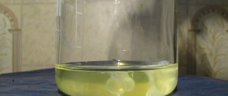
The human bladder is a hollow muscular organ located in the pelvis. Depending on the fullness of urine, its shape changes: when filled, it has a pear-shaped shape; when emptied, the organ becomes saucer-shaped, as its walls collapse after urination. The absence of pathologies on ultrasound is indicated by the symmetry of the organ, clear, even contours and echo-negativity (its structure does not reflect ultrasound waves well). If echogenic formations are found in its cavity, this indicates the development of pathogenic processes. Such an echogenic formation can be a fine suspension or sediment. In this article we will tell you what a suspension in the bladder detected on ultrasound in adults and children means, whether it poses a danger to human health and how to treat it.
What is echogenicity
Often, when conducting ultrasound diagnostics, specialists are faced with hyperechoic formations in the gallbladder, which are not a disease, but only indicate the presence of pathologies in this organ. These are mainly tissues with a calcified structure or fatty deposits. To make an accurate diagnosis, additional examination is required.
On the monitor of ultrasound equipment, echogenicity appears as a white or light spot, which leads to a conclusion regarding the presence of compactions in this area. Structures can be point or linear, and also quite voluminous.
How to accurately determine the diagnosis
Often, after an ultrasound procedure, doctors diagnose “hyperechoic pyramid syndrome,” but additional examination is necessary to make an accurate diagnosis. To do this, you need to notify your doctor about all the symptoms and signs that concern you. If you suspect the presence of kidney stones, your doctor will order a urine and blood test. In case of sclerosis of the renal vessels, surgery is necessary, but before this an MRI study is necessary to accurately determine the location of these vessels.
If a doctor suspects a patient has malignant tumors, the patient is prescribed sonoelastography, a type of diagnostic that detects cancer even at an early stage. This method makes it possible to detect malignant tumors even of such small sizes that they are not visible to the naked eye.
Why does echogenicity appear?
There are many reasons for the occurrence of hyperechoic formations in the gallbladder. They depend mainly on the general condition of the body, the size and area of localization of the formations. Most often, ultrasound reveals calcified structures, which can be single or multiple or have an acoustic shadow.
If microscopic calcifications are found, these are usually malignant tumors. Such pathologies occur with nephritis occurring in the chronic stage, injury and many other diseases. There is no liquid inside such seals, but their acoustic density is very high, so sound is poorly transmitted. Sometimes such structures can act as frame elements in organ tissues.
Ultrasound for cystitis
The most informative methods of examination for cystitis include ultrasound of the bladder with detection of residual urine and ultrasound of the prostate. Before the study, certain preparation is required, the purpose of which is to fill the bladder. To do this, an hour and a half before the procedure, you need to drink about 1 liter of liquid (non-carbonated). If there are urgent indications, the bladder can be filled using the catheterization method (a catheter is inserted through the urethra and saline solution is infused through it).
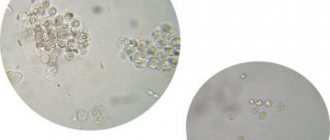
An ultrasound sign of acute cystitis is the presence of small hyperechoic particles in the bladder cavity. This is the so-called “sediment”, which consists of accumulations of epithelial cells, red blood cells, white blood cells and necrotic tissue. It is possible that the walls of the organ may thicken by more than 3 mm, but in general their contours are smooth and without deformation. If the disease becomes chronic, the walls may become deformed, the shape of the bladder often changes, and it becomes asymmetrical. The cavity also contains sediment in the form of hyperechoic (light in the picture) clots. The size of the organ decreases slightly, which indicates fibrous changes in the walls due to prolonged inflammation.
Sometimes round hyperechoic formations are found in the cavity of the bladder, which indicates urolithiasis. In men, it is possible to identify signs of prostate adenoma - an increase in its size, heterogeneity of structure, etc.
Who is at risk
The formation of atypical tissues in an organ, which is detected during ultrasound, is considered an echo-positive structure. The risk group for the occurrence of such seals includes patients with conditions such as:
- acute infections in the body;
- prolonged chemical or thermal effects on the organ;
- inflammation of the gallbladder;
- chronic diseases in the acute stage;
- poor diet and bad habits;
- insufficient physical activity;
- mechanical injury.
All these factors lead to the occurrence of pathology, so there is a need to carry out comprehensive diagnostics, treatment and preventive procedures.
Diagnosis of cystitis
Cystitis is an inflammatory lesion of the bladder caused by bacteria (much less commonly, viruses and fungi). The disease can be provoked by the following factors and pathologies:
- hypothermia;
- urolithiasis disease;
- sexual infections;
- pyelonephritis;
- adenoma and prostate cancer with difficulty in urine outflow;
- abuse of alcohol, spicy and fatty foods;
- weakened immunity.
There are chronic and acute types of cystitis. The main symptoms of the disease are:
- pain in the groin area;
- pain, burning when urinating;
- increased urination;
- unpleasant odor of urine;
- unusual color of urine, blood, pus or mucus.
- This symptomatology manifests itself most clearly during the acute course of the disease or exacerbation of the chronic form.
Main symptoms
In the presence of a hyperechoic formation, symptoms largely depend on the degree of damage to the mucous membranes. Among the main signs of pathology, the following should be highlighted:
- fever, weakness, headache;
- weight loss, sleep problems;
- dizziness and severe irritability.
In addition, pain on the right side under the ribs, swelling, nausea and vomiting may appear. If such symptoms are present, diagnosis and treatment are required.
Ultrasound diagnosis of squamous cell carcinoma of the bladder
SonoAce Ultrasound Magazine
Medical Journal of Ultrasonography - Free Subscription
(for ultrasound doctors).
The incidence of bladder cancer around the world is showing a steady upward trend. According to the World Health Organization, bladder cancer accounts for about 3% of all malignant neoplasms. In terms of prevalence, it is second only to tumors of the stomach, esophagus, lungs and larynx. Among all oncological urological diseases, bladder neoplasms occupy the second place in incidence after prostate cancer. More than 150 thousand new cases are registered annually in the world. In terms of prevalence in Europe, bladder cancer ranks fifth in men and 11 in women among all forms of this disease [1]. In 1999, 11,267 cases of bladder cancer were first detected in Russia, of which only 2.1% were detected during preventive examinations [2]. Of all morphological forms, transitional cell carcinoma is the most common, accounting for up to 90%. Less than 10% are adenocarcinoma, squamous cell carcinoma and squamous cell carcinoma.
It has been established that the carcinogenic agent is present in the urine and that the epithelium of the bladder mucosa is predisposed to proliferation. Under the influence of certain types of irritation, the epithelium undergoes changes both morphologically and biologically, which ultimately can lead to neoplasm [4]. More often it occurs in the area of the triangle and the neck of the bladder, which differ in structure from the rest of it.
Among the main etiological factors leading to the appearance of bladder tumors are chemical irritants, mainly aniline products, functional liver disorders, viruses, impaired metabolism of microelements (copper, silver, zinc, manganese, etc.), previous chronic diseases of the bladder (interstitial cystitis, granular cystitis, ulcers, bladder leukoplakia, stones, diverticula, etc., as well as chronic cystitis caused by parasites, in particular schistomatosis), smoking, stagnation of urine, high activity of lactate dehydrogenase [4,5].
At the very beginning of the disease, the clinical manifestations of bladder cancer are scanty and largely depend on the stage of the disease, the presence of complications, and concomitant diseases. The main symptoms of epithelial bladder tumors are hematuria (70%) and dysuria (15-37%). As the tumor process progresses, patients experience pain in the suprapubic region, which is constant. The pain intensifies at the end of urination. The intensity of pain depends on the location and nature of tumor growth. Exophytic neoplasms can reach large sizes without causing pain. Endophytic growth is accompanied by constant, dull pain above the womb and in the pelvic cavity. If the tumor grows into the bladder wall and spreads to the paravesical tissue and neighboring organs, symptoms of pelvic compression may occur, manifested by swelling of the lower extremities, scrotum, phlebitis, pain in the perineum, lumbar region, and genitals.
Descriptions of cases of ultrasound diagnosis of squamous cell carcinoma of the bladder are extremely rare in the literature. That is why in this observation we want to share our experience.
Patient A., born in 1930, was referred by a urologist for an ultrasound scan of the kidneys, bladder and prostate gland with a preliminary diagnosis of prostate adenoma and chronic pyelonephritis. From the anamnesis it is known that over the past 5-6 months. He noted dysuria (frequent urge to urinate, accompanied by a burning sensation when urinating, pollakiuria). Later, the process of urination became painful, pain appeared in the suprapubic and left lumbar regions. On examination: condition is satisfactory. The physique is asthenic. The skin and visible mucous membranes are in satisfactory condition. The physique is asthenic. The skin and visible mucous membranes are pale. Breathing is vesicular, no wheezing. Heart sounds are muffled. Pulse 82 beats per minute, satisfactory filling. BP=140/85 mmHg. The tongue is moist and covered with a white coating. Pasternatsky's sign is weakly positive on the left. In a general urine test taken on the day of the study: specific gravity 1025, dark orange color, cloudy urine, acidic reaction, protein 1.12 g/l, leukocytes 7-8 in p.s., red blood cells 15-20 in p.s. sp., mucus, bacteria in moderate quantities.
Ultrasound revealed the following picture: the right kidney is bean-shaped, with a smooth, clear contour, dimensions 110x55 mm, parenchyma thickness 13 mm, single dilated calyces up to 8 mm are located. The left kidney is oval in shape, with a smooth, clear contour, dimensions 115x58 mm, parenchyma thickness 11 mm, the collecting-pelvis system is expanded, calyces up to 12 mm, pelvis 25x12 mm. The sinuses of both kidneys have unevenly increased echogenicity, corticomedullary differentiation is difficult, the parenchyma has small echo-positive inclusions up to 2 mm without an acoustic shadow. After emptying the bladder, the ultrasound picture of the FLS of both kidneys was unchanged.
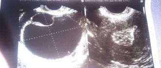
Bladder: anterior-posterior size 8 cm, transverse - 7 cm, superior-inferior - 7 cm, volume 188 cm³, wall - 4 mm, contents anechoic. On the left side wall an echo-positive formation of irregular shape is visualized, with uneven, bumpy contours, heterogeneous structure, with areas of higher echogenicity along the contour facing the bladder cavity, measuring 52x35x36 mm. The wall of the bladder closer to the mouth of the left ureter is not clearly differentiated and is blurred. The residual volume of the bladder is 102 ml. (Fig. 1 a,b). Prostate gland: oval, symmetrical, with an even, unclear contour, increased echogenicity, anterior-posterior size 48 mm, transverse - 35 mm, superior-inferior - 38 mm, heterogeneous structure, with small areas of reduced and increased echogenicity without clear contours, with echo-positive areas up to 3 mm without an acoustic shadow and with a slight acoustic shadow. Ultrasound of the inguinal lymph nodes: on the right - without features, on the left - a single hypoechoic formation of an oval shape, with clear contours, homogeneous structure, dimensions 15x7x8 mm was located, retroperitoneal lymph nodes - without features. Conclusion: diffuse changes in the parenchyma and sinuses of the kidneys. Pyeelectasia on the left. Ultrasound picture of a bladder mass with signs of wall infiltration. Increased volume of residual urine. To clarify the diagnosis, cystoscopy is recommended. Diffuse changes in the prostate gland. A single enlarged lymph node in the groin area on the left.
Hyperechoic formations are inclusions that are visible on ultrasound. Their main characteristic is the reflection of ultrasound stronger than from adjacent normal tissues. On the monitor they appear as light or white formations; their edges can be even, equal, or have no clear boundaries. The size, shape and number of such formations serve as an important diagnostic sign.
Hyperechoic inclusions reflect ultrasound waves much more strongly than normal tissues
In this article you will learn:
Features of formation in the gallbladder
Detection of an area with increased density may indicate that it is:
- stone;
- cholesterol polyp;
- bile sludge
The stone is located in the lumen of the organ and provides an acoustic shadow. He is often agile when turning and taking deep breaths. However, it may be a hyperechoic nondisplaceable mass in the gallbladder.
A cholesterol polyp grows from the wall of the organ. It has increased density. This is a parietal hyperechoic formation in the gallbladder, which has a small diameter and even outline.
Bile sludge is the accumulation of thick bile sediment at the bottom of the organ. In this case, you need to conduct an additional ultrasound after taking choleretic drugs.
What is a hyperechoic formation: about the danger of changing organ structures in simple words
A hyperechoic formation in an ultrasound image indicates tissue compaction. If such a symptom is detected, the doctor prescribes an additional examination. Compaction can be a sign of a tumor, stones, or calcifications.
The concept of echogenicity
The ultrasound method is based on the use of ultrasound. It is able to pass through the tissues of the human body and be reflected from them. This property is called echogenicity. Each organ has its own echogenicity.
In the ultrasound image, the internal organs range in color from white to black. Most are gray, which indicates uniform echogenicity. Dense tissues reflect ultrasound quickly, which is why they are white in the image. Normally these are cartilage and bones. The property is called hyperechogenicity.
Soft tissues and blood vessels reflect ultrasound slowly and appear dark gray in the image. The liquid does not reflect ultrasonic waves at all and turns black.
What are hyperechoic tissue inclusions?
Hyperechoic inclusions are formations in tissues that appear white in the image. They are similar in density to cartilage or bone. A healthy person should not have such inclusions. Hyperechoic formations are:
- tumors;
- stones;
- sand;
- calcifications.
Hyperechoic structures can appear in any organ. They are clearly visible because they differ in density from the surrounding tissue. Particularly dense inclusions have an acoustic shadow, also determined by ultrasound. The shadow appears due to the sharp transition from very dense fabric to soft one. It is located on one side of the pathological formation and looks like a dark gray spot.
Reasons for education
Hyperechoic formations appear due to pathological processes:
- inflammation;
- rebirth;
- accumulation of salts;
- stone education.
One disease can have several causes.
Features depending on the organ
Hyperechoic formations appear in various organs of the human body. They are found in soft tissues or inside cavities. Formations are distinguished according to the following characteristics:
- form;
- size;
- contours;
- intensity of brightening in the image.
Based on these criteria, the specialist suggests a particular disease.
Liver
The hyperechoic neoplasm here is a tumor; more often it is malignant. Represented by a light spot of irregular shape.
Completely clear liver tissue combined with a decrease in size is a sign of cirrhosis. This is a disease in which normal liver tissue is replaced by scar tissue.
Liver cysts can be detected due to the reverberation effect. Once in the cavity, ultrasound is reflected several times from its walls and attenuates. The image shows the light walls of the cyst with a darkening in the center.
Read a detailed article about formations in the liver.
In the video you will see the hyperechoic walls of the liver portal system:
Gallbladder
Hyperechoic formations of the gallbladder are located inside it or on the wall.
- Sludge is a thick hyperechoic suspension of bile at the bottom of the bladder. Represented by a white stripe along the bottom contour.
- Polyp. Formed by cholesterol deposits, it grows from the wall of the organ. It has a wide base and elongated shape. Size up to 4 mm.
- The stone is compressed bile salts. It looks like a bright white spot at the bottom of the bubble. There is always an acoustic shadow. The shape of the stones is often round.
It is easy to distinguish sludge or stone from a polyp. The stones move when the body position changes, but the polyp remains in place.
Uterus
A hyperechoic focus in the uterine muscle is a fibroid or malignant tumor. With fibroids, an increase in the organ itself and its deformation are simultaneously observed.
A hyperechoic formation may be a uterine polyp or a menstrual blood clot. To confirm this, an ultrasound should be done 3-4 days after menstruation.
If a hyperechoic formation is detected, shaped like the letter T, this is an intrauterine contraceptive. It has clear contours and is located in the middle of the organ.
Breast
Dense neoplasms in the mammary gland occur against the background of hormonal imbalances or inflammation:
- fibrous mastopathy - rounded light formations with an acoustic shadow;
- mastitis - linear areas of clearing or nodes;
- malignant tumor is a formation with unclear contours.
To distinguish between neoplasms, mammography and puncture are performed.
Bladder
Most often, stones are found here - small light inclusions with an acoustic shadow. They have different shapes and sizes. At the initial stage of urolithiasis, sand is detected - a hyperechoic suspension.
The bladder is affected by the infectious disease schistosomiasis. Many pinpoint hyperechoic inclusions are found in its wall.
Kidneys
Hyperechoic formations are often stones. Small white inclusions located in the cups provide an acoustic shadow. They have a round or coral shape.
A kidney tumor is usually located on its surface. It is represented by a light, rounded growth. A benign tumor has an even lighter rim - the capsule.
Thyroid
The hyperechoic area is a tumor of the thyroid gland. A light, round formation with clear contours. Inside there may be even denser areas - calcifications.
Ovaries
A dense formation in the ovary with unclear contours and a dark border around it is cancer. A tumor with a capsule around it and a heterogeneous structure is a teratoma. This is a congenital ovarian cyst containing fat, hair, and teeth.
Ultrasound of the fetus
When examining pregnant women, the fetus sometimes reveals a GEF - a hyperechoic focus - in the left ventricle of the heart. Most often, it does not matter for the further development of the fetus. The GEF in the heart is an accumulation of calcium salts or an additional chord - an outgrowth of the ventricular wall.
But sometimes it is a sign of chromosomal abnormalities. If a woman has risk factors, she undergoes additional examination. A needle is inserted into the amniotic sac and a small amount of water is taken for analysis.
The same examination can be done in the placenta. The analysis identifies any genetic abnormalities.
Watch a video about fetal GEF:
Conclusion
Ultrasound examination reveals even minor changes in tissues. But with the help of ultrasound, one can only guess the nature of the pathological changes - in shape, size, density.
The final diagnosis is made by the doctor taking into account other examinations. To obtain the most accurate result, you need to prepare for the examination.
Acoustic access, that is, the passage of ultrasound, is difficult due to flatulence and feces in the intestines. To cleanse the intestines, a diet and carminative drugs are prescribed. Preparation for ultrasound of the thyroid gland and kidneys is less strict.
Leave comments on the article and share useful information with friends on social networks. All the best.
Source: https://uziman.ru/metodika/giperehkhogennoe-obrazovanie
Formations with acoustic shadow
During diagnostics, various foreign inclusions may be detected. Quite often, after an ultrasound examination, the report states “hyperechoic formation with an acoustic shadow.” It is formed from stones, dense and connective tumors.
Many people are interested in what a hyperechoic formation means. This is a fairly dense structure that reflects ultrasonic waves. In addition, when conducting a study, the specialist must find out whether there is an acoustic shadow behind the formation. Its presence indicates that the object under study is so dense that it does not transmit ultrasonic waves at all.
If the doctor detects a solid mass followed by an acoustic shadow, the first thing he thinks about is a gallstone. It is dense enough that ultrasonic waves do not pass through.
A shadow forms at the border of tissues that reflect ultrasound well. During exploration and passage through such very dense structures, the ultrasonic beam is almost completely interrupted and a reflection is formed. That is, such fabrics have high acoustic density.
Types of benign bladder tumors
In order to determine a therapeutic strategy, it is first necessary to establish the type and nature of the tumor. In urology, all neoplasms are usually divided into two broad groups: epithelial and non-epithelial. Moreover, the vast majority (about 95%) of formations belong to the first group. The undisputed leader in incidence among all epithelial tumors of the bladder is cancer of this organ. However, if a patient hears from a specialist about the presence of an epithelial tumor, this does not mean that the tumor is malignant. The following formations are considered benign:
- Bladder polyps.
These growths face the lumen of the bladder and in the structure of the covering tissue do not differ from the surrounding healthy urothelium. A distinctive feature of papillary formations of this type is the presence of a fibrovascular base, which can be either thin or quite massive. - Bladder papillomas.
This group includes exophytic neoplasms. Most often, the development of papilloma occurs as a result of the proliferation of integumentary tissue. In this case, the tumor has a light pink tint and a velvety surface, and its consistency is soft. Multiple papillomas are often found.
Heterogeneous formation
This may indicate the presence of malignant or inflammatory processes in the body. A heterogeneous hyperechoic formation generally indicates that cysts are forming inside the organ. Tumors can change the structure of the gallbladder at any stage. To determine what a hyperechoic formation with a heterogeneous structure means, a comprehensive diagnosis is required.
It may also indicate inflammation. In a healthy person, the echostructure of the gallbladder is homogeneous and even. When performing an ultrasound examination, it is possible to determine the peculiarities of the functioning of this organ.
Diagnostics
As mentioned earlier, to detect a hyperechoic inclusion, the patient is prescribed an ultrasound examination. But still, in order to further determine its specific origin and nature of the darkening, the doctor prescribes a number of additional diagnostic procedures to the patient. If the diagnosed formation in the kidney has dense edges, as well as clear and even outlines, and fluid is diagnosed inside it, then most likely it is a cyst.
If we are talking about a tumor, then such a formation, on the contrary, has blurry and uneven outlines that have a heterogeneous structure. Additional symptoms that indicate a developing tumor are enlarged lymph nodes behind the peritoneum.
In order for the doctor to make an accurate diagnosis of the patient, in addition to ultrasound examination, the following procedures are prescribed:
- it is necessary to take a blood and urine test;
- undergo MRI and CT;
- Dopplerography;
- apniography;
- biopsy of a section of kidney tissue.
The patient must undergo blood and urine tests
Only after all the results of the above studies are available to the doctor, will he be able to make an accurate diagnosis and, based on it, make the right decision on the method of getting rid of the pathology.
Formation with anechogenic inclusions
The echogenicity of tissues depends on the ability to absorb and reflect ultrasound passing through them. This is directly related to the characteristics of the organs. The less liquid is contained inside the object under study, the higher the degree of echogenicity. The absence of fluid indicates the lowest density, that is, anechogenicity.
A hyperechoic formation with inclusions generally indicates malignant processes in the body. Additional research may be required to more accurately indicate the type of echostructure and its danger.
What do hyperechoic inclusions in the kidneys mean and why are they dangerous?
Hyperechoic structures can be detected during ultrasound diagnostics, the result of which is the visualization of a black and white image.
Lighter areas of the image indicate the presence of areas with increased acoustic density (hyperechoic).
Abnormally “hard” formations in the kidneys, especially those accompanied by pathological symptoms, require immediate medical attention.
Hyperechoic inclusions - what are they and where can they be found?
When performing an ultrasound examination, the result is formed based on the ability of tissues to reflect ultrasonic waves emitted by the sensor. Moreover, the coefficient of this ability will vary depending on the tissue density in each individual section of the organ.
It is for this reason that ultrasound diagnostics are of little information for organs in which there are cavities containing air (intestines, esophagus - due to the fact that air does not provide an acoustic picture) and requires preparation for the bladder or ureters (filling the organs with fluid).
There is a relationship between the echogenicity of tissues and the fluid content in them - the more of the latter, the lower the echogenicity will be.
Hyperechoic structures are areas within an organ whose increased echogenicity stands out against the background of neighboring formations.
They can be detected when performing diagnostic procedures to examine:
- organs in the abdominal cavity - liver, spleen, gallbladder and bile ducts, pancreas, kidneys. Sometimes an ultrasound of the stomach is performed (with adjacent organs - the duodenum and esophagus), although this study is considered uninformative;
- organs of the genitourinary system - the bladder and ureters, the prostate gland in men, the ovaries and uterus in women.
Hyperechoic objects are denser than neighboring areas; these can be cysts, hematomas, areas of tissue degeneration or regressive age-related changes, kidney stones.
The interpretation of the result will depend not only on the echostructure of the organ being studied, but also on the “picture” provided by those adjacent to it.
For example, hyperechogenicity of the pancreas occurs if its tissues on the monitor look “whiter” than the structures of the liver.
For this and many other reasons, hyperechogenicity does not always characterize pathology, but if the disease still exists, the study can provide the doctor with a lot of diagnostic information.
Types of hyperechoic structures
During an ultrasound examination, a diagnostician can detect echo-dense structures that appear on the screen as light, almost white spots. The equipment also makes it possible to differentiate these objects based on the presence of an acoustic shadow. It occurs in cases where the area is so dense that it does not allow a sound wave to pass through it.
Hyperechoic inclusions, based on how they are visualized on the monitor and the presence/absence of an acoustic shadow, are divided into three groups:
- Significantly sized formations with the presence of an acoustic shadow. This picture is typical for stones and macrocalcifications. Sometimes this is what areas of sclerosis (connective tissue that has replaced functional tissue as a result of a necrotic process) or hematoma (bleeding in the kidney tissue) look like, in which a large amount of calcifications have been deposited over time.
- Large, moderately dense inclusions with no acoustic shadow. This is what benign neoplasms can look like on ultrasound - fibromas, oncocytomas, hemangiomas, as well as fluid-filled cavities - cysts (dense). Sclerotic tissue areas, accumulations of very small kidney stones (sand), or abscess areas may also have medium density.
- Small inclusions of white color that look like luminous dots on the monitor. These can be either very dense, but small calcifications, or psammoma bodies - round-shaped formations up to 3 mm in size, having a layered structure. The latter are based on protein-lipid complexes. Such inclusions are denser than any tissue of the human body; their presence in the kidneys can be a symptom of a malignant neoplasm.
In real conditions, changes in the ultrasound picture are rarely homogeneous; more often, inclusions contain a combination of hyperechoic structures; for example, an area of sclerosis may also contain calcifications.
What diseases are inclusions typical for?
An increase in the echogenicity of an organ area suggests the presence of pathologies such as:
Often, oncological pathology in the early stages is characterized by an asymptomatic course. But you can find out about the presence of other diseases by paying attention to the following signs:
- sharp, rapidly increasing pain in the kidneys (colic);
- chills, feverish manifestations;
- pain of various types in the abdomen or lower back;
- nausea or vomiting, weakness (signs of intoxication);
- disruption of the urination process (cramps, changes in the volume of diuresis per day);
- pain in the abdomen or groin area (paroxysmal or aching);
- change in the color and/or consistency of urine;
- disturbances in intestinal function.
Based on the type of hyperechoic inclusion and the patient’s complaints, further examinations of the body are prescribed.
Carrying out diagnostics
The main method for detecting a hyperechoic formation is ultrasound diagnostics. If such a compaction is detected, the cause of its occurrence must be determined. It is important to take into account the general condition of the body, as well as existing symptoms. To make an accurate diagnosis, additional studies may be required, which are prescribed by the doctor.
After the diagnosis, the doctor draws up a treatment plan. Treatment may be medication or surgery. If a small parietal hyperechoic formation is detected, which is not accompanied by pathological symptoms, constant monitoring of its condition through regular ultrasound is indicated.
Consequences of the disease
If you do not diagnose a hyperechoic formation in the kidney in a timely manner or try to get rid of it yourself, then the pathological process can provoke the development of serious and life-threatening complications. As soon as a patient is diagnosed with this type of disease, it is very important to find out the cause of its occurrence, since the treatment tactics and prognosis for recovery will completely depend on this. If the cause is a congenital pathology, then it seems possible to get rid of the disease only through surgery.
If a patient is diagnosed with a cyst, then such a formation can provoke suppuration, twisting or necrosis. Purulent formations, in turn, are dangerous because they can lead to the spread of the inflammatory process throughout the kidney, and the infection can enter the blood and provoke its infection. In some cases, purulent formations can lead to the development of sepsis. The same danger awaits a person if inflammation of the hematoma occurs.
The riskiest option is considered to be a tumor, which in most cases is malignant. This indicates the development of cancer in the human body. It is possible to get rid of such a formation only at an early stage of development, until metastases appear.
The appearance of even minor symptoms indicating a hyperechoic formation requires immediate diagnosis. To make an accurate diagnosis, the patient must undergo a full range of examinations. Only in this case can you make an accurate diagnosis and find out the cause of the development of the pathological process and, accordingly, decide on the most optimal and effective method of treatment.
Regardless of what hyperechoic inclusions are diagnosed in the kidneys, this is a disease that requires timely diagnosis and professional treatment. Only in this case can you get rid of the pathology without harm to your body and increase the survival rate.
Features of treatment
Treatment of hyperechoic formation in the gallbladder is selected depending on the results of the study. If calcifications are detected, the doctor prescribes medications that help remove them. In particularly complex and advanced cases, surgery is indicated.
In the presence of compactions caused by various diseases, inflammatory and infectious processes, antibiotics are required. Benign and malignant tumors are treated with surgery and may also require chemotherapy. For multiple hyperechoic avascular formations, medical supervision is indicated.
Treatment of the disease
Depending on the diagnosis, each patient is prescribed treatment on a purely individual basis.
If a patient is diagnosed with an anomaly in the structure of the kidney, then in most situations a decision is made to monitor the patient’s condition and monitor all changes. If we are talking about disturbances in the functioning of the kidney, then the only way out of this situation is surgical intervention.
Very often, patients experience hemorrhages or hematomas as a result of injuries; surgical intervention, of course, is not prescribed for the treatment of such formations, the only exception being situations where there is a real threat to the normal functioning of the kidney.
Medicines are prescribed individually by a doctor
Diagnosed purulent processes are treated mainly with medications and, in extreme cases, removed surgically. In this case, the decision is made exclusively by the attending physician separately in each case.
As for formations such as cysts, each case is also considered separately. If there is no threat of its rupture, degeneration or suppuration, then observational tactics are chosen, otherwise an operation is performed to remove it.
Tumors are rightfully considered one of the most dangerous formations. Treatment decisions are made only after the type, size and location have been determined. If a patient is diagnosed with a malignant tumor, then it can only be gotten rid of surgically and then only with timely access to a medical facility. In an advanced stage, malignant tumors in almost all cases lead to death. In the process of getting rid of malignant tumors, either part of the organ or the entire kidney can be removed. Metastases and lymph nodes are also removed together.
If a patient is diagnosed with a malignant tumor, the only way to get rid of it is through surgery.
Regardless of what kind of formation develops in the kidney, all of them can be gotten rid of without harm to health, only if you contact a medical facility in a timely manner and strictly follow all the recommendations of your doctor.
Drug therapy
Treatment largely depends on the underlying cause that provoked the appearance of a hyperechoic formation in the gallbladder. Medicines should be selected only by a doctor, taking into account the patient’s condition. If inflammation is observed in the body, then anti-inflammatory drugs are required, in particular, such as Ibuprofen or Ketorol.
Ibuprofen is an anti-inflammatory drug with an analgesic effect. Used for inflammation. The dosage and duration of administration depends on the severity of the pathology. It is worth remembering that the medicine has contraindications and can cause side effects.
"Ketorol" is an anti-inflammatory drug with analgesic and anti-inflammatory effects. It is used for fairly intense pain and inflammation in the body.
Additionally, you may need to take vitamin complexes that help strengthen the immune system and help fight various diseases.
Hyperechoic formation: causes of development and characteristic signs
Hyperechoic formations are inclusions that are visible on ultrasound. Their main characteristic is the reflection of ultrasound stronger than from adjacent normal tissues.
On the monitor they appear as light or white formations; their edges can be even, equal, or have no clear boundaries.
The size, shape and number of such formations serve as an important diagnostic sign.
Hyperechoic inclusions reflect ultrasound waves much more strongly than normal tissues
The concept of hyperechogenicity
Ultrasound uses the ability of human body tissue to reflect ultrasound, which is expressed differently in different tissues. Normoechogenicity is the natural reflection of ultrasonic vibrations for a certain tissue. Hyperechogenicity is an excess of this indicator. It indicates that a certain area is denser than neighboring tissues.
The normal echogenicity index varies for different tissues - for bones it will be higher than for muscles, for lungs it will be lower than for liver. Accordingly, the concept of hyperechogenicity changes for different tissues.
It should be clarified that hyperechogenicity is not a disease or a syndrome, but only an indicator of changes in the physical properties of the tissue. In order to find out the reasons for such changes, additional examinations are required.
Where are formations located, their types
The localization of pathological formations may be different. Most often they are found in the liver, kidneys, female and male genital organs, endocrine glands, heart, digestive organs, and less often - bones. In general, they can be found in any organ.
Types of hyperechoic inclusions:
- small formations with pronounced boundaries. There may be several of them; on the monitor they are displayed as bright inclusions in the main fabric;
- large formations with clear boundaries, less bright than the previous group;
- formations of especially large sizes with an acoustic shadow. Most often they do not have clear boundaries;
- heterogeneous formations - having a different structure, inclusions, changes in density.
Hyperechoic formations can be detected in any organs
Each type, as a rule, has its own causes of origin, but making a diagnosis only based on ultrasound results without additional examinations will be inaccurate.
Possible reasons
The reasons for the appearance of such formations are processes leading to compaction of organ tissue. Among them may be:
- consequences of inflammatory diseases;
- deposits of calcium salts and similar substances;
- proliferation of connective tissue in glandular organs;
- high density foreign body;
- presence of stones;
- benign and malignant tumors;
- artificial structures (for example, in bone tissue).
Hyperechoic formations may indicate the presence of stones in the organ
Such conditions can cause hyperechogenicity, but this is not always a sign of disease. However, based on the typical location, one can assume a possible pathology in the patient. Thus, hyperechoic formations in the gallbladder most often turn out to be stones, and the same formations in the thyroid gland are a possible tumor.
How they show themselves
Symptoms of this pathology vary significantly depending on the location and causes. Some of them may not manifest themselves in any way for many years. In any case, the symptoms are not associated with hyperechogenicity itself, but with the processes leading to it. The main characteristics are shown in the table.
LocalizationVarietyPossible causesSymptoms
| Liver | Small affected areas without clear boundaries | Tumors, in particular hemangioma | Signs of liver dysfunction - loss of appetite, heaviness in the right side, decreased performance, “liver laziness” |
| Gallbladder | Large, bright formations with clear boundaries | Stones, anatomical defects | Digestive disorders, biliary colic. May remain asymptomatic for a long time |
| Kidneys | Volume or point hyperechoic inclusions | Stones, vascular pathologies, tumors | Urinary disorders, general malaise, unpleasant odor of sweat, changes in the amount and type of urine, lower back pain |
| Bladder | Large, with clear edges | Passing stones, anatomical pathologies | Pain when urinating, difficulty urinating, blood in the urine |
| Uterus | The density is close to normal, there are no clear boundaries | Pathological formations in the uterus, consequences of an incorrect abortion or curettage | Pain during menstruation, menstrual irregularities, infertility or recurrent miscarriage, bleeding from the genitals |
| Prostate | Small point inclusions | Stones in the prostate, consequences of prostatitis, prostate adenoma | Impaired potency, pain in the groin area, urinary disorders |
| Pancreas | Various inclusions – large and small, with varying degrees of boundaries | Pancreatitis, tumors and metastases, calcifications, fibrosis, necrotic changes | Digestive disorders, abdominal pain, signs of diabetes, severe deterioration in health |
| Heart | Formations without clear edges, elongated or diffusely located, linear hyperechoic inclusions | Consequences of heart attack, myocarditis, cardiosclerosis, necrotic changes | Chest pain, disturbances in heart function, signs of heart failure - decreased physical endurance, deterioration of resistance to emotional and physical stress |
| Thyroid | Nodular formations | Tumors or metastases | Various hormonal disorders |
Diagnostics
A functional diagnostician evaluates the results he sees on the screen and ultrasound images.
They clearly show the various characteristics of the formation - its size, clarity of boundaries, contrast with surrounding tissues, the number of such formations, their location and similarity to each other.
Sometimes this data is enough to make a full diagnosis, for example, with cholelithiasis or urolithiasis.
Ultrasound is not the only method for diagnosing hyperechoic formations. If such an examination reveals abnormalities, the patient needs a number of additional tests.
Depending on the area in which the formation is found, these may be thyroid hormones or sex hormones, a biochemical blood test, urine tests, a biopsy of the affected organ, functional tests that reveal the extent of the damage.
From the video you will learn what a hyperechoic focus in the fetal heart is and what it looks like:
Physiotherapeutic techniques
For hyperechoic round formations, physiotherapeutic techniques may be prescribed for treatment. They imply an effect on the body through:
- electric current;
- heat or cold;
- laser, infrared, ultraviolet radiation;
- ultrasound;
- magnetic field.
It is also possible to use hirudotherapy and massage. The main advantage of such methods is that they are effective and safe. This will help reduce the period of therapy for various pathologies and activate biochemical processes. As a result, recovery is accelerated.
Cryotherapy is based on the effect of fairly low temperatures on the body. It relieves pain, stimulates the immune system, eliminates inflammation and swelling.
Laser therapy is a biostimulation method based on the effect of laser on living tissues. It activates the most basic biochemical processes occurring in the body, promotes the regeneration of cells and tissues. Laser therapy improves blood microcirculation, accelerates the healing process of various lesions and eliminates inflammation.
Magnetotherapy is a modern method of influencing the body using a magnetic field. It promotes overall health, normalizes blood circulation and oxygen saturation. In addition, this technique improves the functioning of internal organs.
If a person has echogenic inclusions in the form of stones, then electrophoresis with antibiotics is often used to treat them. During the procedure, medications quickly penetrate the affected tissues, normalize blood circulation and promote the resorption of tumors.
What does the presence of renal hyperechoic inclusions indicate?
Volumetric or linear hyperechoic inclusions in the kidneys may indicate the presence of urolithiasis, and areas with increased acoustic density are renal calculi (stones). The absence of shadow echoes in this case excludes urolithiasis.
Kidneys with small hyperechoic inclusions, if they are streak-shaped, are not considered by doctors as a pathology, since these can also be vessels. In the worst case, these are foci of fibrosis.
To exclude the possibility of developing cancer, doctors must prescribe additional tests:
- blood test for tumor markers;
- kidney tissue biopsy;
- daily urine analysis for the presence of mineral salts;
- general blood analysis.
The final diagnosis can be established only by the doctor, comparing the ultrasound results with the clinical manifestations of the disease and additional laboratory tests.
urolog.pw
Folk remedies
They are used to reduce the size of echostructures and prevent their growth, help relieve inflammation and help accelerate metabolic processes.
Take 20 g of rosehip rhizome, pour boiling water and leave for 5-7 minutes. Drink 50-70 ml before meals. Positive results are observed with regular use of the product for 6 months.
Take 10-15 g of birch bark and pour 200 ml of boiling water. After 30 minutes, filter, add water and 10 ml of lemon juice. Drink the infusion 3 times a day before eating. If a stone has been discovered, then to treat it you need to mix dandelion roots, larkspur, St. John's wort, knotweed and violet in equal proportions. Take 5 tbsp. l. mixture, pour 1 liter of boiling water and leave until completely cool. Drink 250 ml 2-3 times a day.
Diseases caused by hyperechoic inclusions of the kidneys. Treatment
In most cases, hyperechoic renal inclusions appear as:
- inflammatory process: carbuncle, kidney abscess.
- cyst-like growths (usually containing liquid).
- hemorrhages in the kidney (a kind of hematoma).
- kidney tumors (benign or malignant).
If the doctor suspects the above diseases, he sends the patient for a comprehensive examination using MRI. In some severe cases, a kidney biopsy is required.
Hyperechoic inclusions are not easy to cure, but it is possible. Stones are removed in two main ways. The first method is based on frequent urination, for which special diuretic herbs or medications prescribed by the doctor are used. The second method is removing stones using laser beams when they are crushed. Using the first method, small stone formations, no more than 5 mm, can be treated. In case of advanced disease, the kidney is removed, then chemotherapy is prescribed in order to remove the remaining formations. In such radical situations, constant adherence to the diet is necessary.
Remember: only a specialist can make an accurate diagnosis. Based on the kidney ultrasound and test results, he will prescribe appropriate treatment. Never self-medicate - this can make the situation worse.
po4ku.ru
Carrying out the operation
A radical method of getting rid of echo-positive formations is to perform surgery. It is indicated for large calcifications that are localized in various tissues and organs. The operation can be open or using laparoscopy.
Surgery is required if there are many small stones with sharp edges. Such compactions are considered very dangerous, since there is a risk of injury to tissues and organs due to the movement of these inclusions. It is also possible to remove malignant tumors. The operation is combined with drug therapy and various physiotherapeutic techniques.
Prevention of discharge pyramidal syndrome in the kidneys
Few people know about the echogenicity of their organs, and even fewer think about it. Meanwhile, the hyperechogenicity of the kidneys indicates the presence of dangerous inclusions, which indicate the syndrome of isolated pyramids developing in the body.
Sharp outlines or white spots in an ultrasound image of the kidneys are signals of ongoing pathologies and even insufficient body weight. In any case, everyone needs to know about the condition of their kidneys.
Briefly about the syndrome
Echogenicity is characterized by the degree of reflection of sound from the tissues and fluid of internal organs, and the appearance of strong reflections on ultrasound signals the presence of various foreign inclusions.
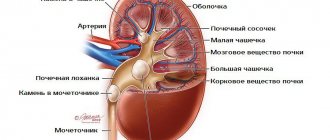
Most often, hyperechoic formations form in the renal pyramids and parenchyma. Pyramids are the triangle-shaped areas of the kidneys through which, after filtration, urine exits into the pyelocaliceal system, and parenchyma refers to the tissue that fills the organ from bone and cortex. Any extra element of the structure, being a consequence of the pathology occurring in the organ, introduces dissonance into its functioning, and often developing on one, soon affects the second kidney.
The degree of negative impact of hyperechoic inclusions directly depends on their size, composition and the possible development of malignant cancer processes in them.
All excess elements are divided into calcium stones, stones, sand and neoplasms. The last group includes formations of various types:
- Small formations appear as white dots during ultrasound. The absence of an acoustic shadow indicates their safety;
- The following type of inclusions is characterized by a large size, which can be either benign or malignant, cancerous tumors. They are extremely rare and require constant monitoring by doctors;
- Large light spots on ultrasound indicate cancerous inclusions, which, unlike other formations, necessarily have an acoustic shadow, areas of sclerosis, and also contain calcifications and psammoma bodies.
8 main causes of hyperechogenicity
Hyperechoic renal pyramid syndrome is not an independent disease, but an accompanying ailment, as it is a consequence of a pathology developing in the body. The cause of their appearance may be the following pathological renal processes:
- Polycystic disease;
- Sclerosis of blood vessels;
- Injuries;
- Bleeding;
- Purulent inflammation of tissues;
- Accumulation of several abscesses and boils in one place;
- Oncological processes;
- Fatty formations in the pyramids of the kidney.
Symptoms and diagnosis
Hyperechoic pyramid syndrome can be suspected by the following characteristic clinical manifestations that occur against a general background of weakness and fatigue: high temperature (up to 39°C), dark brown or red urine, stabbing pain in the kidney area; spasms in the abdomen and groin, vomiting, nausea, nagging pain in the groin.
The symptoms of the syndrome are characteristic of many renal diseases, however, with the help of ultrasound, a specialist will immediately diagnose the presence of hyperechoic inclusions, and will also assess the condition of the kidney parenchyma against the background of prominent pyramids.
When a diagnosis of “hyperechoic pyramid syndrome” is made during diagnosis, additional procedures are prescribed to identify the root causes of the process and prescribe appropriate therapy. Thus, general blood, urine, and stool tests are required.
Therapy and prescriptions
Treatment of hyperechoic pyramid syndrome is complex and is aimed at relieving pain syndromes and eliminating pathological preconditions.
Timely detection of illness involves drug therapy, during which a urologist or nephrologist prescribes antibiotics, analgesics, antispasmodics, as well as drugs that have anti-inflammatory and antibacterial effects to the patient. A change in routine and diet is required, including proper rest, drinking plenty of fluids, eating low-fat foods, fresh vegetables and fruits.
When the disease passes into the chronic stage, physiotherapeutic procedures and homeopathic medicines are recommended.
The folk way to cleanse the kidneys! Our grandmothers were treated using this recipe...
Cleaning your kidneys is easy! You need to add it during meals...
Advanced cases require a more individual approach and surgical intervention.
Prevention of hyperechoic syndrome
To prevent the occurrence of hyperechoic inclusions in the tissue and pyramids of the kidneys, it is necessary to promptly identify and treat all diseases of the body’s urinary system, for which it is recommended to undergo regular preventive examinations.
Of great importance in the prevention of renal pathologies is a balanced, proper diet, which does not impede, but stimulates the functioning of the organ:
- Constant consumption of fermented milk products;
- Drink plenty of clean water, berry compotes and fruit drinks, jelly and weak tea;
- Steam cooking;
- Preference for fresh homemade seasonal products: fruits, vegetables, berries;
- Predominant consumption of pasta and various cereals.
The syndrome of protruding renal pyramids, which is not life-threatening, only indicates an ongoing pathological process, which, if neglected, can develop into a cancerous tumor, chronic or acute stage of renal failure. Only timely and regular examinations will get rid of looming problems and prevent minor formations from developing into serious diseases.
Forecast
Identification of hyperechoic areas during ultrasound diagnostics is not a diagnosis. However, in any case, you need to undergo repeated diagnostics or resort to auxiliary instrumental techniques. The prognosis directly depends on the results of the examination.
If the compacted areas are small and do not affect other organs and tissues, then you can simply monitor them and visit a doctor periodically. If stones, calcifications, or tumors are detected, then it is imperative to carry out the therapy prescribed by the doctor. Treatment can be conservative and surgical.
Classification of changes in the kidneys
Depending on the visualization of changes in organs, they are divided into 3 types of formations of increased density:
- volumetric inclusions creating an acoustic shadow. Large formations or a sclerotized lymph node due to inflammation are reported;
- volumetric structures without acoustic shadow. Usually they inform about the formation of: cysts, atherosclerotic vascular pathologies, sand in the kidneys, fatty tissue of the sinus organ, benign or malignant tumors;
- small, pinpoint, hyperechoic formations without acoustic shadows. Such echo responses are considered a sign of calcifications or the presence of psammoma bodies. Observed in diffuse sclerosing, cancerous types of neoplasms.
Thanks to ultrasound diagnostics, it is possible to detect stones as small as 2 mm.
Smaller changes are more difficult to distinguish, since they have to be differentiated from other hyperechoic structures of the body. A mass formation in the kidney up to 3 cm can also be a sign of:
- small benign cyst;
- pseudotumors;
- abscess;
- renal cell carcinoma;
- adenomas;
- oncocytomas;
- angiomyolipomas;
- tumor metastasis to the kidneys;
- lymphomas.
There are 3 types of volumetric renal inclusions:
- cystic - smooth, round formations with clear boundaries that transmit echo signals well through the posterior wall;
- solid – characterized by an irregular shape with uneven edges, lack of clear visualization between the volumetric inclusion and the kidney;
- complex - abscesses, areas of necrosis, calcification or hemorrhage form within the detected structure.
Morphological variants of hyperechoic renal formations:
- fibrous-sclerotic areas – accumulations of calcareous salts (70%);
- calcifications - grouping of calcium salts (30%);
- psammoma bodies – protein-lipid deposits (50%).
Psammoma bodies in renal masses are a sign of a malignant tumor, since they are never present in benign tissues. The latter usually have fibrous-sclerotic areas as their main composition.
The presence of calcium salts reflects the age of the pathology. Calcifications take several months to deposit. They most often accumulate in areas of inflammation or damaged tissue.
Prevention
There are many reasons for the appearance of echostructures in tissues and internal organs. Their prevention consists in preventing provoking factors, that is, possible diseases. Prevention measures include:
- timely treatment of chronic diseases;
- proper nutrition and sufficient physical activity;
- consumption of vitamins.
Particular attention should be paid to the treatment of infectious and inflammatory processes, which often provoke pathological changes. Proper nutrition and sufficient physical activity are very important to maintain beauty and health. Diet therapy and drinking regimen are required. They help keep the body in good shape and give a boost of vivacity and energy.
It is important to regularly consume foods and vitamin supplements containing vitamins A and E. They improve immunity and protect the body from bacteria and infections.
In addition, it is imperative to undergo periodic preventive examinations with a doctor. If pathological symptoms occur, you should not self-medicate, but immediately seek medical help.
Reasons for the formation of tumor formations
Scientists have not yet identified an unambiguous etiotropic factor that provokes the appearance of a neoplasm. However, the most important role is played by stasis (stagnation) of urine in the bladder, which can be a consequence of many pathological conditions, such as urolithiasis, strictures and diverticula of the urinary tract, prostate adenoma, as well as prostatitis and prostate cancer. Since most of the above conditions are exclusively “male” diseases, neoplasms are much more often diagnosed in the stronger sex. At the same time, the risk of establishing a tumor process increases with age and reaches a critical value at about 50 years.
Urine is retained in the bladder much longer than in any other organ of the urinary system, which explains the prevalence of this tumor localization. As a result of the accumulation of breakdown products of amino acids such as tryptophan, proliferation of the epithelium lining the organ cavity (urothelium) can develop. In the future, this process is compensated by the formation of growths, which experts call neoplasms. In the development of this pathological mechanism, the chemical factor also plays an important role. For example, statistics show that both benign and oncological formations are diagnosed much more often in individuals who have frequent and prolonged contact with aromatic amines, such as gasoline and benzene.
Sometimes the etiology of benign tumors is associated with the lack of rational and timely treatment of cystitis, as well as with the presence of trophic and ulcerative lesions of the organ lining.