How to identify and treat kidney tumors in children
- 4 minutes to read
Kidney tumor in childhood is a disease that is diagnosed in extremely rare cases. Nephroblastoma (Williams tumor) is considered one of the most common types of malignant pathology among children under 5 years of age.
Content
The formation of kidneys in intrauterine development occurs at a rapid pace. As a result, in some cases the cellular differentiation of the organ is disrupted. Some remain incompletely formed even after the birth of the child. It may happen that by the age of four, the growth of these cells gets out of control, against which the oncological process begins.
Causes and prerequisites for the occurrence of nephroblastoma in children
In modern medicine, there are no specific reasons why kidney cancer occurs in children. In this case, carcinogenic substances are also not detected. However, it is still worth noting that some conditions and factors can become decisive in the process of disease formation. So, here are the main ones:
- If the baby has a hereditary predisposition to cancer, then there is a chance of developing a tumor;
- If any of your bloodline relatives suffered from diseases such as Bourneville-Pringle disease or Hippel-Lindau syndrome, then there is a possibility of a tumor forming in the baby;
- If your child has a weak and insufficient immune system;
- Smoking has a negative effect on the development of the disease, and passive smoking has an even greater effect;
- Long-term exposure to radiation increases the risk of developing kidney cancer.
One of the common types of tumors in children is the mixed type, which is named after Williams - in honor of the discoverer. This is one of the most popular types of disease, which occurs in many children, especially under the age of 5 years. It is worth noting that this disease is also observed in infants.
Any embryonic tumor is a failure of intrauterine development. Germ cells, being the basis for the growth and development of the fetus, after birth should gradually disappear in the first years of the child's life. Most often, the causes of tumor pathology can be:
- late age of the woman bearing the fetus (over 40 years);
- viral diseases in the expectant mother, especially in the 1st trimester of pregnancy;
- hereditary kidney pathology in the family;
- fetopathy in the fetus (intrauterine development defects);
- multiple congenital pathology.
Most often, Wilms tumor is an unpredictable situation: a pregnant woman safely carries and gives birth to a healthy baby, who is diagnosed with nephroblastoma 1-3 years after birth.
The development of Wilms tumor is believed to be caused by changes in genes responsible for the normal development of the genitourinary system. Examples of common congenital anomalies associated with Wilms tumor are:
- Cryptorchidism.
- Double urine collection system in the kidneys.
- Horseshoe kidney.
- Hypospadias.
Environmental exposure, although considered a risk factor, has virtually no effect on tumor growth.
In the early 1970s, Nadson proposed a genetic model for the development of Wilms tumor. The WT1 gene, the first suppressor gene on chromosome arm 11p13, has been identified as a direct culprit in the development of nephroblastoma, as well as other genitourinary anomalies and mental retardation. Characterization of this novel suppressor gene has provided insight into the mechanisms underlying normal renal development and Wilms tumorigenesis. A second gene that predisposes to Wilms tumor development has been identified but has not yet been cloned, a telomere from WT1 on arm 11p15.
The results of analysis in large pedigrees with familial transmission of predisposition to Wilms tumor suggest the existence of an additional genetic background.
Types of tumors
Kidney cancer in childhood is classified into two types:
- tumor neoplasm with favorable histology;
- tumor with unfavorable histology.
On this topic
Adenoma and carcinoma
- Natalya Gennadievna Butsyk
- December 9, 2020
The latest type of oncological process indicates that the cells that have undergone changes have impressive sizes and a different structure compared to unaffected kidney cells. This phenomenon in medical terminology is called anaplasia. The higher the concentration of anaplastic cells, the less likely it is to completely cure the disease.
Depending on the stages of development, a kidney tumor goes through several stages:
- first - the neoplasm is localized inside the organ and does not extend beyond its limits, and there is no growth into the sinuses of the kidney;
- second - nephroblastoma can extend beyond the kidney and grow into nearby organs and vessels;
- third – metastasis to the abdominal cavity and lymph nodes is noted;
- fourth - the tumor affects organs distant from the kidneys;
- the fifth is bilateral nephroblastoma in any of its variants.
If a child’s neoplasm is diagnosed in the early stages, then treatment of the disease will be effective.
Tumors in the kidney
A tumor in the kidney, like in any other place in the body, is a pathological process. It is characterized by uncontrolled tissue proliferation, and in the case of a malignant course, by the complete loss by cells of the typical structure of the tissue from which they originated.
Classification based on the nature of growth, the presence of a membrane, and the ability to form metastases divides tumors into cancerous (malignant) and non-cancerous (benign).
Oncological formations
Kidney cancer is a disease in which the cells of the organ become malignant and grow uncontrollably. Most often, kidney tubular cells are susceptible to this process, and the disease is called renal cell carcinoma.
It is difficult to reliably establish the causes of kidney cancer, but there are a number of conditions that increase the risk of its occurrence:
- Smoking.
- Male gender. Women are half as likely to get kidney cancer.
- Obesity. Excess weight indicates a disruption in the hormonal system, which also increases the risk.
- Some painkillers.
- Long-term hemodialysis.
- Genetic diseases.
- Hereditary predisposition.
- The influence of certain substances, for example, asbestos, cadmium, benzene, organic solvents or some herbicides.
- High blood pressure.
- Presence of lymphoma.
Wilms tumor
A striking example of the uncertainty of the nature of the appearance of neoplasms can be called Wilms tumor. This form of cancer occurs in young children.
In an unborn child, the kidneys are formed from primitive cells. Over time, these cells become more specialized and mature and organize into normal kidney structures. Sometimes clusters of these cells remain in a primitive form, and when they begin to multiply after birth, they can eventually form a large mass of abnormal cells. The specificity of tumor formation leads to damage to both the right and left kidneys.
This pathology often occurs in patients with other birth defects. Their manifestations are the absence of the iris, enlargement of half the body (hemihypertrophy), some congenital malformations of the urinary tract and genital organs.
How can it manifest itself?
Unfortunately, the disease often occurs without any symptoms at first. However, over time, the symptoms of a kidney tumor increase:
- blood appears in the urine;
- sensation of a lump in the abdominal cavity;
- loss of appetite;
- pain in the side or abdomen that is constantly present;
- weight loss for an unknown reason;
- a fever that lasts for weeks and is not due to a cold or other infection;
- extreme fatigue;
- anemia;
- swelling.
In addition, if the process spreads to other organs, then the following symptoms may occur:
- shortness of breath;
- cough streaked with blood;
- bone pain.
How to recognize cancer
The diagnosis of a malignant kidney tumor is made after a thorough medical examination, based on the medical history and test results.
During the examination, the doctor can palpate the abdomen and identify unusual lumps and nodes in it. Next is the turn for laboratory tests and tests.
Diagnostics should help distinguish cancer from polycystic disease, carbuncle or abscess of the kidney, tuberculosis of the organ:
- Urine tests check for the presence of red blood cells or other signs of illness.
- Blood tests show how well the kidneys are working (creatinine, urea concentration).
- Intravenous pyelogram - with the help of a radiopaque substance, any neoplasms are clearly visible in the images.
- Ultrasound allows you to determine the structure of the formation - homogeneous or filled with liquid.
- The gold standard examination is computed tomography. A series of photographs of the kidney allows you to obtain a detailed image of the tissues of the organ with a tumor.
- Renal arteriogram - a test used to evaluate the blood supply to the tumor.
- Biopsy is the only method that gives a clear answer to the question of the nature of the tumor - benign or malignant.
If the result is a diagnosis of kidney cancer, then a full examination of the body is additionally prescribed to determine the presence of metastases to other organs and tissues.
Prospects
The prognosis of the disease depends on your overall health and the stage at which the cancer is detected. The higher the level, the further the disease has progressed.
Stage I – formation is less than 7 centimeters, localized exclusively in the kidneys.
Stage II – location of the kidney, the size of the formation is more than 7 cm.
Stage III – The tumor is in the kidney and at least one of the nearby lymph nodes.
Stage IV – the cancer has spread beyond the kidney, metastases are found in adjacent tissues.
Treatment
There are several standard treatments for patients diagnosed with kidney cancer.
Types of surgical intervention:
- Partial nephrectomy – for small tumors. The tumor and some surrounding tissue are removed.
- Simple nephrectomy - removal of the entire organ.
- Radical nephrectomy - the kidney, adrenal gland and some adjacent lymph nodes are removed. Most surgeons, playing it safe, prefer this type of operation, especially since today it can be done less traumaticly - using a laparoscope.
People can fully exist with one kidney. If there is a need to remove both organs, then the patient will subsequently undergo a dialysis procedure.
If the cancer cannot be removed through surgery, there are several other methods:
- Cryotherapy uses extreme cold to destroy the tumor.
- Radiofrequency ablation uses high-energy radio waves to cause destruction.
- Arterial embolization is an artificial blockade of the afferent renal blood vessels. This allows you to deprive the tumor of nutrition and somewhat restrain its growth before surgery.
- Immunotherapy is the fight against metastases by administering interferon alpha or interleukins-2. These biologically active substances help the immune system fight abnormal cells.
- Targeted therapy - specific multikinase and tyrosine kinase inhibitors inhibit enzymes that are involved in the growth of cancer cells.
- Radiation therapy – used in patients who cannot tolerate chemotherapy. For this purpose, high-energy x-rays are used to kill cancer cells.
- Chemotherapy is the use of aggressive drugs to stop the growth and reproduction of abnormal cells. Effective against a certain type of cancer represented by spindle cells. The most famous drugs are dactinomycin, doxorubicin, vincristine and cyclophosphamide.
Causes
To date, the causes of the development of kidney tumors in childhood have not been established. However, experts identify some predisposition to the disease:
- Girls are more susceptible to tumor damage than boys.
- Nephroblastoma can be caused by certain congenital . Malignant mutations can develop with congenital enlargement of some organs, with the complete or partial absence of the iris, or with underdeveloped genitals of the child.
- Children relatives have a history of cancer are more susceptible to the disease.
In every 20th case, bilateral organ damage is detected.
Complications
If kidney cancer is detected early in a child, the prognosis is quite positive. However, as the disease progresses, the following complications are possible:
- excessive bleeding when using the toilet,
- chronic inflammation in the genitourinary system,
- restriction of the child’s capabilities in everyday life,
- deterioration of condition and appearance after chemotherapy,
- metastasis to other organs,
- psychological problems,
- death.
Symptoms
Young children, as a rule, are not able to explain exactly what is bothering them. Moreover, the majority of kidney tumors develop without any symptoms. Often, a tumor is discovered completely by accident, during a routine medical examination or during the examination of another disease.
On this topic
How to improve urination with prostate adenoma
- Olga Vladimirovna Khazova
- November 29, 2020
Occasionally, the disease may be accompanied by symptoms such as:
- underweight or sudden weight loss;
- presence of blood in the urine;
- disorders of stool and urination;
- pain in the back or side;
- increased blood pressure, which is not typical for children.
Timely diagnosis of tumor changes in the kidneys is complicated by the lack of clear accompanying symptoms.
Classification
There are several classifications of kidney cancer in children. The most common types are renal cell, transitional cell and Wilms tumor. In addition to these three species, there are others, but much less frequently. Below are all the currently known types of kidney cancer.
- Renal cell carcinoma usually occurs in the upper layers of the kidney. One of the most common today. 75–80% of all patients with this disease suffer from this type. This tumor is the most aggressive of all; it is prone to spreading metastases, which cause tumors of the liver, uterus and other internal organs.
- Transitional cell carcinoma of the kidney. It is the second most common. 10–15% of all patients with kidney cancer. It arises from the renal pelvis. This type has the same symptoms as a bladder tumor. Tests taken from the patient are not able to show which organ is affected: the kidneys or the bladder. The main cause of occurrence: smoking, both active and passive. This type is curable if detected early in 90% of cases.
- Wilms tumor. In most cases it occurs in children. It is believed that the cause of nephroblastoma, also called Wilms tumor, is associated with genetic diseases. Most often occurs in children aged 1 to 6 years. The survival rate, if treated on time, is up to 90–95%. As a rule, such a tumor does not relapse.
- Kidney sarcoma. Perhaps the rarest type of kidney cancer, 1% of all cases of cancer of this organ. It manifests itself in the same way as renal cell disease, namely: pain in the side, abdominal tightness and other symptoms. This tumor forms on the connective tissues of the kidney. It is almost impossible to distinguish sarcoma from renal cell carcinoma without instrumental studies. An accurate diagnosis can only be determined after a CT or MRI. This type of cancer, if left untreated, spreads metastases to neighboring organs, which can result in oncology of the liver, bladder, uterus and other vital structures of the human body.
Diagnostics
To establish a diagnosis, it is necessary to undergo certain types of examination. The main ones include:
- Visual inspection and palpation of the affected area. These actions make it possible to determine the development of malignant formation.
- General urine analysis. Necessary for determining the color of a liquid and its quantitative composition.
- Ultrasonography . The procedure is performed to determine the size and structure of the tumor.
- Detailed urine analysis. It is carried out to establish the concentration of platelets, leukocytes, red blood cells and hemoglobin.
- X-ray examination of the abdominal cavity. Existing pathological processes and internal organs are visualized.
- CT scan. Necessary to determine the location and tissue composition of the tumor neoplasm.
- Biopsy. Helps in establishing a final diagnosis.
Stages
The classification of kidney cancer also includes stages of the disease. A kidney tumor, like all other types of oncology, has four stages or, as they are also called, degrees.
- At the first stage, the size of the tumor can reach 7 centimeters in diameter. There are no metastases. The location of the tumor depends on the type of cancer. It does not yet extend beyond the pelvis in the case of transitional cell carcinoma. If the cancer is renal cell, the tumor is still located in the upper tissues of the kidney. If the diagnosis is made in time, recovery is possible in 80–90% of cases. A relapse is not excluded. The chances of survival are the highest. The consequences for the body are minimal.
- At the second stage, the tumor size exceeds 7 centimeters, but metastases still do not appear, so there is no threat to other internal organs: liver, uterus, lungs, bladder and others. Renal cell and other types of cancer are still treatable at this stage. The tumor can be removed with surgery. Even if treatment is successful, relapse is possible. Doctors can give a prognosis for full recovery with 50% confidence; the individuality of each organism plays a huge role.
- In the third stage, metastases begin to spread throughout the body, causing cancer of the bladder, adrenal glands, ovaries and uterus in girls and women. If before this the tumor was localized in the pelvis, in the case of transitional cell carcinoma or within the upper layers of the kidney, now it spreads throughout the body. Of course, there are chances of recovery, but they are very low. And even in case of complete recovery, relapse can occur immediately. It’s difficult to say how long people live with this stage; everything is decided by the individual qualities of the body. The most common prognosis is death.
- At the fourth stage, the tumor has grown through the renal capsule. Metastases are raging throughout the body. The consequences are unpredictable. Pain occurs in the area of the kidneys and nearby organs, which may not stop. The prognosis is not good. How long people live with the fourth stage of cancer depends on the measures taken, and the individuality of the sick child’s body also plays a huge role.
All stages of kidney cancer must be carefully treated. After all, cancer that is not completely cured will lead to a relapse. Treatment is also necessary at the fourth stage (the only one that is practically incurable). Of course, the prognosis at this stage is disappointing, but there are cases of stable remission. Treatment will also reduce pain.
Treatment
The main objectives of treating kidney tumors in children are:
- partial or complete removal of the affected organ;
- preserving the life and health of the child;
- in a long course of therapy to prevent the spread of metastases to other organs and systems.
Specialists use several types of therapeutic actions.
Drug therapy
The use of medications is a necessary condition in the treatment of the disease. Drugs can be prescribed in two types of targeted therapy:
- postoperative – helps prevent relapses during partial kidney resection;
- preoperative – when taking medications helps to reduce the tumor growth, which makes it possible to completely remove it during surgery.
Treatment of kidney cancer with folk remedies
Treatment of kidney cancer with folk remedies these days is acquiring new aspects; new treatment methods, previously unknown to medicine, are appearing.
Our readers recommend
For the prevention of diseases and treatment of the kidneys and urinary system, our readers recommend Cirrofit Drops. which consist of a set of medicinal herbs that enhance each other’s actions. Drops can be used to cleanse the kidneys, treat urolithiasis, cystitis and pyelonephritis.
Before starting treatment, it is necessary to undergo a course of examination, and if the diagnosis is confirmed, only then should the course of treatment be started. Traditional medicine does not recognize traditional medicine and always refutes traditional methods of treatment. But I would like to note that cancer is a disease for which any treatment method is good. According to traditional medicine, the most effective treatment for this disease is surgery and complete removal of the affected kidney.
I would like to note that kidney cancer: the metastases it can cause often spread to other organs, affecting them.
In conclusion, I would like to note that nutrition for kidney cancer should be specialized, that is, a diet for kidney cancer excludes the consumption of spicy, fatty, spicy foods, and the consumption of alcohol should also be excluded.
Source: https://opochkah.ru/rak-pochki-simptomy-i-prichiny-zabolevaniya.html
Causes
The causes of the disease in newborns are poorly understood, but the main reason, according to doctors, is hereditary and genetic disorders. The formation of a malignant tumor begins during the intrauterine development of a child in those cells that participate in the formation of organs.
Sometimes, the development of malignant tumors is accompanied by a disorder of the urinary system in newborns, and the disease is equally likely to occur in both boys and girls.
It is worth noting that kidney cancer in newborns occurs mainly on one side and is very rarely diagnosed in both kidneys, and the tumor can reach quite large sizes. Kidney cancer in children is usually unilateral and very rarely found in both kidneys.
Among some perinatal tumors in a newborn, it is worth noting ossifying tumor, rhomboid tumor, and renal cell carcinoma.
Causes of tumor
A malignant tumor grows from embryonic buds due to disruption of the development of the primary and secondary kidney. In scientific circles there is an assumption that this disease is familial. A significant role in the occurrence of nephroblastoma is played by a genetic factor - chromosomal abnormalities that are inherited. Therefore, in children, Wilms tumor is often combined with other malformations of intrauterine development. It has been noted that the older the mother, the greater the likelihood of giving birth to a child with nephroblastoma.
Advances in medical genetics have made it possible to identify 2 specific genes associated with this disease.
When deciding to get pregnant after 40 years of age, a woman is at greater risk of giving birth to a child with Wilms tumor than younger mothers.
Symptoms
Symptoms of the disease are quite difficult to identify, especially in the early stages; the disease, as a rule, proceeds without symptoms, but as the malignant tumor grows, they appear. In newborns it manifests itself with the following symptoms:
- Palpation of the lump is the most common method;
- Detection of blood in a child’s urine;
- Maintaining high temperature for a long time;
- Bloating in a child, loss of appetite;
- Increased blood pressure in a child associated with weight loss.
It is important to understand that the symptoms may be similar in various diseases and it is impossible to give an unambiguous picture of the disease, so you need to consult a doctor for a full examination and establish an accurate diagnosis.
Diagnosis of kidney cancer in a newborn
It is worth noting that timely diagnosis of the disease allows you to increase the likelihood of the baby’s recovery and it is necessary to monitor the health of your child, and if you suspect that he has an illness, carry out diagnostics, which consists of:
- Collecting urine and blood from a child for testing;
- Performing an ultrasound examination aimed at identifying neoplasms in the child’s organs and capable of identifying even small changes;
- Performing computed tomography and magnetic resonance imaging, which determines the size of the tumor itself, its location and the presence of metastases;
- In some cases, it is possible to perform urography, which has now been replaced by computed tomography;
- Performing a chest x-ray to determine metastases;
- If the diagnosis is clarified, a biopsy may be performed.
How is diagnostics carried out?
The presence of the above-described symptoms and complaints and a careful study of the medical history should direct the pediatrician’s attention to a thorough examination of the child’s kidneys. It should include:
- external examination of the body;
- palpation of the abdomen;
- lab tests;
- hardware research on special instruments.
Palpation
Palpation of the kidneys in children is carried out very carefully and carefully. And in children under three years of age, to obtain the most accurate results, palpation of the abdominal organs should be performed under anesthesia.
Blood and urine tests
The presence of a Wilms tumor is indicated by the following abnormalities in laboratory test results.
- decreased hemoglobin levels (anemia);
- significant leukocytosis;
- acceleration of erythrocyte sedimentation (increased ESR);
- increasing the concentration of polypeptide-specific antigen.
The latter indicator is a reliable marker of the malignancy of the oncological process. In healthy people and in patients with benign tumors, the concentration of this substance in the blood is certainly lower than in children with adenosarcoma.
- increased number of red blood cells (hematuria);
- proteinuria (the presence of protein that should not normally be present);
- leukocyturia and bacteriuria (with infection).
Ultrasound of the kidneys and abdominal organs
An ultrasound scan is the first instrumental examination to which a doctor refers a child with suspected Wilms tumor. In addition to its high information content, it has other significant advantages. Thus, the method is harmless and painless and does not require the introduction of any drugs or instruments into the body, which is especially important when examining small patients.
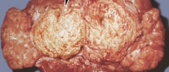
Ultrasound sonograms reveal a voluminous tissue node in the kidney, which compresses the pyelocaliceal complex of the organ and deforms the contours of the latter. A small nephroblastoma is usually shown on images as an echo-negative (dark) formation, free of internal structures. In large tumors, the internal contents are polymorphic. Next to the dense structure, multiple echo-negative formations are determined, caused by hemorrhages and necrosis.
So, ultrasound can detect:
- adenosarcoma of the kidney;
- fragment of a tumor in the inferior vena cava of the organ;
- liver metastases;
- enlarged retroperitoneal lymph nodes.
In addition to all this, using ultrasound scanning you can get an idea of the extent and stage of the oncological process.
Wilms tumor on an ultrasound sonogram is defined as a polymorphic round formation (indicated by arrows)
Video: what Wilms tumor looks like on ultrasound scan
Excretory urography
Further clarification of the nature of the disease and assessment of the functional state of both kidneys is carried out using x-ray examination - intravenous excretory urography. On urograms, the outlines of the kidneys and changes in their shape are clearly visible. The method makes it possible to establish the characteristic signs of nephroblastoma: a defect in the filling of the pelvis and calyces with urine stained with a contrast agent, as well as deformation of the hollow organ system and disruption of its structure.
If there is absolutely no kidney function on the affected side, this indicates complete occlusion of the pelvis by the neoplasm or significant replacement of parenchymal tissue by it. Or about a high degree of thrombosis by a tumor clot of the renal vein.
This excretory urogram of a child shows that the pyelocaliceal system of the left kidney is sharply expanded and deformed, but the excretory function of the organ is preserved
Computed and magnetic resonance imaging
CT and MRI are the next stages of examining the kidneys of a sick child. These are methods that have very high diagnostic capabilities. They allow you to install:
- presence of tumor formation;
- its location and size;
- condition of regional lymph nodes;
- the relationship of the tumor with the pyelocaliceal system and the kidney capsule;
- the degree of its germination into other organs and tissues;
- the presence or absence of metastases in the liver and lymph nodes.
Wilms tumor is visible on tomograms as a space-occupying lesion. The latter has a homogeneous or varied internal structure, can deform the hollow system of the kidney and spread to the perinephric tissue.
A computed tomogram of the right kidney shows the polymorphic structure of Wilms tumor (indicated by arrows)
Angiography
In the case of a large nephroblastoma, especially if it affects both kidneys, angiography may be useful to study the relationship between blood vessels and the tumor.
The method consists of fluoroscopic examination of blood vessels using a contrast agent injected into them.
On aortograms of the kidneys with Wilms tumor, typical signs of the disease are visualized:
- the characteristic vascular network of the neoplasm comes from the branches of the renal artery;
- the vessels are shortened, unevenly tortuous, and have narrowing of the lumen;
- in some of their areas, arterial sinuses and accumulations of radiocontrast agent in the form of small spots or lacunae are identified;
- At the border between adenosarcoma and healthy renal parenchyma, a dense network of transformed renal arteries is visible in places.
Angiography can also reveal indirect signs of nephroblastoma:
- displacement of the renal artery and aorta;
- bringing the intrarenal branches of the artery closer together or further apart from each other;
- shift of the affected kidney to an anatomically incorrect position;
- changes in the size and contours of the organ.
Signs of Wilms tumor: This renal angiogram shows that the aorta is displaced and the vessels are empty and have uneven lumen
Scintigraphy
Nephroscintigraphy makes it possible to see defects in the accumulation of radiopharmaceuticals, indicating the presence of a tumor, and to analyze the performance of each kidney. The absence of an image of an organ on the tomograph screen indicates a complete loss of functionality.
Skeletal scintigraphy will help see bone metastases.
Differential diagnosis
When embryonal nephroblastoma is detected, the doctor is faced with the task of distinguishing it from other tumor and non-tumor pathologies with similar symptoms.
Differential diagnosis is carried out with the following diseases of the kidneys and abdominal organs that occur in children:
- solitary cyst;
- neuroblastoma;
- developmental anomalies of the urinary organs;
- angiolipomyoma;
- splenomegaly (pathological enlargement of the spleen);
- polycystic kidney disease;
- hydronephrosis, etc.
X-ray examination methods, as well as MRI, will help you avoid making a mistake in diagnosis. Neuroblastoma is differentiated from Wilms tumor by special laboratory tests of the child’s 24-hour urine and blood serum.
Treatment
What can you do
Self-treatment in case of detection of a disease such as kidney cancer in a newborn is excluded. The main goal of parents is to timely identify an illness in their child and consult a doctor for diagnosis and, if necessary, surgical treatment.
What does a doctor do
The choice and purpose of treatment is determined depending on the type of malignant tumor that appears and the stage at which the disease is located. As a rule, surgical intervention is used, which involves removing the damaged organ along with the tumor. Sometimes it is possible to perform an operation in which the child’s kidney can be saved. In cases where the tumor has already metastasized and affected other organs, certain chemotherapy is used, prescribed in accordance with the individual characteristics of the body and depending on the stage of the disease.
It is worth noting that surgery is the only treatment method that can achieve greater results, in contrast to drug treatment, which is not as effective. The basis for surgery is the appearance of accompanying symptoms or an increase in tumor size.
At the same time, the use of drug treatment can be effective in some cases, but it requires careful monitoring with the use of x-rays over several months.
Symptoms of kidney cancer
As you know, there are two kidneys in a healthy human body. They are formed by a dense system of tubules that filter the blood. The tubules cleanse the blood of harmful components and form urine. Cancer develops suddenly when a tumor forms from diseased epithelial cells, which gradually impairs kidney function more and more. Every year, tens of thousands of people around the world die from this relatively rare disease.
Many cancers do not manifest themselves in any way at the earliest stages of their inception. Kidney cancer also hides symptoms for some time. In the vast majority of cases, a completely accidental discovery of a disease such as kidney cancer occurs. Symptoms and manifestations of the disease may not yet occur, and only when examining any organ of the abdominal cavity using infrasound, alarming images appear on the screen. Thus, having come with suspicions of one disease, the patient can find out about the onset of another.
However, a person may feel some manifestations of the disease, although he may not know their meaning. That is why it is important to know the symptoms not only to the doctor, but also to all other people, and if they appear, do not be lazy to go to the hospital for examination. By starting treatment on time, there is a chance to pass the test of the disease with minimal losses. Remember that cancer progresses very quickly. Several months of illness can lead to irreversible consequences.
Many internal diseases require great qualifications and experience to accurately determine them.
Infectious diseases and skin diseases, for example, are almost immediately noticeable. Here everything is different.
An insidious disease, kidney cancer, shows only minor symptoms at first. Sometimes people start to self-medicate incorrectly. Let's look at the symptoms characteristic of a disease such as kidney cancer. The manifestation of some of them can save your life.
Prevention
Prevention of kidney cancer in a child is carried out through periodic examination of the kidneys using ultrasound, monitoring the condition of the child’s blood and urine. It is important to understand that the appearance of the first symptoms of the disease must be monitored by a urologist or pediatrician and in no case should be ignored.
During examinations during pregnancy, doctors can identify abnormalities in the development of the child’s kidneys and conduct additional studies to refute or confirm the suspected diagnosis.
Kidney cancer prognosis
After treatment for kidney cancer, regular monitoring and examination by a urologist is indicated. The prognosis of kidney cancer is determined mainly by the stage of the tumor process. With early detection of tumors and metastases of kidney cancer, one can hope for a favorable treatment outcome: the 5-year survival rate of patients with stage T1 kidney cancer after nephrectomy is 80-90%. at T2 stage 40-50%, at T3-T4 stage the prognosis is extremely unfavorable - 5-20%.
Prevention of kidney cancer consists of maintaining a healthy lifestyle, giving up bad habits, and timely treatment of urological and other diseases.
Source: https://www.krasotaimedicina.ru/diseases/zabolevanija_urology/renal-cancer
How and why kidney cancer develops in children, the risks of complications
Kidney cancer in children accounts for approximately 20 - 50% of all tumors. In addition, mixed tumors are often diagnosed in children.
With the development of renal oncology, one or several tumor nodes are formed. Oncological cells are capable of spreading outside the organ along with the bloodstream or lymph. In this regard, secondary tumor nodes appear in the body - metastases. Metastases are mainly found in the liver, in the bones, in the lungs, in the lymph nodes near the aorta or the inferior vena cava. Cancer can line up differently, depending on whether the cancer cells resemble normal kidney tissue.
There are several types of oncology:
- Highly differentiated.
- Moderately differentiated.
- Undifferentiated.
The lower the degree of differentiation of oncology, the faster the neoplasm grows, and the faster the metastatic lesions of the body develop. The prognosis of treatment also worsens.
Forecast
The prognosis regarding the effectiveness of the treatment, as well as the child’s life expectancy, directly depends on the degree to which the child’s body perceives the therapy. In addition, the stage at which the tumor was diagnosed is important. In the later stages of development of the malignant process, the prognosis is less favorable.
If doctors receive a response from the child’s body to chemotherapy, then the probability of a complete recovery is about 80%. In the presence of foci of metastasis, life expectancy is affected by the location of metastases and their size. The five-year survival rate for malignant kidney tumors in children is 40-45%.
Only timely diagnosis and complex therapy of the malignant process give the child a chance for a favorable prognosis regarding recovery and vital functions.
Types of kidney cancer in children
- Neuroepithelial tumor neoplasm is a very rare lesion; it is typical for adults, but sometimes manifests itself in adolescence. The tumor is characterized by rapid growth.
- Clear cell sarcoma.
- The primary form of synovial sarcoma of the kidney is mainly diagnosed in adolescence.
- Adenosarcoma is the most common type of cancer, otherwise known as Wilms tumor.
Typically, the tumor is detected in 90% of cases of the disease before the age of five years. Sometimes diagnosis is carried out in newborns and even in the fetus. The disease affects both boys and girls with equal frequency.
It is important! Typically, the pathology affects only one side and can damage any area of the organ. As the tumor grows, it puts a lot of pressure on the tissue and causes atrophy. The tumor grows, affects the capsule and affects the fiber in the space behind the peritoneum and the peritoneum itself. Over time, the tumor can reach large sizes.
The main reasons for tumor formation:
How and with what to effectively treat kidney hydronephrosis in a newborn
In the first month of life, all children are prescribed an ultrasound of the kidneys and abdominal cavity.
It helps to find birth defects that, for some reason, were not previously detected. It is almost impossible to detect hydronephrosis in other ways, since a healthy kidney takes over all the functions of the affected one. If renal hydronephrosis is detected early, conservative treatment can be performed in the newborn. The operation should be performed only in complex cases when the damage is too extensive. The folk way to cleanse the kidneys! Our grandmothers were treated using this recipe...
Cleaning your kidneys is easy! You need to add it during meals...
Clinical signs of tumor formation
In the early stages, there are almost no symptoms of kidney cancer in children.
But as the tumor grows, the first signs become noticeable. Children typically develop the following symptoms:
- Feeling a tumor in the abdominal area.
- Presence of blood in the urine.
- Prolonged increase in body temperature.
- Pain in the lower back and abdomen.
- Bloating, lack of appetite and vomiting.
- Loss of body weight, severe fatigue, increased blood pressure.
The listed signs may indicate other pathologies, so careful diagnostic measures are necessary.
Clinical picture
Like other cancers, at an early stage the disease is asymptomatic. As the malignancy grows, the first signs appear. In children, the tumor manifests itself with the following symptoms:
- The most common sign is palpation of a space-occupying formation in the abdomen
- Appearance of blood in urine
- Prolonged increase in temperature
- Pain in the abdomen or lower back
- Bloating, loss of appetite, vomiting
- Weight loss, fatigue, emaciation, increased blood pressure
But the listed symptoms can also appear with other diseases, so it is necessary to conduct a thorough examination of the child.
Implementation of diagnostics for suspected kidney tumors
It is important to remember that early diagnosis increases the chances of a speedy and full recovery. The following diagnostic methods are implemented:
- Urine and blood tests.
- Ultrasound examination, which helps to detect even a small tumor.
- MRI and computed tomography help to identify the size of the tumor, its exact location, the presence of metastases and the specifics of blood circulation in the kidneys.
- Urography of the kidneys.
- X-ray examination of the chest organs to determine the extent of the lesion.
- Sometimes a biopsy is performed to clarify the diagnosis.
It is important! The main method of diagnosis is ultrasound examination - it is completely harmless to the child’s body and helps to accurately identify the presence of even small tumors.
Sometimes excretory urography is also performed, but at present it is almost completely replaced by MRI and computed tomography, which are more suitable for diagnosing oncology. All this helps to accurately determine the location, size, damage by metastases to other organs - all this plays an important role in choosing the optimal treatment. To assess the functioning of the opposite kidney, dynamic nephroscintigraphy is used.
Manifestation of a kidney tumor
Have you been trying to cure your KIDNEYS for many years?
Head of the Institute of Nephrology: “You will be amazed at how easy it is to heal your kidneys just by taking it every day...
Read more "
A kidney tumor is a pathological formation that develops as a result of the proliferation of qualitatively changed tissues. A benign kidney tumor, if left uncontrolled, can develop into cancer. The disease develops for various reasons, some of which are not fully understood.
Why does a tumor appear?
The reasons for the appearance of tumor formations in the kidneys are not fully understood. Only some of the main factors that cause the disease are known. These include:
- negative effects of carcinogens;
- long-term use of certain medications, for example, painkillers based on phenacytin (Sedalgin, Citramon, Askofen);
- exposure to radiation rays;
- diabetes;
- arterial hypertension;
- overweight;
- aggravating heredity;
- pathological structure of the organ;
- smoking;
- weak immune system.
Neoplasms are malignant and benign in nature. Oncological tumor formations are diagnosed in most cases; they require urgent treatment. Benign lesions require regular monitoring.
Benign tumor
A benign kidney tumor often does not cause discomfort and is not accompanied by pain. Its dimensions are usually small. This tumor grows very slowly. And yet, its development must be regularly monitored and treated.
A benign formation may be accompanied by the following symptoms:
- weakness;
- pain in the lumbar region;
- swelling of the lower extremities;
- increased body temperature;
- weight loss.
The seal can be felt by palpation. To confirm the diagnosis, you will need to undergo an examination. Diagnostic procedures include laboratory tests and ultrasound diagnostics.
Benign quality does not mean that the disease cannot be treated. Don't put off visiting your doctor. The disease can be successfully treated, but if left untreated and without appropriate therapy, it can develop into a malignant formation. For kidney tumors, symptoms and treatment are the responsibility of a nephrologist.
Types of benign tumors
There are different types of tumors. Some of them are absolutely harmless, but there are also those that over time can develop into malignant ones. Classification of benign kidney tumors:
- adenoma is the most common variant, a tumor of this kind is small in size, the reasons for its appearance are not fully understood, the risk of developing into an oncological formation is high;
- lipoma develops in the fatty tissues of the kidney and can reach impressive sizes, reaching up to 25 cm. In most cases, the cause is poor heredity, and the disease can also develop due to excessive alcohol consumption;
- angioma affects the blood and lymphatic vessels of the kidney, there is a risk of progression to oncology;
- angimyolipoma is a rare type of tumor, its cause lies in the genetic characteristics of the body, and is often not accompanied by any symptoms;
- fibroma appears due to an excess of fibrous tissue; women most often suffer from this disease.
Benign formations, even in the absence of painful symptoms, require regular monitoring.
Malignant formations
Tumors in the kidneys in the vast majority of cases are malignant. A cancerous tumor affects epithelial tissue. The rate of growth of the pathological neoplasm in this case will be different. The disease can progress quickly, but there are also cases when the disease develops over several years.
The disease in most cases affects men. Smokers are also at risk. Most often the right kidney is affected, less often the left one, and extremely rarely the tumor develops on both kidneys.
Kidney cancer has several types. The following types are found:
- renal cell carcinoma ranks first among all malignant tumors; it is localized in the inner layer of the renal tubules;
- transitional cell carcinoma begins in the pelvis, most often, it is caused by excessive smoking, the disease can be treated at the initial stage;
- nephroblastoma is a rare type of malignant neoplasm, most often this disease affects a child between the ages of two and five years, the cause of the disease lies in a genetic mutation;
- sarcoma affects connective tissues and can spread to nearby organs; such a tumor can only be eliminated surgically.
Cancerous tumors are accompanied by lower back pain, blood may be found in the urine, and a person quickly gets tired and loses weight.
Kidney tumor in children
Kidney tumors in children are most often malignant. A childhood tumor includes Williams disease or, as it is also called, nephroblastoma. A malignant tumor can develop in any part of the organ and reach enormous sizes.
The first symptoms are usually noticed by the age of two years. The disease affects both boys and girls. This type of tumor develops on the right or left kidney. The bilateral option is extremely rare.
Early diagnosis gives a chance for recovery. Currently, this disease is treatable at the initial stage of development. It is not always possible to suspect a disease at an early stage. Most often, symptoms appear when the seal begins to be clearly palpable.
The disease can only be treated surgically. The type of surgery will depend on how affected the organ is. In some cases, the diseased kidney is completely removed. If the second kidney also suffers, only the tumor formation and surrounding tissue are removed. When malignant cells are also present in the surrounding structures, the adrenal gland and ureter are removed.
Diagnostic methods
If pain occurs in the lumbar region, you should consult a doctor and undergo an examination. The main diagnostic methods include ultrasound and computed tomography.
Additional examination methods are used:
- magnetic resonance therapy;
- aortography;
- radiography;
- angiography;
- arteriography;
- puncture biopsy.
The patient will also need to undergo urine and blood tests. A kidney tumor leads to the appearance of blastoma cells in the urine. An accelerated erythrocyte sedimentation reaction is observed in the blood.
If cancer was discovered during the examination, the prognosis of the disease depends on the stage of development of the disease. At an early stage, the disease is cured in 80% of cases. If the diagnosis showed that the renal veins are affected, then the prognosis worsens to 60%. When lymph nodes are affected, the risk of death increases, the prognosis worsens to 25%. Late stages are in the vast majority of cases incurable.
A patient with a severe form of cancer usually lives no more than five years. The earlier treatment is started, the higher the chance of recovery. Therefore, at the first symptoms you need to consult a specialist.
Video: Removing a kidney tumor
How is kidney cancer treated in a child?
The selection of treatment will depend on the type of malignant neoplasm and the stage of development of the pathology. There are several main treatment methods, namely:
- Surgery.
- Radiation therapy.
- Immunotherapy.
The operation is considered the only effective method of ridding a child of renal oncology. There are several types of operation:
- Complete removal of the organ - radical nephrectomy.
- Partial removal of an organ - kidney resection.
- Palliative nephrectomy - is implemented as preparatory measures before immunotherapy, chemotherapy when distant metastatic lesions are detected or when the tumor grows into nearby organs and tissues.
If the situation allows, the specialist tries to preserve the organ. Increasingly, laparoscopic surgery is performed for kidney cancer.
Currently, there is no definitive opinion on the need for radiation therapy for renal cancer. Nowadays, radiation therapy is currently implemented only if there are contraindications to surgery in order to prolong the patient’s life. Immunotherapy often complements surgery or is also performed if there are contraindications. The correct treatment regimen helps to achieve positive results in 46% of cases and significantly prolongs life.
Prognosis for kidney cancer depends on the stage of the tumor process at which treatment began. With timely surgery, the child recovers completely.
Kidney cancer treatment
Surgical treatment is the main and most effective method in most cases of kidney cancer; it is used even for regional and distant metastases and can increase the survival period and quality of life of patients. For cancer, kidney removal (radical and extended nephrectomy) and partial nephrectomy are performed. The choice of treatment approach is determined by the type of kidney cancer, the size and location of the tumor, and the predicted survival of the patient.
Kidney resection is performed to preserve the organ in patients with a local form of cancer and a tumor size of less than 4 cm in the case of: a solitary kidney, a bilateral tumor process, dysfunction of the second kidney. During kidney resection, an intraoperative histological examination of tissue from the edges of the surgical wound is performed to determine the depth of tumor invasion. After resection, there is a higher risk of local recurrence of kidney cancer.
Radical nephrectomy is the treatment of choice for all stages of kidney cancer. Radical nephrectomy involves surgical excision in one block of the kidney and all nearby structures: perirenal fatty tissue, renal fascia, adrenal gland and regional lymph nodes. Removal of the adrenal gland is performed when the tumor is located in the upper pole of the kidney or pathological changes are detected in it. Lymphadenectomy with histological examination of removed nodes helps to establish the stage of kidney cancer and determine its prognosis. In the absence of kidney cancer metastases in the lymph nodes (according to ultrasound, CT), lymphadenectomy may not be performed. Performing radical nephrectomy for cancer of a single kidney requires hemodialysis and subsequent kidney transplantation.
During extended nephrectomy, tumor tissue that has spread to surrounding organs is excised. When a tumor grows into the lumen of the renal or inferior vena cava, thrombectomy is performed; if the tumor affects the vascular wall, a marginal resection of the inferior vena cava is performed. In the case of advanced kidney cancer, in addition to nephrectomy, surgical resection of metastases in other organs and lymphadenectomy is mandatory.
Arterial tumor embolization can be performed as a preoperative preparation to reduce blood loss during nephrectomy, as a palliative method for the treatment of kidney cancer in inoperable patients, or to stop bleeding in cases of massive hematuria. As an additional to surgical (and in inoperable patients, the main) treatment for kidney cancer, conservative methods are used: immunotherapy, chemotherapy, targeted therapy.
Immunotherapy is prescribed to stimulate antitumor immunity in advanced and recurrent kidney cancer. Monotherapy with interleukin-2 or alpha-interferon, as well as combination immunotherapy with these drugs, is usually used, which allows achieving partial tumor regression (in approximately 20% of cases), long-term complete remission (in 6% of cases) in patients with kidney cancer. The effectiveness of immunotherapy depends on the histotype of kidney cancer: it is higher in clear cell and mixed cancer and extremely low in sarcomatoid tumors. Immunotherapy is ineffective in the presence of kidney cancer metastases to the brain.
Targeted therapy for kidney cancer with the drugs sorafenib, sunitinib, Sutent, Avastin, and Nexavar allows blocking tumor vascular endothelial growth factor (VEGF), which leads to disruption of angiogenesis, blood supply and growth of tumor tissue. Immunotherapy and targeted therapy for advanced kidney cancer may be given before or after nephrectomy and resection of metastases, depending on the intractability of the tumor and the patient's general health.
Chemotherapy (vinblastine, 5-fluorouracil) for metastatic and recurrent kidney cancer gives minimal results due to cross-drug resistance, and is usually carried out in combination with immunotherapy. Radiation therapy in the treatment of kidney cancer does not provide the required effect; it is used only for metastases to other organs. In case of widespread kidney cancer with invasion of surrounding structures, extensive metastases to the lymph nodes of the retroperitoneum, distant metastases to the lungs and bones, only palliative or symptomatic treatment is possible.
Diagnosis of the disease
The initial diagnosis of kidney sarcoma includes an external examination of the patient and laboratory tests:
- urine (to detect hidden hematuria);
- general blood test (will indicate the presence of anemia, thrombocytopenia and other common abnormalities);
- detailed biochemical blood test. It is necessary in order to find out which substances are in excess and which are lacking. This helps determine how the kidneys, liver and other organs are working.
To clarify the location and size of a kidney tumor, the degree of its invasion in the kidney tissues and surrounding structures (including lymph nodes and abdominal organs), a set of studies is used:
- Ultrasound;
- CT (computed tomography);
- MRI (magnetic resonance imaging).
These methods are also used to detect metastases in other parts of the body: lungs, liver, bones, etc.
On CT and MRI, a mesenchymal tumor appears as a well-defined mass of varying density and signal intensity, just like carcinoma. Therefore, no imaging methods can answer the question: is it cancer or sarcoma.
A diagnosis indicating the type of tumor is made only after histological analysis of its tissues. They are taken using aspiration biopsy (through a syringe). The information obtained is used to establish the stage of the disease and determine the tactics of its treatment.
Treatment of relapse
After resection and chemotherapy, an examination is carried out to find out the result of the treatment. If the doctors managed to completely destroy the cancer focus and there are no signs of sarcoma on the CT scan, then they talk about remission. It can last several years, and in the worst case, several months. The kidney tumor then develops again, which is called recurrence.
Sarcomas have a high relapse rate: they occur in 50-60% of cases! For this reason, it is very important to visit a doctor every 3 months for examination after treatment. This will help to detect recurrence of kidney sarcoma as early as possible and carry out effective therapy.
A second tumor occurs again in the kidney or in the form of metastases in other organs. Depending on its location, the treatment option is selected. A person may be offered the same methods that were used initially, or some new approaches (for example, targeted or immune therapy).
In some cases, this gives a positive result and allows you to achieve remission for another couple of years, but more often, recurrent renal sarcomas behave aggressively and quickly lead to the death of the patient.
Diagnosis of tumors
https://www.youtube.com/watch?v=ytcopyrightru
An important element of diagnosis is an external examination, during which the heart and lungs are listened to, certain reflexes are checked to assess the type or obtain indications of the course of the disease, and organs (liver or spleen) and lymph nodes are palpated. To obtain a medical history, symptoms of kidney cancer are recorded.
To make a diagnosis today it is impossible to do without research methods in accordance with modern technologies: ultrasound, MRI (magnetic resonance imaging), CT (computed tomography) to determine the type of tumor and its size, the degree of spread throughout the body with an accuracy of 55%.
To clarify the diagnosis, the following is carried out:
- X-ray examination and CT scan of the chest;
- scintigraphy with MIBG;
- kidney urography;
- examination of a general analysis of urine and blood;
- determination of blood markers;
- before a course of chemotherapy - echocardiogram (EchoCG);
- audiometry (testing hearing);
- kidney testing using nuclear medicine;
- dynamic nephroscintigraphy to determine the functioning of the second kidney.
After initial preoperative chemotherapy, which lasts 4-6 weeks, a tumor sample taken during surgery is examined under a microscope for biopsy. The analysis can be verified by histological and molecular genetic methods. Exceptional cases require a biopsy using a thin needle to obtain a sample of tumor tissue before surgery.
As an auxiliary diagnostic method before surgery, in addition to standard laboratory tests, blood cells are counted to assess the extent of anemia, and creatinine levels are determined to assess kidney function.
Features of the therapeutic course for tumors
The course of therapy is prescribed by the doctor depending on the stage of the disease and its characteristics.
Modern medicine does not stand still, and new discoveries and invented drugs never cease to please. It would seem that just yesterday cancer was a virtual death sentence, however, at the moment it is possible to overcome the disease or significantly reduce its harmful effect on the child’s body. It is best for treatment if foci of development and formation of the disease are detected at the first stage. The course of therapy is prescribed by the doctor depending on the stage of the disease and its characteristics. In this case, symptoms must be taken into account, as they may differ among patients.
Causes of prolapsed kidney: symptoms and treatment
After establishing an accurate diagnosis, the doctor must make a general conclusion about the child’s health condition. If these are the first stages of the disease, then surgical intervention is used, namely tumor removal. This cancer therapy keeps organs intact. In other situations, in order to avoid unforeseen difficulties, other treatment methods are prescribed, including:
- In some cases, a procedure such as cryoablation is performed;
- Embolization is also very popular;
- Popular treatments for pediatric tumors include chemical therapy, radiation therapy, and targeted therapy.
So we examined all the features of the formation and development, as well as the elimination of pathology, such as a tumor of the kidney organs in children.
Types of pediatric oncology
Cancer can be differentiated, moderately differentiated and undifferentiated. A tumor with a lesser degree of differentiation grows faster, as does the spread of metastases.
Wilms tumor (adenosarcoma or nephroblastoma) is a solid, highly malignant renal tumor. Mutated primitive tissue of various types becomes the basis for it even during the intrauterine development of the fetus. Most often, the precursor cells of kidney tissue and immature precursor cells of muscle, cartilage and epithelium tissue become oncogenic. Therefore, nephroblastoma is a mixed tumor.
Its rapid growth and early metastases are dangerous, therefore, in 10% of cases, diagnosis is carried out when the signs of kidney cancer in children become obvious and metastases are determined. First, they are found in the lymph nodes adjacent to the kidneys, then in the lungs and liver. Some children suffer from nephroblastoma of two kidneys at once. It grows from immature embryonic nephroblastomatous kidney tissue.
Signs of kidney cancer (Wilms tumor) appear most often in children with the presence of:
- developmental anomalies (defects);
- changes in the WT1 gene on chromosome eleven, which is normally responsible for kidney development without pathology;
- changes in other specific genes and DNA chromosomes;
- signs of congenital cancer syndrome.
Overall, Wilms tumor accounts for 5.5% of all cases of childhood cancer. Of this amount, 16% affect children from birth to one year and 68% from one to 5 years, since Wilms tumor is embryonic. With congenital malignant syndrome and abnormal development, kidney cancer, symptoms, manifestation is possible in 90% of children. With heredity, Wilms tumor can affect both kidneys in 1% of affected children.