| Bladder | |
| 1. Human urinary system: 2. kidney 3. renal pelvis 4. ureter 5 . bladder 6. urethra. 7. adrenal gland Vessels: 8. renal artery and vein 9. inferior vena cava 10. abdominal aorta 11. common iliac artery and vein Other: 12. liver 13. colon 14. pelvis | |
| Male bladder | |
| Latin name | vesica urinaria |
| Blood supply | superior vesical inferior vesical umbilical vaginal artery |
| Venous drainage | cystic venous plexus |
| Innervation | vesical plexus |
| Lymph | external and internal iliac lymph node |
| Precursor | genitourinary sinus |
| Catalogs |
|
Bladder, urea
(Latin vesica urinaria, ancient Greek κύστις) is an unpaired hollow organ of the excretory system of vertebrates, located in the small pelvis.
The bladder acts as a reservoir for urine from which it is expelled; in other words, it serves to accumulate urine flowing from the kidneys and periodically excrete it through the urethra, regulated by the detrusor.
General information
Bladder cancer occurs in 70% of cases of all neoplasms of the urinary organs encountered by specialists in the field of clinical oncourology. In the structure of general oncopathology, the proportion of neoplasia of this organ is 2-4%. Among malignant tumors of various localizations, bladder cancer ranks 11th in women and 5th in men in terms of frequency of development. The pathology is more common in residents of industrialized countries; The age of the patients is predominantly older than 65-70 years.
Causes
There is no generally accepted hypothesis regarding the etiology of bladder cancer. However, certain risk factors are known that significantly contribute to the development of a malignant tumor:
- Rare urination
. A number of studies indicate an increased likelihood of neoplasia with prolonged urine stasis in the bladder. Various metabolites contained in urine in high concentrations have a tumorigenic effect and cause malignant transformation of the urothelium. - Diseases of the genitourinary area
. Long-term retention of urine in the bladder can be caused by various urogenital pathologies: prostatitis, prostate adenoma and cancer, bladder diverticula, urolithiasis, chronic cystitis, urethral strictures, etc. - Infections
. The question of the role of human papillomavirus infection in the etiology of neoplasm remains controversial. Parasitic infection - genitourinary schistosomiasis significantly contributes to carcinogenesis. - Occupational hazards
. A correlation has been proven between the incidence of bladder cancer and occupational hazards, in particular, prolonged contact with aromatic amines, phenols, phthalates, and anticancer drugs. The risk group includes drivers, painters, designers, artists, workers in the leather, textile, chemical, paint and varnish, oil refining industries, and healthcare workers. - Other carcinogens
. Tobacco smoking has a high carcinogenic potential: smokers suffer from bladder tumors 2-3 times more often than non-smokers. The consumption of chlorinated drinking water has an adverse effect on the urothelium, increasing the likelihood of neoplasms by 1.6-1.8 times. - Heredity
. In some cases, neoplasia may be genetically determined and associated with a family predisposition.
Functions of the ureter
The ureter serves to transport fluid to the bladder
. The organ has motor autonomous specific functioning.
The rhythm of contractions is provided by a pacemaker, which is located at the apex of the pelvis of the ureter. The cyclicity, speed, and frequency of the rhythm depend on the volume of fluid accumulation, the position of the person’s body, his physical activity, the state of the nervous system, and irritation of the urinary tract.
The urge to contract is determined by calcium concentration
in the fibrous structure of the ureter.
Classification
Bladder cancer varies in histological type, degree of cell differentiation, growth pattern, and tendency to metastasize. Taking these characteristics into account is extremely important when planning treatment tactics. According to morphological characteristics, the most common are transitional cell (80-90%) and squamous cell tumors (3%), adenocarcinoma (3%), papilloma (1%), sarcoma (3%). Based on the degree of anaplasia of cellular elements, low-, moderate- and highly differentiated neoplasias are distinguished.
Of practical importance is the degree of involvement of various layers of the organ wall in the tumor process, and therefore they speak of low-stage superficial cancer or high-stage invasive cancer. The neoplasm can have a papillary, infiltrative, flat, nodular, intraepithelial, or mixed growth pattern. According to the international TNM system, the following stages of neoplasia are distinguished:
- Ta - non-invasive papillary carcinoma
- Tis – flat carcinoma in situ
- T1 – tumor invasion affects subepithelial tissue
- T2 – cancer spreads to the muscle layer (T2a - superficial, T2b - deep)
- T3 – paravesical tissue is involved in the process
- T4 – invasion affects adjacent organs (vagina, uterus, prostate gland, abdominal wall)
- N1-3 – metastasis is detected in one (N1) or many (N2) regional lymph nodes or in the common iliac lymph nodes (N3).
- M1 - metastasis to distant organs is detected
Symptoms
An early manifestation of bladder cancer is the release of blood in the urine - microhematuria or gross hematuria. Minor hematuria leads to a pinkish coloration of urine; it may be episodic and not recur for a long time. In other cases, total hematuria immediately develops: the urine becomes bloody in color, and blood clots may be released. Prolonged or massive hematuria sometimes causes the development of bladder tamponade and acute urinary retention, a progressive decrease in hemoglobin and anemia of the patient.
As the tumor grows, patients begin to experience dysuric symptoms and pain. Urination, as a rule, becomes painful and frequent, with an imperative urge, and sometimes difficult. Pain is noted in the pubic area, groin, perineum, and sacrum. At first, pain occurs only against the background of a full bladder, then, as the muscle wall and adjacent organs grow, they become permanent.
Many symptoms of bladder cancer are not specific and can occur with other urological diseases: cystitis, prostatitis, urolithiasis, tuberculosis, prostate adenoma, sclerosis of the bladder neck, etc. Therefore, patients in the early stages are often treated conservatively for a long time and ineffectively. In turn, this delays timely diagnosis and initiation of treatment, worsening the prognosis.
Complications
Compression of the orifice of the ureter causes disruption of the outflow of urine from the corresponding kidney. Hydronephrosis, an acute pain attack similar to renal colic, develops. When both orifices are compressed, renal failure increases, which can result in uremia. Some types of cancer with infiltrating growth are prone to disintegration and ulceration of the cystic wall. Against this background, urinary infections (cystitis, pyelonephritis) easily occur, urine becomes purulent and has a foul odor. Germination of neoplasia into the rectum or vagina leads to the formation of vesico-rectal and vesico-vaginal fistulas, accompanied by corresponding symptoms.
Diagnostics
To detect cancer and determine the stage of the oncological process, a comprehensive clinical, laboratory and instrumental examination is required. The laboratory diagnostic standard includes a general urine test to determine hematuria, a cytological examination of the sediment to detect atypical cells, a bacteriological urine culture to exclude infection, and a test for a specific VTA antigen. Blood tests usually confirm varying degrees of anemia, indicating bleeding.
- Ultrasound of the bladder.
Detects tumor formations with a diameter of more than 0.5 cm, located mainly in the area of the lateral vesical walls. To detect neoplasia in the cervical area, transrectal scanning is most informative. Sometimes transurethral endoluminal echography is used, performed using a sensor inserted into the bladder cavity. - Tomographic diagnostics.
The most valuable and informative methods are CT and MRI of the bladder. They allow one to assess the depth of spread of the tumor process and identify small tumors that are not accessible to echographic visualization. - Endoscopy of the bladder.
A mandatory imaging diagnostic method is cystoscopy, which clarifies the location, size, appearance of the tumor, and the condition of the ureteric orifices. Endoscopic examination can be supplemented by a biopsy, which allows for morphological verification of the neoplasm. - X-ray diagnostics
. Among the methods of radiation diagnostics for bladder cancer, cystography is performed, which reveals a filling defect and deformation of the contours of the bladder wall and allows one to judge the nature of tumor growth. Pelvic venography and lymphangioadenography are performed to identify the involvement of the pelvic veins and lymphatic system.
Bladder cancer treatment
Radical treatment can only be performed surgically. In this case, the method and type of operation correlates with the stage of the oncological process. Types of surgical interventions for bladder cancer:
- TUR of the bladder.
For muscle-invasive cancer, endoscopic surgery is performed - transurethral resection of the bladder wall with the tumor. During a TUR, the tumor is removed using a resectoscope through the urethra. - Laser en-bloc resection.
The most modern method is laser thulium en-bloc resection. This method allows you to remove the tumor en bloc along with the muscle layer, which is very important for histological examination to assess the degree of invasion. - Cystectomy.
Resection of the bladder (open, laparoscopic, robot-assisted) has been used less and less in recent years due to the high percentage of relapses, complications and low survival. In most cases of invasive bladder cancer, radical cystectomy is indicated. During this operation, the bladder is removed en bloc with the prostate gland and seminal vesicles in men; appendages and uterus in women. At the same time, part or all of the urethra and pelvic lymph nodes are removed.
The following methods are used to replace a removed organ:
- implantation of ureters into the skin - ureterocutaneostomy
- diversion of urine into the sigmoid colon - a method of urine diversion according to Bricker
- formation of an intestinal reservoir according to Studer (orthotopic bladder) from the tissues of the small intestine, stomach, and colon. Radical cystectomy with intestinal plastic surgery is optimal because it preserves the ability to retain urine and urinate.
Surgical treatment can be supplemented with external or contact radiation therapy, systemic or intravesical immunotherapy.
Ureter
The ureter, ureter, is a paired organ located in the retroperitoneal space and subperitoneal tissue of the small pelvis. Accordingly, it is divided into the abdominal region (pars abdominalis) and the pelvic region (pars pelvina). The length of the ureter in men is 30-32 cm, in women - 27-29 cm.
In the same subject, the right ureter is shorter than the left by about 1 cm. About 2 cm of the length of the ureter is in the intravesical part, and the ratio of the length of the intramural and submucosal segments is 1:2. Throughout the rest of the length, the ureter is divided almost equally between the abdominal and pelvic sections.
There are three narrowings in the ureter, the location of which is important when a stone passes through the ureter: at the junction of the pelvis with the ureter in the ureteropelvic segment (UPS), at the intersection with the iliac vessels at the entrance to the pelvis and near the bladder. The lumen of the ureter in narrowed areas has a diameter of 2-3 mm, in expanded areas - 5-10 mm.
The projection of the ureter on the anterior abdominal wall corresponds to the outer edge of the rectus abdominis muscle, on the lumbar region - the line connecting the ends of the transverse processes of the vertebrae. The ureter is surrounded by fiber and layers of retroperitoneal fascia; through the fascia it is quite closely connected to the parietal peritoneum by connective tissue bridges.
In the retroperitoneal space, the ureter lies on the psoas major muscle with its fascia; above the middle of this muscle, the ureter crosses the testicular vessels in men and the ovarian vessels in women, located posterior to them. At the terminal line of the pelvis, the right ureter crosses the external iliac artery, the left ureter crosses the common iliac artery, located anterior to them.
Above this intersection, the ureters with their posterior surface come into contact with the genital femoral nerve, which innervates the skin of the groin and perineum, where pain in renal colic can radiate. Inward from the right ureter is the inferior vena cava, outward - the inner edges of the ascending colon and cecum, in front and above - the descending part of the duodenum, in front and below - the root of the mesentery of the small intestine.
Medial to the left ureter is the abdominal aorta, lateral to the inner edge of the descending colon, in front and above is the small intestine, in front and below is the root of the mesentery of the sigmoid colon and the intersigmoid recess of the peritoneum. In the pelvic region, the ureter, adjacent to the side wall of the male pelvis, crosses the iliac vessels, then the obturator vessels and the nerve and is 2.5 cm from the side wall of the rectum.
Approaching the bladder, it bends anteriorly and inwardly, passes between the posterior wall of the bladder and the anterolateral wall of the rectum outward from the vas deferens, crossing the latter at a right angle, then goes between the bladder and seminal vesicles and pierces the wall in the bottom area bladder from top to bottom and from outside to inside.
Located on the lateral surface of the female pelvis, the ureter goes anterior to the internal iliac artery and the uterine artery extending from it, then at the base of the broad ligament of the uterus at a distance of about 1.5-2.5 cm from the cervix it once again crosses the uterine artery, passing behind the uterine artery. On average, the distance between the ureter and the cervix is 2.3±0.8 cm (from 0.1 cm to 5.3 cm), if it is less than 0.5 cm, which is observed in 12% of women during surgical interventions on the uterus with ligation of the uterine arteries increases the incidence of ureteral damage.
The ureter then goes to the anterior wall of the vagina and flows into the bladder at an acute angle. The upper wall of the ureter at the confluence is a fold lined on both sides with mucous membrane, which, due to the presence of muscle fibers in its thickness, is capable of contracting, closing the lumen of the ureter and playing the role of a valve.
Prognosis and prevention
For non-invasive cancer, the 5-year survival rate is about 85%. The prognosis is much less favorable for invasively growing and recurrent tumors, as well as bladder cancer that gives distant metastases. Quitting smoking, eliminating occupational hazards, drinking purified drinking water, and eliminating urostasis will help reduce the likelihood of tumor development. It is necessary to conduct a preventive ultrasound, urine examination, timely examination and treatment by a urologist for symptoms of urinary tract dysfunction.
In 1892, the first classification of bladder cancer , which was used by urologists and surgeons for a long time. Subsequently, over the course of half a century, various additions and changes were made to the classification and at the same time new classification options were proposed. Of these, the Christeller and Broders classifications were widely used in their time. According to the latter, 4 stages of the cancer process in the bladder were distinguished, taking into account the depth of invasion of blastomatous cells and their anaplasia.
The existence of a significant number of classifications of bladder cancer has created great complexity and often introduced confusion in assessing the type and stage of the disease, establishing indications for various types of treatment and assessing the results achieved. Significant progress in creating a more advanced classification based on new advances in oncological morphology was the classification of Jewett and Strong (1946), modified by Marshall (1952) and Wallace (1956). This classification is based on two pathomorphological features of bladder tumors:
- The stage of the tumor, indicated by the depth of infiltration of the bladder wall and adjacent organs by tumor elements, as well as by the presence or absence of metastases;
- The degree of differentiation and mitotic activity of tumor cells; With the exception of typical fibroepitheliomas, the rest of the tumors have a low or high degree of malignant activity.
It was noted that the malignancy of the cells and the depth of tumor infiltration of the bladder wall progress in parallel to each other. To establish these features, in addition to cystoscopy and general clinical research methods, biopsy results are needed.
In most countries of the world, oncology institutions use the Jewett and Strong classification, modified by Marshall. According to this classification, there are 7 stages of bladder cancer, which can ultimately be reduced to 3-4 main categories.
The stages of low malignancy include 0, A, B15, the stages of major malignancy - B2, C and the metastatic stage - D1; D2.
The Congress of the Society of Urologists of the Mediterranean Latin Countries, held in Rome in 1959, comprehensively discussed the classification of bladder tumors and recommended the adoption of the modified Jewett and Marshall classification for international use. In addition to it, it was recommended to include a designation of the degree of malignancy in the histological interpretation according to Cavazzana (1960):
a) high degree of malignancy;
b) low degree of malignancy.
The International Union Against Cancer (Union Internationale Contre le Cancer - UICC) paid special attention to creating the most advanced classification of bladder cancer.
His subcommittee was entrusted with the task of finally preparing a classification of bladder neoplasms most suitable for clinical purposes. The UICC subcommittee proposed that all urologists around the world implement the following international classification into daily practice. This classification includes 3 components that reflect the extent of the blastomatous process in the bladder and beyond.
The degree of tumor infiltration is indicated by conventional symbols, which facilitates statistical recording and practical use of the classification. Thus, the presence of a tumor in the bladder is denoted by the letter T (Tumor), the depth of blastomatous infiltration - by the numbers 1, 2, 3, 4, damage to the regional lymph nodes by the tumor process - by the letter N (Nodulus), the presence of metastases - by the letter M (Metastasis), absence tumors or absence of metastases and damage to lymph nodes - number 0, for example TO, N0, MO.
So, the symbols T, N, M represent the categories of malignant neoplasm of the bladder. Let us briefly explain the designations of these 3 categories and their degrees.
T—primary bladder tumor. Tx means the presence of a tumor that invades the mucous membrane and infiltrates the subepithelial connective tissue, destroying the basement membrane 1 ;
Notes
- Konstantinov, Naumov, Shatalova, 2012, p. 38.
- Dzerzhinsky, Vasiliev, Malakhov, 2014, p. 125, 139.
- Konstantinov, Naumov, Shatalova, 2012, p. 131, 167, 232.
- Dzerzhinsky, Vasiliev, Malakhov, 2014, p. 212, 326.
- Konstantinov, Naumov, Shatalova, 2012, p. 339.
- Dzerzhinsky, Vasiliev, Malakhov, 2014, p. 406.
- Sapin and Bilic, vol. 2, 2009, p. 175.
- Manski, Dirk.
Urodynamik (Harnblasendruckmessung): Zystometrie.
// Website www.urologielehrbuch.de
(23.10.2015). Retrieved June 23, 2020. - ↑ 1 2 Agadzhanyan N. A., Smirnov V. M.
Normal physiology. - M.: Medical Information Agency, 2009. - 520 p. — ISBN 978-5-9986-0001-2. — P. 387. - Sapin and Bilic, vol. 2, 2009, p. 175-176.
- Sapin and Bilic, vol. 2, 2009, p. 177.
- ↑ 123
Histology, cytology and embryology, 2004, p. 693. - ↑ 1 2 MacLennan GT
Hinman's Atlas of Urosurgical Anatomy. 2nd ed. — Philadelphia: Elsevier Health Sciences, 2012. — xi + 368 p. — ISBN 978-1-4160-4089-7. - P. 240-241. - ↑ 1 2 Shirokorad V.I., Minaev I.I., Demin D.I., Dolgikh V.T.
Method of surgical rehabilitation of patients after combined operations on the pelvic organs // Siberian Scientific Medical Journal. - 2003. - T. 23, No. 4. - P. 82-87. - Clark PE, Stein JP, Groshen SG, Cai Jie, Miranda G., Lieskovsky G., Skinner DG
Radical Cystectomy in the Elderly: Comparison of Clinical Outcomes between Younger and Older Patients //
Cancer
, 2005,
104
(1). - P. 36-43. — DOI:10.1002/cncr.21126. - PMID 15912515. - Manski, Dirk.
Harnableitung nach Zystektomie.
www.urologielehrbuch.de
(23 October 2015). Retrieved June 23, 2020. - Kostyuk I.P., Vasilyev L.A., Krestyaninov S.S.
Classification of locally advanced neoplasms of the pelvis and secondary tumor lesions of the bladder // Oncourology. - 2014. - T. 10, No. 1. - P. 39-43. — DOI:10.17650/1726-9776-2014-10-1-39-43. - Atala A., Bauer SB, Soker S., Yoo JJ, Retik AB
Tissue-engineered Autologous Bladders for Patients needing Cystoplasty //
The Lancet
, 2006,
367
(9518). — P. 1241—1246. — DOI:10.1016/S0140-6736(06)68438-9. - PMID 16631879. - Alberti C.
Whyever Bladder Tissue Engineering Clinical Applications Still Remain Unusual even though many Intriguing Technological Advances have been Reached?
// Il Giornale di Chirurgia
, 2020,
37
(1). - P. 6-12. — DOI:10.11138/gchir/2016.37.1.006. - PMID 27142819.
Classification of cancer types
The oncology industry identifies the following types of bladder cancer:
- adenocarcinoma;
- squamous cell tumor;
- transitional cell malignancy.
The latter type is often diagnosed and is characterized by the modification and proliferation of healthy cells that create the wall of the bladder. For this reason, cancer is called transitional. Squamous cell cancer is associated with changes in thin and flat cells as a result of a long-term inflammatory process or frequent irritation of the bladder walls. Adenocarcinoma is a rare type of disease in which changes affect glandular cells.
The macroscopic type distinguishes the following cancer:
- Papillary, in which the tumor has villi located on a wide stalk. This formation often affects the neck or bottom of the internal organ, the opening of the ureter. In this case, necrosis, tissue polymorphism and disruption of cell structure are noted.
- Solid, in turn, is divided into exophytic and endophytic formation. In the first case, a lumpy tumor appears, protruding into the inner part of the bladder. On top, the formation is covered with a purulent membrane. The endophytic subspecies is characterized by rapid growth and affects, in most cases, the bottom and neck of the organ.
Taking into account the depth of damage to the walls of the organ, non-invasive and invasive or infiltrative bladder cancer are distinguished. With non-invasive or superficial cancer, only the mucous and submucosal layer of the organ is injured. In such cases, the chances of survival are much greater. If an invasive formation is diagnosed, then the likelihood of survival is extremely low. This is explained by the fact that cancer cells reach the muscles of the internal organ, making therapy much more complicated.
How the urinary system works
The walls are abundantly supplied with blood vessels through which oxygen is delivered to the organ and waste substances are removed through venous blood. It should also be noted that there is an abundant network of lymphatic capillaries - the immune system must function intensely in this organ, providing protection against infections. Nerve endings also approach the muscular wall of the organ from all sides, some of which are responsible for relaxing the muscles, others for tensing them. Thanks to the coordinated work of the nervous system, there is a clear regulation of the outflow of urine.
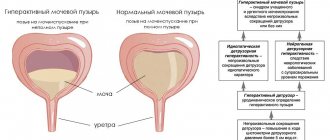
Studies have demonstrated the presence of two phases of the human urinary system. During the transport phase, urine is expelled from the urinary tract under the action of special muscle contraction. These muscles are called detrusors. The second phase of work is retention, translated from Latin as delaying. At this time, tension and compression of the sphincters occurs, the urinary tract cavity closes, and urine accumulates. Both phases constantly alternate with each other and are strictly connected with various departments of the system. When the kidneys are in the first phase, the bladder is in the second and vice versa.
The structure of the bladder in representatives of different sexes is almost the same, but the influence of neighboring organs differs. The fact that the urethra in the female body is shorter and wider than in men increases the risk of infectious diseases. Therefore, women are more likely to experience bacterial cystitis - inflammation of the walls of the bladder. In men, urinary disorders are usually mechanical in nature, due to prostate adenoma; inflammatory processes occur much less frequently. However, it is men who are at risk for urolithiasis; they more often undergo operations on the urinary organs.
Stages of tumor in the bladder
Zero stage
In medicine, the most popular classification of cancer is by stage. At stage zero, the pathological process is just beginning. This stage is not yet characterized by a malignant formation, but is an accumulation of atypical cells that can at any time become malignant. There are no special signs of the disease at this stage. Today in medicine, diagnostic methods have been developed that make it possible to detect the zero stage of bladder cancer. This allows you to get rid of the disease almost painlessly using transurethral cauterization. If the disease is detected in a timely manner and treatment measures are taken, then the chances of recovery are quite favorable, and the chances of a relapse are extremely small.
First stage
The first stage is characterized by the appearance of formation in the mucous and submucosal layers of the bladder. The cancer has not yet spread or metastasized to neighboring tissues. At stage 1 of cancer, a person does not experience any discomfort or distinctive signs of the disease. Only in rare cases are the following symptoms observed:
- bloody discharge when urinating;
- delayed urine output;
- painful sensations during urination.
Due to the lack of pronounced symptoms, it is almost impossible to detect cancer at the first stage. If a person is lucky enough to be diagnosed with the disease in the initial stages, then therapy is carried out in which doctors try to preserve the internal organ. Transurethral resection is often performed using a cystoscope or resectoscope. A malignant formation is removed in this way:
- cryodestruction;
- traditional resection;
- electrocoagulation;
- laser ablation.
The survival prognosis for a patient with stage 1 bladder cancer is quite high. According to statistics, 8 out of 10 patients live more than 5 years after surgical therapy.
Stage 2 cancer
Stage 2 bladder cancer is marked by the transition of the formation to the invasive type and the growth of cells into the muscles. In turn, the disease at this stage is divided into stages 2A and 2B. In the first case, the cancer managed to penetrate the muscles, but did not grow further. If substage 2 B is diagnosed, this indicates that the malignant tumor has completely broken through the muscle layer of the bladder.
Symptoms and treatment of bladder diseases in women
Have you been trying to cure your KIDNEYS for many years?
Head of the Institute of Nephrology: “You will be amazed at how easy it is to heal your kidneys just by taking it every day...
Read more "
What bladder diseases can be in women, symptoms, types, causes, treatment, all this is of interest to women who monitor their health. Illnesses in women, unfortunately, are not uncommon. The development of diseases is directly related to the anatomical structure of the female genitourinary system. An infection can easily penetrate the bladder and cause the development of an inflammatory process. In addition, some diseases can be provoked due to the fact that a woman leads an unhealthy lifestyle, eats poorly, and takes in small amounts of water. It is worthwhile to dwell in more detail on the symptoms of bladder diseases, since many people know how pain syndrome develops, but most women cannot describe the symptoms.
Symptoms of bladder pathologies
Below we will consider bladder diseases and symptoms related to each individual pathology.
Symptoms of bladder neurosis. This bladder disease usually affects older people. The disease is manifested by irritation of the walls of the bladder, loss of elasticity of the tissues of the walls and sphincter. Manifestations of this bladder pathology are as follows:
- frequent urge to go to the toilet;
- urination without result;
- urinary incontinence, suddenly leading to shock.
Symptoms of bladder endometriosis. The development of this disease often occurs as a consequence of ovarian endometriosis or uterine endometriosis. In medicine, there is evidence that endometrial cells move throughout the body and can provoke endometriosis of certain organs. The symptoms of this pathology are as follows:
- heaviness in the lower abdomen, which intensifies before menstruation;
- frequent urination;
- pain during urination;
- dysuria, pain syndrome with transition to the intestinal area.
The signs of a bladder cold are the same as those of an inflammatory process:
- feeling of bladder fullness;
- pain syndrome during the release of the bladder from urine;
- frequent urge to go to the toilet.
Symptoms of a benign or malignant tumor are as follows:
- blood masses in urine;
- pain in the pubic region, spreading to the pelvic region;
- urinary disturbance.
Symptoms of the presence of salts in the bladder. The norm is when salts leave the body with urine, but sometimes salts accumulate and turn into stones. This all happens due to an unhealthy diet. The symptoms are as follows:
- urine becomes opaque;
- the shade changes, up to red;
- pain syndrome in the lumbar region;
- frequent urge to go to the toilet.
Irritable bladder syndrome. The symptoms of the pathology are as follows:
- constant desire to go to the toilet;
- feeling of a full bladder;
- discomfort in the lower abdomen.
The main symptoms of MP tuberculosis are pain when going to the toilet and urine mixed with blood.
Symptoms of cervical sclerosis. The symptoms of the pathology are as follows:
- dysfunction;
- difficulty emptying the bladder;
- symptoms of cystitis and pyelonephritis.
Symptoms of the presence of sand grains in MP. When the bladder contains sand, this may result in the presence of symptoms such as:
- endless cystitis;
- cramps during the period of MP devastation;
- painful urge to go to the toilet;
- pain syndrome in the lower abdomen.
The symptoms of various bladder pathologies are intertwined and similar. However, if the MP ruptures, the woman will feel severe, unbearable pain, which can result in shock.
Causes of inflammation of the genitourinary system
The reasons contributing to the development of inflammation may be different. The most common bladder disease in women is cystitis. The disease develops due to an infection entering the vagina. When the vagina is healthy, it functions normally and contains beneficial bacteria, such as:
- lactobacilli;
- bifidobacteria;
- other beneficial microorganisms.
The normal state for the vagina and the health of the woman is maintained when all these microorganisms are in continuous interaction. But due to various factors, this environment may be disrupted, and then vaginal dysbiosis will develop.
Often inflammation can be triggered by a pathology such as urethritis. This inflammation is localized in the urethra. Do not forget that the urinary system includes:
- kidneys;
- bladder;
- ureters;
- urethra.
The provocateurs of the development of infection that must be treated are parasites, viruses, bacteria, and fungi. Women more often than men suffer from inflammation of the bladder, and at the same time, symptoms do not appear at all in the initial stages, and when the symptoms become obvious, the disease, unfortunately, already has a chronic form. Usually these are pathologies such as:
- cystitis;
- urethritis;
- pyelonephritis.
But men more often develop either urethritis or prostatitis.
Factors in the development of cystitis
A disease such as cystitis can develop due to the following factors:
- presence of a tumor in the bladder;
- presence of stones in the bladder;
- alcohol abuse;
- unhealthy diet, consumption of salty foods, spices, smoked and spicy foods;
- violation of personal hygiene;
- violation of sexual hygiene;
- hypothermia of the intimate area.
Cystitis can be chronic or acute. The acute form of cystitis has symptoms such as pain during urination, small portions of urine, pain in the lower abdomen, which intensifies during emptying of the bladder. In 85% of cases, cystitis in women develops due to infection with E. coli. The symptoms of the chronic form of cystitis are the same as those of the acute form. It may get worse from time to time. The urge to go to the toilet may be very strong, but very little urine may be released. In addition, during urination you will feel pain and severe pain. The cervical form can cause the development of urinary incontinence.
Drug treatment
When bladder cystitis is diagnosed in women, treatment consists of taking medications that eliminate the symptoms of the pathology. Not so long ago, cystitis was treated using alternative medicine methods: women did douching using medicinal herbs. In addition, they practiced proper lifestyle, diet and genital hygiene. Today, this technique can be used, but only as an addition to drug treatment. Still, doctors prefer treatment with antibiotics, since if cystitis is not treated, it will eventually become chronic.
Acute cystitis is treated with antibacterial medications. But before starting treatment with antibiotics, it is imperative to undergo an examination and pass certain tests. We must remember that antibiotics and any other drug can only be prescribed by a doctor. If the form of the disease is very severe, then it is necessary to instill Collargol.
The folk way to cleanse the kidneys! Our grandmothers were treated using this recipe...
Cleaning your kidneys is easy! You need to add it during meals...
After therapy with antibacterial medications is completed, it is necessary to submit urine for bacterial culture.
This will help determine whether the bladder inflammation has resolved. Only a gynecologist can prescribe treatment if a woman is ill, or a urologist if a man is ill. Cystitis therapy is carried out using the following antibacterial medications:
- Monural. This medication is prescribed for inflammation of the bladder, which occurs in an acute form. This medication is prescribed by a doctor; no prior testing is required. The medicine is available in the form of powder, granules or suspension. The medicine has a very strong effect. A powerful antibiotic, but it will not help with chronic cystitis.
- Nolitsin.
- Normax.
- Norbactin.
- Ofloxacin.
- Ciprofloxacin.
- Nitroxoline. This antibacterial medication is prescribed not only for inflammation of the bladder, but also for urethritis and pyelonephritis. This antibiotic belongs to the cheap segment.
- Palin. Fights microbes that infect the urinary tract.
- Furamag, Furagin. These medications have a good effect in fighting the infection that has led to inflammation of the urinary tract.
- Nevigramon. An antibiotic that fights infections of the genitourinary system very well.
- Ampicillin.
- Biseptol.
Unconventional means
Bladder diseases today are treated very effectively and quickly. If this is not a complex internal tumor, when abdominal surgery is required, then therapy is carried out with the help of medications and, possibly, in combination with traditional medicine methods. If a woman is diagnosed with stage 1 cancer, then after productive treatment her health is restored in 95-100% of cases. If the cancer is stage two or three, removal of the bladder may often be necessary. Often, chemotherapy and radiation therapy may be required after removal to avoid metastasis. Recovery occurs in 65% of cases.
It is with regret that we can state the fact that one removal does not guarantee a complete recovery. Relapses occur frequently. Such patients should be monitored by a doctor at all times. It is necessary to undergo examinations in a timely manner and adhere to all instructions of the attending physician.
Alternative medicine can help as a supplement. Medicinal plants have anti-inflammatory, healing, weak diuretic effects. There are several effective recipes:
- You need to take 50 g of herbs: knotweed, St. John's wort, chamomile, bearberry. The herbs should be mixed and poured with two glasses of boiling water for a couple of hours. After the product has infused, it must be strained and taken before meals, three times a day, half a glass.
- You need to take 100 g each: horsetail, corn silk. You also need to take 50 g of: violet, juniper fruits. These components also need to be mixed, and pour 1 spoon of the mixture with boiling water in the amount of two glasses. Next, you need to strain the infusion and take it exactly as in the previous recipe.
- You need to take 100 g each: horsetail, corn silk. You also need to take 50 g each: dandelion, licorice root. All herbs must be mixed. Next, take 3 tablespoons of the mixture and brew it in a thermos with two glasses of boiling water. The infusion should be taken before meals, three times a day, half a glass.
It is necessary to use methods of alternative medicine for a long time, only then can there be a positive result. Traditional medicine will help only when the main treatment is drug therapy. Doctors recommend eating more greens in the summer. In addition to proper nutrition, it is necessary to give up bad habits and monitor genital hygiene. It is necessary to wash yourself at least twice a day, in the morning and evening, before going to bed. If negative symptoms develop, you should not self-medicate; you should consult a doctor who will conduct an examination and prescribe effective treatment.