Kidney ultrasound is a safe, highly informative and accessible diagnostic method for examining the urinary system in a child.
Ultrasound examination (ultrasound) is a non-invasive research method, absolutely painless, allowing you to assess the condition of the urinary system without age restrictions (even in utero). Approximately 5% of children are born with various congenital anomalies of the urinary tract and kidneys, so ultrasound diagnostics of the kidneys was included in the program of mandatory comprehensive screening examination for children 1-1.5 months old. Ultrasound of the kidneys allows you to assess not only the anatomical and physiological features of the child’s urinary system, but also assess the function of the bladder (assessment of residual urine), and also allows you to determine the pathological mobility of the kidneys.
Kidney ultrasound is often performed in older children. Approximately 35–40% of all diseases in pediatrics are disorders of the urinary system, and some of them may not manifest themselves for a long time. They can only be detected using ultrasound.
Normal ultrasound readings of the kidneys in children differ from those in adult patients and change as the child grows older. In this regard, it is advisable to conduct this study in specialized children's clinics. Our ultrasound diagnostics are performed by doctors with extensive experience. You can register your baby for a kidney ultrasound on our website using the online registration form or by phone.
Indications for ultrasound examination of the urinary system in children
Kidney ultrasound is considered a mandatory diagnostic procedure for a newborn at 1-2 months of life. It is necessary to identify congenital pathologies of the genitourinary system.
Most often, sonography is prescribed to newborns with the following symptoms:
Older children are referred for an ultrasound of the kidneys and bladder if they have the following symptoms:
- pulling sensations in the lower back;
- painful urination;
- the presence of blood or mucous clots in the urine;
- abdominal or lower back injuries;
- increase in body temperature for no apparent reason.
About the study
Ultrasound of the kidneys and bladder is a non-invasive method for studying the condition of the urinary system. The features of the anatomical structure and functioning of the links of this system, naturally, cannot be fully assessed only through ultrasound, although such a diagnosis is considered quite complete and accurate. But ultrasound is an integral part of the examination, along with urine and blood tests, if the child has symptoms characteristic of urinary pathologies.
The procedure is completely painless, the child will not experience any discomfort.
Regarding the dangers of ultrasound examinations, medicine gives an official answer - the procedure is safe. Nevertheless, many parents are worried about the possible consequences of ultrasound exposure on the child’s body.
Preparing the child for the procedure
How to prepare children for sonography? Preparation consists of correcting nutrition. Before the procedure, remove from the diet dishes containing foods that provoke excessive gas formation: cabbage, legumes, apples, white bread.
The next stage of preparation is to drink enough fluid. In order for the picture on the screen to be clear, the bladder should be slightly full. Before an ultrasound of the bladder, the child must first go to the toilet - approximately 60 minutes before the diagnosis, and then drink some water. For each age, the amount of fluid is different:
You should not exceed the indicated amount. The liquid will stretch the bladder, and the picture will be distorted, and the baby will not be able to endure the procedure, because he will want to go to the toilet. It is not necessary to drink water; the child can be given compote, juice, tea, or fruit drink. The main thing is not to give him carbonated drinks and lemonade. The infant should be fed in advance so that he does not become capricious during the examination.
Preparation
Whether preliminary preparation for the study is needed is usually determined by the doctor who gave the referral. But even if the doctor forgot to tell the parents about this, the mother must remember like twice two - preparation is needed. And she must be very thorough. It determines how accurate the research results will be. It is necessary to prepare the child for the examination procedure in advance; preparation begins 2-3 days before the date of the visit to the diagnostic room.
- Foods that stimulate gas formation in the intestines should be excluded from the child’s diet. These are fermented milk products, carbonated drinks, bananas and grapes, as well as baked goods, bread and legumes, and white cabbage.
- You should not give your child anything to eat at least three hours before the examination.
- An hour before the examination, the child should be given water to drink. The bladder should be full. This will help the doctor correctly assess the amount of residual urine and understand the condition of the bladder and ureters. Children aged 1 to 3 years are given 100-150 ml of water or fruit drink, children from 3 to 7 years old are offered a glass (250 ml) of liquid, schoolchildren from 7 to 12 years old - at least 400 ml, teenagers older - 600-800 ml .
An infant under the age of one year does not require any special dietary restrictions. The only requirement is that the child should not be fed at the time of the diagnostic procedure. It is best to get examined before the next feeding. Before the ultrasound, give your baby about 50 ml of liquid half an hour before the ultrasound, but no one guarantees that he will retain it. If your baby wants to write, he definitely won’t ask your permission to do so at such a young age.
Procedure for conducting the study
Sonography is performed in a special ultrasound room. Depending on what suspicions the nephrologist has, the sonogram is done in different positions. If the doctor suspects a kidney displacement, then the child should be in a standing position during the ultrasound. To identify other pathologies, he lies on the couch.
The inspection is carried out from 3 positions:
The mother holds the newborn in her arms, and the older child lies down on the couch. At the preparation stage, you should take a diaper or sheet - not all public clinics provide disposable diapers to cover the couch with.
How the research is carried out
Kidney ultrasound is performed for children upon referral from a doctor at any diagnostic center. The procedure is carried out by a sonologist. During an ultrasound examination of the kidneys in newborns, the baby is in the mother’s arms in the desired position. Older children are placed on a couch, lying on their side, back or stomach. If there is a suspicion that the organ is displaced, an ultrasound is performed in a standing position.
The examined area is exposed and treated with a special gel. The specialist uses a sensor to determine the boundaries of the organ and examine the image on the monitor screen in three projections:
- translumbar (from the back);
- transabdominal (through the peritoneal wall);
- frontally (with the patient in the lateral position).
After completing the diagnostic procedure, the results obtained are compared with age norms. The indicators are deciphered by a pediatric urologist or nephrologist.
Interpretation of results, norms of organ sizes by age
After receiving the results of the kidney ultrasound, they should be taken to your doctor. Only a specialist should interpret the results of sonography. First of all, he pays attention to the size of the organ and its thickness, which differ by age.
The table provides information on the sizes of the right and left kidneys in children of different ages:
| Age | Right kidney, dimensions in mm | Left kidney, dimensions in mm | ||||
| Thickness | Width | Length | Thickness | Width | Length | |
| Up to 3 months | 16-27,3 | 13,7-29,3 | 36,9-58,9 | 13,7-27,4 | 14,2-26,8 | 36,3-60,7 |
| 3-6 months | 19-30,6 | 17,2-31 | 47-72 | 19,1-30,3 | 18,2-31,8 | 45,6-70 |
| 1-3 years | 20,4-31,6 | 20,9-35,3 | 54,7-82,3 | 21,2-34 | 19,2-36,4 | 55,6-84,8 |
| 5-7 years | 23,7-38,5 | 26,2-41 | 66,3-95,5 | 21,4-42,6 | 23,5-40,7 | 67-99,4 |
| 7-10 years | 23,9-39,5 | 24,5-44,9 | 67,7-103,3 | 27-41 | 26-44 | 71,2-103,6 |
| 10-14 years | 25,5-43,1 | 28-48,7 | 74,4-113,6 | 27-46,3 | 27,2-47,7 | 74,4-116 |
Norms
Decoding the conclusion is a matter for professionals. Independent conclusions are inappropriate in this case. But if you really want to check the data with existing standards, especially if the doctor was taciturn and did not tell the mother everything he knows, then we present the standards in the table:
| Age | Left kidney, mm | Right kidney, mm | Parenchyma thickness (mm) | Pelvis width (mm) |
| 0-28 days | 48-51.0 x 20.5-21.2 | 47.5-50.0 x 20.3-24.6 | 15-21 | No more than 10 |
| 1-6 months | 52.3 -53.8 x 22.9-23.8 | 52.7-56.9 x 26.1-28.2 | 15-21 | No more than 10 |
| 7-12 months | 61.8 x 24.6 | 60.6 x 29.7 | 15-21 | No more than 10 |
| 1-4 years | 69.6-76.0 x 27.6-30.2 | 68.3-75.4 x 31.2-32.7 | 15-21 | No more than 10 |
| 4-10 years | 82.5-86.8 x 31.9-34.6 | 80.5-85.4 x 34.5-36.3 | 15-21 | No more than 10 |
| 10-14 years | 95.5-114.79 x 37.8-45.5 | 94.5-113.1x 37.9-41.0 | 15-21 | No more than 10 |
| Over 14 years old | 116.7 x 46.8 | 115.2 x 42.1 | 15-21 | No more than 10 |
Reasons for deviations from standard indicators
What abnormalities can an ultrasound detect? First of all, these are the sizes of organs that do not correspond to age, heterogeneity of the parenchyma, enlargement of the renal pelvis, narrowing of the ureters.
The following kidney pathologies may cause the changes:
The following pathologies of the bladder can be seen on ultrasound:
- Chronic cystitis is inflammation of the bladder. Sonography shows uneven thickening of the walls of the organ.
- Neoplasms. These can be oncological diseases and benign tumors.
Treatment based on diagnostic results
Usually, after deciphering the ultrasound results, the attending doctor prescribes adequate therapy. This could be a special diet, medication, physical therapy, or surgery. Minor disorders of the urinary system in children are treated by prescribing compulsory drinking plenty of fluids, following a dietary diet and taking preventive supplements.
Diseases with a pathological effect on the structural characteristics of organs cause great difficulties. If the disease is uncomplicated, treatment may be prescribed at home; more serious pathologies are treated only in a hospital.
uzimetod.ru
Where to do it and how much does it cost?
How to get a referral for an ultrasound, and where can it be done? If you suspect kidney problems, you first need to make an appointment with your pediatrician - he will give you a referral to a urologist or nephrologist. After examination and questioning, the specialist will send the little patient for ultrasound diagnostics.
An ultrasound can be performed at any clinic that has the necessary equipment. If your baby is being treated at a regional public clinic, you can also get a referral for an examination there and undergo it for free.
Sonography can be done at a private medical center. As a rule, there are convenient work hours, there are offices that are open in the evenings or on weekends, and there are practically no queues. However, you have to pay for such conveniences. The cost of the procedure depends on the pricing policy of the medical center and averages 1000 rubles.
The results of kidney ultrasound in children performed in a private clinic are accepted in government institutions. The baby can be treated in a state clinic, and undergo tests and diagnostics in a private center.
Proper preparation of an ultrasound of the bladder in children allows one to accurately determine disturbances in the functioning of the urinary system and identify possible inflammatory processes.
Indications
Sometimes mothers are surprised to receive a referral for such an ultrasound from a pediatric doctor when the child has no problems with urination. The study is not always indicated only for children with such pathologies. Quite often it is recommended for children who were born prematurely in order to assess the functioning of the system and exclude possible complications due to early birth. The study is also recommended for children whose parents suffer from diseases of the urinary system - quite often pathologies are inherited, but do not appear immediately.
In what other cases does a child need an ultrasound of the kidneys and bladder:
- when the color or amount of urine changes, or when an unpleasant, pungent odor appears;
- when crying during urination in newborns or infants or complaints of pain when emptying the bladder in older children;
- with a small amount of urine or, conversely, with increased diuresis;
- impurities are visible to the naked eye in the liquid - flakes, pus, blood;
- the child has anemia, pale skin, blue circles under the eyes;
- pain in the lower back, side;
- closed blunt abdominal injuries that a child can receive when falling on their stomach.
Also, an indisputable basis for prescribing such a diagnosis is a change in the composition of urine at the biochemical level.
If you and the child have no complaints, and the doctor considers the urine tests to be bad, he is obliged to send the child for an ultrasound scan of the kidneys and bladder to understand whether there are grounds for concern and treatment or whether there are no such grounds and a laboratory error occurred.
Indications for the study
The most common disease of the urinary system in children is cystitis. For cystitis in girls, the doctor will prescribe ultrasound diagnostics. The disease can occur as a result of chronic kidney disease, hypothermia, congenital pathologies, insufficient hygiene, wearing tight clothing, or infection.
The pediatrician prescribes a study in cases where the following symptoms are present:
- urination is accompanied by pain;
- change in the color of urine, the presence of turbidity, sediment or flakes in it;
- feeling of pain in the lower abdomen;
- weakened emptying;
- indicators of stool and urine analysis differ from the norm;
- the presence of oxalates or leukocyturia in the urine;
- increased body temperature;
- there is a suspicion of a pathological disease of the organ;
- confirmation or refutation of the presence of an inflammatory process or infection;
- frequent urge to urinate;
- feeling of discomfort in the lumbar region;
- examination for preventive purposes.
An ultrasound of the bladder in a child helps to identify:
- congenital pathologies and developmental anomalies;
- tumors (malignant and benign);
- the presence of stones, polyps and sand;
- narrowing of the urinary canal;
- circulatory disorders;
- inflammatory processes occurring in the bladder.
In addition, ultrasound determines important parameters of the organ: size, contours, volume, etc.
Ultrasound of the kidneys: preparation and conduct of the procedure
Abnormalities in urine tests, swelling, high blood pressure, pain in the lumbar region are indications for an ultrasound examination of the kidneys. In addition, surgical operations on the urinary organs and their transplantation require comprehensive monitoring before and after surgery; a number of necessary examinations for these purposes include an ultrasound scan of the kidneys. In our article we will tell you how to prepare for an ultrasound of the kidneys, how long an ultrasound examination of the kidneys lasts, how to eat and drink correctly so that the ultrasound diagnosis of the kidneys goes without problems and the results are accurate.
Why perform a kidney ultrasound?
Ultrasound of the kidneys is a diagnostic measure carried out one of the first to determine pathological changes in the urinary system.
Ultrasound of the kidneys is a diagnostic measure carried out one of the first to determine pathological changes in the urinary system. This examination is also prescribed for children in the first year of life to determine possible anomalies in the structure of internal organs.
Ultrasound examination of the kidneys, as a rule, is done in conjunction with examination of other urinary organs, so it began to be included in the list of standard preventive examinations. During the procedure, the condition of the adrenal glands, ureters and bladder is also assessed, and the functioning of the renal vessels and blood flow (Doppler) are studied. During the same procedure, the digestive organs and reproductive system of a person can be examined along with the kidneys.
Patients go to a medical facility to have an ultrasound of the kidneys done when they feel pain in the lumbar region, pain when urinating, even without the results of urine tests. During this study, the location of organs, their shape, contours and sizes are assessed, and the structure and condition of the parenchyma are also studied. The presence of formations is also diagnosed.
Advantages of the ultrasound method
Ultrasound examination is one of the most popular types of instrumental diagnostics today.
Ultrasound examination is one of the most popular types of instrumental diagnostics today. Although it is a young type of research, it has a number of advantages:
- High information content;
- Requires minimal preparation;
- It is considered a safe method that can be performed repeatedly;
- The procedure is carried out without pain symptoms;
- There are no side effects;
- Well tolerated by the patient;
- The study does not require the introduction of a contrast agent.
For renal diseases, among instrumental methods, ultrasound diagnostics takes a leading position.
Ultrasound diagnostics of the kidneys has two types:
- Ultrasonography is a method based on the reflection of ultrasonic waves from tissues of different densities. This type allows you to examine the renal parenchyma, detects various neoplasms and conglomerates and disturbances in the topography (for example, inflammation, tumors, cysts, stones, etc.);
- Doppler ultrasound (USDG) is a method that allows you to visualize various lesions of blood vessels (for example, stenoses, injuries, blood clots) and assess the state of blood flow and its direction in the renal vessels.
For reference: in 1979, the American Ultrasound Institute declared that there were no negative consequences after undergoing an ultrasound procedure; no adverse biological effects have been identified to this day. There are also no restrictions on how often this diagnosis can be done.
When examining with ultrasound, there is no negative impact at the point of contact with human skin; this technique does not use magnetic waves or radiation. All possible risks and individual reactions depending on the health status of the patient should be discussed with your doctor. Conditions that make it difficult to examine the kidneys with ultrasound include:
- Presence of gases in the intestines. This is explained by the fact that ultrasound waves do not pass through voids, which greatly distorts the real picture. Therefore, it is important to know how to prepare for a kidney ultrasound;
- The presence of a significant amount of fatty tissue;
- The presence of barium in the intestines (usually after a barium test the day before).
Preparing for an ultrasound of the kidneys
The attending physician will answer the question of whether you need to take laxatives or sorbents before an ultrasound.
Preparing the patient for a renal ultrasound plays an important role in obtaining accurate examination results. It’s worth mentioning right away that preparing for a kidney ultrasound in women is no different from how it is done to prepare for a kidney ultrasound in men. For kidney ultrasound, preparation for the study begins several days before the procedure. During this time, it is necessary to eliminate gas formation in the intestines, since ultrasound does not pass through gases and voids. For this purpose, preparation for the procedure necessarily includes a special diet; you can only drink still water.
Note! The attending physician will answer the question whether you need to take laxatives or sorbents. Before an ultrasound of the kidneys is performed, it is sometimes necessary.
In order for adults and children to properly prepare for the procedure, the diet before a kidney ultrasound stipulates that the following foods are excluded from the diet for 3 days: potatoes, cabbage, raw vegetables and fruits, various sweets, brown bread, dairy products, and baked goods. You can eat your last meal before the kidney ultrasound the night before, no later than 18:00. If only the kidneys are examined, then you can eat on the day of the examination. And if the entire abdominal cavity is examined, then eating is not allowed on this day; the examination is carried out on an empty stomach.
One hour before the procedure, as far as possible, you need to drink 1 liter of still water. In extreme cases, the patient drinks enough to fill the bladder. It is better not to use diuretics.
Note! Not all medical institutions provide disposable sheets and napkins for the procedure, so it is better to take them with you. And due to the fact that ultrasound gel does not wash well, you need to dress in appropriate clothing.
Indications for ultrasound of the kidneys and urinary system
Mandatory indications for ultrasound examination of the kidneys are frequent headaches that cannot be treated with medication.
Mandatory indications for ultrasound examination of the kidneys are:
- Dispensary observation of patients with various diseases of the urinary system (for example, pyelonephritis, cysts and tumors, urolithiasis, etc.);
- Swelling of the extremities associated with high blood pressure;
- Preventive examinations related to professional activities;
- Puffiness of the face;
- Various endocrine diseases;
- Injuries to nearby tissues and the kidneys themselves;
- Anomalies in the structure and congenital diseases of the genital organs;
- Pain in the lumbar region, renal colic;
- Infectious infection;
- Urinary incontinence;
- Impaired urination;
- Abnormalities in urine tests indicating diseases of the urinary system.
To clarify the picture and make an accurate diagnosis, the attending physician may ask you to take additional blood and urine tests.
Conditions and diseases detected during ultrasound diagnostics:
- Acute pyelonephritis, chronic pyelonephritis;
- Stones in the kidneys;
- Congenital pathologies of the urinary organs;
- Cysts;
- Tumors are malignant and benign;
- Narrowing of the lumens of the ureters;
- Inflammation of the renal vessels;
- Abscesses;
- Inflammation;
- Dystrophic changes in organ tissues;
- Kidney prolapse;
- Air in the kidney;
- Accumulation of fluid around the kidney;
- Diverticula in the bladder;
- Rejection of a transplanted organ.
All these changes can be detected using ultrasound. Many clinics today have equipment that provides high diagnostic accuracy that can compete even with computed tomography.
How is the kidney ultrasound procedure performed?
Diagnostics is carried out using a transducer that sends high-frequency ultrasonic waves
Diagnostics is carried out using a transducer that sends high-frequency ultrasonic waves. These waves cannot be heard; the human ear does not perceive them. The sensor, which is applied to the surface of the skin, sends ultrasound to the desired organ, the waves are reflected from it and return to the transducer, like an echo, then displayed as a picture on the doctor’s monitor.
A water-based gel, which is applied locally to the surface of the skin, improves the glide of the sensor and eliminates the presence of air between the device and the patient's skin. Ultrasound moves very slowly through air voids, and fastest through bone tissue. Special supersonic waves make it possible to evaluate and examine blood flow in the kidneys using Doppler examination. If the signals are weak or absent, there is an obstruction in the blood vessel.
Note! Ultrasound can be performed on pregnant women, as well as people with allergies to contrast agents used in other types of instrumental diagnostics.
In the ultrasound room
Before going through the procedure, it is a good idea to understand the entire process. In the ultrasound room you will be asked to:
- Remove jewelry and clothing that may interfere with the procedure;
- Depending on the clinic, you may be asked to wear a special gown and given a disposable sheet and napkins;
- During the examination, the position of the body, at the request of the doctor, can be changed: on the back, on the stomach, on the side, standing;
- A gel is applied to the desired area of skin and a sensor is applied, which the doctor will move to obtain a high-quality image. After the procedure, the gel must be wiped off with a disposable napkin;
- The doctor may ask you to take a deep breath and hold your breath while expanding your belly;
- First, the bladder and ureters are examined, then the kidneys are examined;
- When examining Doppler ultrasound, you may hear a characteristic noise and whistle;
- The procedure does not cause pain or discomfort;
- The examination of the kidneys takes about 10-15 minutes, the entire urinary system - a little longer, and when examining all the organs of the abdominal cavity - even longer (exactly how long your examination takes can be clarified at the reception desk or with the attending physician);
- For those undergoing a bladder examination, you should be prepared to be asked to empty your bladder and then return to the office to continue the procedure.
Those who have undergone the procedure are given ultrasound results. The printout contains an inscription indicating the date of the examination and your personal data; below you can see the doctor’s conclusion based on the results of the examination. It indicates the detected pathologies, which are also marked in the photo attached to the conclusion. In some clinics, a video recording of the examination is attached to the results.
How to Prepare Based on the Type of Procedure
Ultrasound of the urinary organs in children is performed primarily using the external method. The procedure takes 15-20 minutes. Diagnosis is carried out with a full bladder. Before the examination, the child needs to take liquid 1-1.5 hours. Infants should be given something to drink 20 minutes before the ultrasound.
For the study to be informative, it is necessary to free the intestines from gases in advance.
Children over 2 years of age are not recommended to consume foods that cause gas for 3 days before the test:
- legumes;
- black bread;
- rich baked goods made from wheat flour;
- fresh fruits and vegetables.
At least 18 hours must pass from the last meal to the examination. At night, it is recommended to insert a glycerin suppository into the anus or do an enema. Diagnosis is carried out only with a full bladder, so urinating before the procedure is not allowed.
The technique is intended for males. The sensor is inserted into the urethra, while the patient is in a supine position. Using this examination, the doctor can evaluate damage to the urethra.
Disadvantages of the study include:
- need for pain relief;
- risk of damage to the genitourinary system by the sensor.
The examination is prescribed in rare cases.
Ultrasound diagnostics occurs through the anterior wall of the abdomen. The study is suitable for girls and boys.
Before diagnosis, the child should undress to the waist. Then you need to lie on the couch on your back. In the area above the pubic bone, the doctor applies a gel to the patient’s skin, along which the sensor will move. The only unpleasant sensations may be the pressure exerted by the device on the outer wall of the abdomen, or the cooling effect of the gel. The child should be warned about this in advance. From the first minutes of diagnosis, an image of the examined organ will begin to be displayed on the monitor.
The doctor will evaluate the shape, thickness of the walls and their size, examine the condition of the mucosa and identify possible pathologies (narrowing of the canal, the presence of stones, helminthic infestations, etc.).
The method is applicable only to girls who have already had sexual intercourse. An examination is carried out using a vaginal sensor. The procedure is done in a supine position. No preliminary preparation is required before the study. Diagnostics allows us to identify diseases of the reproductive organs.
The examination is carried out through the rectum. The patient removes clothing below the waist and lies on the couch in a lateral position. Diagnostics is used for both sexes. The method is highly informative when compared with the abdominal method.
How to perform an ultrasound of the kidneys and bladder in children
During the procedure, the child is placed lying on the couch (on his back, stomach or side) or standing (if it is necessary to exclude nephroptosis). Sometimes the doctor asks you to take deep breaths and exhalations. He moves the sensor over the skin, which has been previously lubricated with a transparent, light, odorless gel that cannot cause skin irritation. This gel is needed to make the sensor glide better and to increase the conductivity of ultrasonic waves. The ultrasound machine converts ultrasonic waves into electrical waves, and they are displayed on the monitor in the form of an image. The doctor takes the necessary measurements, records the data obtained and makes a conclusion based on them.
Ultrasound is a type of high frequency sound (1–12 MHz) that cannot be detected by the human ear. Ultrasound can be found in nature in many places: in the sounds made by some types of fish, dolphins, individual insects, bats, in the noise of waves and wind. Ultrasound diagnostics is safe for the child’s body.
Drinking fluids before diagnosis
The liquid consumed should not be carbonated.
Before the procedure you can drink:
Coffee, milk, fruit drinks (cranberries, lingonberries) and alcohol are prohibited for consumption before ultrasound diagnostics.
The amount of fluid directly depends on the child’s weight. For 1 kg of body weight, you should consume 5-10 ml of liquid.
Infants are examined under the same conditions. The newborn needs to drink water, breast milk or formula at the same rate.
Older girls can take another 0.3-0.5 liters of still water half an hour before diagnosis.
Minimum fluid intake:
- children under 2 years old - at least 0.1 l;
- up to 7 years – at least 0.2 l;
- 8-11 years – at least 0.3 l;
- over 11 years old - at least 0.4 liters.
Diagnostic procedure
When performing an ultrasound of the kidneys in infants, the mother holds the child in the desired position. Older children can undergo the examination procedure on their own. The ultrasound begins with the preparation of the abdominal and lumbar region; for this it is necessary to free the abdomen and lower back from clothing. Then the doctor applies a gel to the skin to improve contact with the sensor. An ultrasonic sensor emits ultrasound and detects reflected signals. A complete picture of the examination is created by examining the organ in three projections: transabdominal, translumbar and frontal.
A person who does not have special knowledge will not be able to understand the images displayed on the screen. But a specialist will accurately determine the slightest discrepancy with the age norm, how enlarged the kidneys are, and recognize structural changes in the organ.
Decoding and norm
Decryption is based on the following data:
- Organ shape. Normally, it can be oval, narrowed at the top (pear-shaped). After emptying, the shape of the organ takes on a round saucer-like shape. A change in the outline of the bladder may indicate cystitis and neoplasm.
- The walls normally have the same size, their thickness does not exceed 2-4 mm. Thickening of the walls (5 mm or more) may indicate diseases such as:
- cystitis;
- closure of the lumen of the organ (stones, tumor);
- parasitic infestations;
- tuberculosis of the urinary tract (tuberculous granulomas were found on the walls).
- Structure. On the monitor, the organ appears as dark outlines (echo-negative structure).
- The organ cavity normally does not contain sediment, flakes or stones. Flakes (leukocyte and epithelial cells) in the urine are present in cystitis; they appear on the monitor screen as light spots (hyperechoic structure). If the sediment is formed by salts (phosphates), this may indicate the development of urolithiasis.
- Organ integrity. In a healthy state, the walls are intact and free of polyps, stones and injuries.
- Filling speed. In a healthy state, the filling rate is 50 ml/h.
The performance of the urinary system, as well as its structure, is assessed by a complex non-invasive method - a study that identifies the amount of residual urine.
Depending on the age of the patient, the norms for the amount of contents in the bladder differ.
| Child's age | Amount of residual urine volume |
| Up to 1 month | ˂ 3 |
| Up to 12 months | ˂ 5 |
| Up to 4 years | ˂ 7 |
| Up to 10 years | ˂ 10 |
| Up to 15 years | ˂ 20 |
A congenital anomaly is urethral obstruction (obstruction of urine flow). On ultrasound diagnostics, this pathology looks like a thickening of the bladder wall along the entire perimeter.
If there is an accumulation of residual urine above normal after bowel movement and flakes are found in urine streams during urination, then the following pathologies may be detected in the patient:
- prostatic hyperplasia - the growth of the prostate is pathological, which causes compression of the urethra and makes it difficult to empty;
- pathology of bladder innervation;
- the presence of calculi (sediment, flakes, stones) in the organ;
- abnormal development of urinary tract valves (diagnosed in children in newborn age).
If the volume of residual urine is reduced compared to the norm, this may indicate a decrease in the normal size of the organ.
- invasion by schistosomes in the last stage;
- consequences of surgery;
- frequent manifestations of cystitis.
When interpreting the results of an ultrasound examination, the specialist compares the data obtained with generally accepted standard values. The guideline for a specialist when interpreting an ultrasound is the patient’s age; this factor is key.
Diagnosis of the urinary system in a child using ultrasound provides a safe, quick and high-quality assessment of the organ. Experts recommend diagnostics from 3 months (for both boys and girls) to identify possible pathologies, and if they are detected, begin timely treatment.
The level of information content and safety of widely used ultrasound allows it to be used in the diagnosis of diseases of the genitourinary system at a young age. Ultrasound of the bladder in children is the most important method for diagnosing possible health problems.
Indications for children
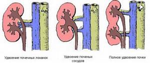
Screening ultrasound of the abdominal cavity and kidneys is recommended for all newborns at the age of 1 month for early detection of asymptomatic abnormalities of the urinary system. Among congenital defects in childhood, there are anomalies in quantity (agenesis, accessory kidney), position (dystopia of the kidney, nephroptosis), size (hypoplasia, aplasia), relationship (fusion of the kidneys, horseshoe kidney), structure (duplication, polycystic kidney, spongy kidney) . Ultrasound of a child’s kidneys allows not only to detect various malformations, but also to carry out dynamic monitoring as often as the clinical situation requires.
Children of preschool and school age often suffer from urinary tract infections, the leading place among which is pyelonephritis. Echographic signs of pyelonephritis (differences in the size of the kidneys, changes in the echo density of the parenchyma, deformation of the maxillofacial joint, etc.) are clearly visible during ultrasound of the kidneys in a child.
An ultrasound of the child’s kidneys may be required to clarify the diagnosis for pain in the abdomen and lower back, edema, increased temperature of unknown origin, injuries to the lumbar region, dysuric disorders, changes in urine tests indicating kidney dysfunction. Kidney ultrasound is included in the mandatory examination algorithm for a child with enuresis and urinary incontinence, dysmetabolic nephropathy, arterial hypertension, glomerulonephritis, kidney stones, renal colic, acute and chronic renal failure, etc.
Kidney ultrasound is necessary for children who have recently had a streptococcal infection - sore throat, pharyngitis, scarlet fever, pneumonia, etc. to exclude secondary kidney damage. To assess the parameters of renal blood flow, an ultrasound scan of the renal vessels is performed as a separate stage of an ultrasound scan of the child’s kidneys.
Indications for an ultrasound of a child's kidneys are usually determined by a pediatric urologist, pediatric nephrologist, pediatrician or pediatric surgeon. Before an ultrasound of the kidneys, the child is recommended to undergo a laboratory examination: OAM, OAC, biochemical analysis of urine and blood.
www.krasotaimedicina.ru
Residual urine
The functional capacity of the urinary system and its anatomical structure allows us to evaluate a complex non-invasive method - ultrasound with determination of the amount of residual urine. For children of different age groups, there are norms for the amount of contents in the bladder:
| Child's age | Amount of residual urine, ml |
| Up to 1 month | Preparation To prepare for a bladder examination, your child needs to drink a certain amount of fluid. The volume of liquid is calculated as follows: 10 ml per kg of weight. Taking liquid or mixture is necessary 20 minutes before the start of the procedure. While examining the condition of the bladder, an examination of the kidneys is usually carried out. Before the ultrasound procedure, children over 2 years old are not recommended to eat legumes, baked goods made from wheat flour, or fresh fruits and vegetables. 1 hour before the examination, the child needs to be given something to drink (the calculation is the same: 10 ml per kg of weight). Older girls can drink 300-500 ml of still water in 30-45 minutes. |
Rules for preparing for ultrasound
In order for ultrasound diagnostics to show the correct results, it is necessary to follow the preparation rules. The last meal should occur 10-12 hours before the procedure. This is important because when food enters the body, the biliary system activates its activity, secreting bile, and the gallbladder must be full at the time of the ultrasound.
The best time for manipulation is 10-11 am. This is the optimal period to examine the condition of the stomach and duodenum. The time after noon will no longer be so informative, since even in the absence of food entering the body, the stomach begins to secrete gastric juice, which, in turn, will change the diagnostic picture.
48 hours before the ultrasound, it is better to stop taking medications, especially painkillers and antispasmodics (with the permission of the attending physician), and in the morning - from using chewing gum and smoking.
Additional recommendations:
- when examining the condition of the kidneys, you should drink 1500 ml of liquid over 40-60 minutes;
- when diagnosing the biliary system, spleen, pancreas, dinner the day before should consist of easily digestible foods;
- for bloating, medications are prescribed (Espumizan, Polysorb, activated carbon);
- To cleanse the intestines, Fortrans, Guttalax, and a regular enema are used.
Preparing children
Conducting an ultrasound of the abdominal organs in children also requires preparation:
- if the patient is an infant, you need to refuse one feeding before diagnosis and not give liquids 60 minutes before the ultrasound;
- a preschool child is not given liquid an hour before the procedure, food - 4 hours;
- Children from 3 years of age are prepared for the procedure in the same way as adults.
Sources used:
- https://o-krohe.ru/uzi/pochek-mochevogo-puzyrya/
- https://uran.help/surveys/radiology/uzd/uzi-mochevogo-puzyrya-u-detej.html
- https://ultra-sonographi.ru/uzi-pochek-rebenku.html
- https://medigid.com/uzi/organy/malyj-taz/mochevoj-puzyr/u-detej.html
- https://idiagnost.ru/uzi/kak-provesti-podgotovku-uzi-mochevogo-puzyrya-u-detej
- https://uzimetod.com/brushina/uzi-pochek-rebenku.html
- https://detki.shukshin-net.ru/uzi-mochevogo-puzyrja-i-pochek-podgotovka-u-detej/
How do they do it?
Before the ultrasound begins, the child is placed on the couch using a disposable diaper. For the examination, it is necessary to free the abdominal area from clothing. A special gel is applied to the skin above the pubic bone in the projection of the bladder.
Using an ultrasonic sensor, which makes tangential movements, an image of the organ is projected onto the monitor. Using this image, the doctor assesses the condition of the mucous membrane, the shape, size and thickness of the walls of the bladder, the presence of pathological changes (diverticulum, narrowing of the canal, neoplasms, stones, sedimentary components, helminthic infestation).
Research methodology
Ultrasound diagnostics is based on the principle of reflection of a sound wave from an obstacle. Ultrasound of a given length, penetrating through the tissue of the organ under study, is reflected. With the help of certain technologies, the reflected wave is converted and a visual picture is created on the monitor screen showing the condition of the organ. The equipment used for examining children does not differ from “adult” diagnostic devices.
Today, all clinics and medical centers have ultrasound diagnostic rooms, where you can freely conduct examinations of the child’s internal organs. Typically, such private institutions are equipped with modern and accurate equipment, which allows them to obtain the most reliable results. Ultrasound examination is a completely harmless diagnostic method. In this regard, repeated use of ultrasound is allowed, even when examining infants.
Ultrasound of children's organs and systems is now performed in many modern clinics. This research method is as safe as possible and can be used repeatedly
Decoding and norm
When interpreting ultrasound readings, the data is compared with generally accepted standard indicators. The guideline for decoding is the patient’s age; this factor is key.
To make a diagnosis, important indicators are the size of the kidneys, their length, thickness and width.
The left and right kidneys are different from each other in these indicators.
If the examination results do not go beyond the standard, then there is no cause for concern.
Readings are recorded in millimeters.
Ultrasound of the child's abdomen and kidneys
It is important to know that ultrasound examination of the abdominal cavity and kidneys is often performed separately. But in some cases, joint diagnostics are prescribed. This is necessary if the child suffers from the following clinical symptoms:
- Pain in the side, abdomen and lumbar region;
- A feeling of bitterness in the mouth and frequent nausea;
- Digestive disorders.
During the procedure, the doctor may also ask you to hold your breath. The best time for diagnosis is in the morning on an empty stomach.
Get a free doctor's consultation
Pathologies
Bladder diseases in infants are common due to the abnormal location of the organ, changes in its shape or parameters.
An ultrasound examination in children can reveal the following pathologies:
- cystitis in acute or chronic form;
- metabolic nephropathy;
- malformations (mismatch of shapes or sizes, diverticulum, narrowing of the urinary canal).
Inflammation of the bladder (cystitis) affects girls more often. The reason for this phenomenon is the physiological structure of the urethra, which facilitates the penetration of pathogenic flora from the vagina.
The structure of the organ is such that on the surface of the mucosa there are receptors that are tropic to this pathogen. The number of these receptors determines the predisposition to the disease. More often, cystitis is diagnosed in an organ with a significant number of such receptors.
Violation of hygiene requirements can lead to the development of this disease.
For example, long-term stay in a filled diaper creates conditions for acute inflammation of the mucous membrane.
The cause of cystitis can be an allergic reaction to a certain type of hygiene products (diapers, napkins). In this case, the pathology develops ascending from vaginitis.
In infants, cystitis can develop due to complications after vaccination, influenza or ARVI. Clinical manifestations of the disease in infants include restlessness, crying when urinating, and changes in the color of urine.
An obstruction to the outflow of urine - obstruction of the urethra - is caused by the formation of a valve, which is a congenital anomaly; on ultrasound it looks like a thickening of the organ wall along the entire perimeter. In this case, there may be an accumulation of residual urine and the presence of flakes in the urine during the act of urination. This pathology most often requires surgical intervention.
Indications
If a child complains to parents about various unpleasant symptoms or discomfort, the best decision in this situation would be to visit a local doctor. Many people put off visiting a specialist, mistakenly thinking that they themselves will be able to diagnose and treat the baby. As a result, precious time is lost, and the disease takes on an acute or chronic form.
Pediatricians advise performing an ultrasound of the bladder in children with the following conditions:
- The child often goes to the toilet to relieve himself;
- The patient is bothered by painful sensations;
- After a general clinical examination of urine, oxalates or multiple leukocytes were detected in the biological material;
- Sometimes there are no obvious symptoms, but there is hyperthermia;
- The baby complains of pain in the pubic area and around the lower back.
Ultrasound screening for such symptoms will most likely help the doctor make the correct diagnosis and prescribe the necessary treatment. During the examination, inflammation, stones, polyps, sand, changes in structures, narrowing of the urinary canal, tumors, malformations, and impaired blood circulation can be detected.
How to eat properly before the procedure?
Changing your diet and switching to a diet before undergoing an ultrasound is necessary due to the fact that ultrasound waves are not able to pass through gas accumulations in the intestines. The presence of gases significantly complicates diagnosis.
Changing your diet and proper drinking regimen can not only prevent the formation of gases, but also eliminate existing accumulations.
Prohibited Products
During the three days preceding the test date, the patient must exclude from the diet foods that increase gas production.
Prohibited:
- potato;
- milk;
- raw vegetables;
- fatty foods, including soups and broths;
- fried and smoked foods;
- spicy and salty foods;
- fruits;
- black bread;
- sweets and confectionery;
- alcohol;
- carbonated and strong drinks.
If you follow the correct diet, the formation of gases will stop, and their remains will gradually leave the intestines.
Allowed foods
Meals during the preparation period should be light, not complicating intestinal function and not provoking the formation of gases.
The following products are recommended for use:
- hard cheese with low fat content;
- porridge made from pearl barley, oatmeal or buckwheat;
- lean meat - beef, chicken, boiled rabbit;
- boiled low-fat sea fish;
- dried white bread;
- eggs - one per day.
Immediately on the day of the procedure, you should refuse to eat, especially if you plan to examine the entire abdominal cavity. The last meal is allowed no later than 8 hours before the ultrasound. Dinner on the eve of the event should be light, consisting of dietary products.
Do I need to drink water before a kidney ultrasound?
The drinking regimen during the preparation period for a kidney ultrasound consists of drinking boiled water and weak unsweetened tea. On the day of the event, it is recommended to increase the volume of fluid taken to half a liter - this volume must be drunk within an hour before the start of the ultrasound.
Drinking carbonated water on the day of the ultrasound is strictly prohibited.
At the same time, it is better not to rush to visit the toilet before the end of the study - doctors insist on filling the bladder, as this improves the passage of ultrasound and increases the information content of the procedure.
The essence of the study
Ultrasound is a method of non-invasive visualization of internal organs, which is based on the use of high-frequency ultrasonic waves. They penetrate well through biological tissues and are partially repelled from them (this indicator depends on the density). Then the broken part is caught by a special sensor, the information from which is transmitted to the computer and displayed on the screen in the form of an image.
A significant advantage of ultrasound of the kidneys for a child is availability and price. The study can be carried out for a fee not only in Moscow and large cities, but in almost every hospital and clinic. In terms of price, it is significantly cheaper than computer or magnetic resonance therapy.
How is an ultrasound of the kidneys performed for children: features of the procedure
When examining the kidneys, the doctor pays attention to the following criteria:
- localization of the kidneys (exclusion of nephroptosis);
- transverse and longitudinal dimensions of the kidney;
- characteristics and condition of the parenchyma (homogeneity, presence of areas of degenerative changes or neoplasms);
- condition of the renal pelvis (size, presence of dilations, absence of concretions or stones);
- state of the blood supply to the kidneys in Doppler mode (arterial patency, presence/absence of vascular development abnormalities, dysplasia).
After the diagnosis is completed, the gel is removed from the baby's skin using disposable towels. When conducting research, you should avoid pressing the sensor hard, since it is much easier to provoke the development of uncomfortable sensations in children.
The duration of the procedure is 5-15 minutes.
Preparatory stage
Let's look at what an ultrasound of the kidneys is and preparation for examining a child.
- A prerequisite is the presence of a full bladder during the examination. To do this, you need to drink a volume of liquid approximately half an hour before the procedure in accordance with the baby’s age. Still water or tea without sugar are ideal for this purpose. Filling the bladder is necessary to obtain better visualization of the organ. In addition, thanks to the liquid, the conductivity of the ultrasonic wave is significantly improved, hence the opportunity to carefully examine the structure of the organ. It happens that the baby is not able to wait until the appointed time, then it is allowed to empty the bladder slightly, but then be sure to make up for the missing volume. The fact is that a bubble that is too full can also distort the ultrasound readings.
Required volume of liquid:
- for a two-year-old toddler - about 100 ml;
- for a child from two to seven years old – 250;
- a child over seven to eleven years old - approximately 400 ml;
- from eleven to fifteen years - half a liter.
- It is also important that the child does not have increased gas formation in the intestines at the time of the study. The fact is that the accumulation of air reduces the conductivity of the ultrasonic wave. Therefore, if there are such problems, it is necessary to take carminative medications.
Ultrasound of the gallbladder for a child
Such a study is carried out in order to draw a conclusion about how the organ itself and its ducts function. In addition, the dimensions of the bladder walls, thickness, condition of the surrounding tissues and the degree of patency are studied.
When is such a diagnosis indicated for a child?
- Bitterness in the mouth;
- Diarrhea or white stool;
- Suspicion of cholelithiasis;
- Blockage of the biliary tract;
- Pancreatitis and jaundice;
- Pain under the ribs on the right and painful sensations in the abdomen.
Preparing a child for an ultrasound of the gallbladder includes the following measures:
- You cannot eat eight hours before the diagnosis;
- Diagnosis should be carried out on an empty stomach;
- Three days before the test, you need to reduce gas formation in the intestines by eliminating foods that contribute to this and by taking enterosorbents;
- A few hours before the test, the child needs to cleanse the intestines;
- You can’t overeat or eat fatty foods the day before.
How do diagnostic doctors conduct such a study? The child can lie on his back, holding his breath. A lying or standing position is also possible. The price for research usually starts from eight hundred rubles. If parents want to do it for free, they need to get a referral for diagnostics from a gastroenterologist or pediatrician.
“Ultrasound (gallbladder): normal (children).” What can be said about deciphering the results of research in children?
According to ultrasound, the dimensions of the gallbladder in children should be as follows:
- The diameter of the gallbladder should not be more than 3.5 cm. The length of the organ should not exceed 7.5 cm. The volume of the organ in older children should not normally exceed 200 ml;
- An echogram may demonstrate the common and cystic ducts of the gallbladder, but it is almost impossible to distinguish between them. That is why it is commonly called the common duct of the gallbladder. The normal size of the duct is 4 mm. The width should ideally not exceed 8 mm. Ducts within the liver should not be visualized. If they are visible, this indicates the appearance of cholestasis or obstructive jaundice;
- The walls of the gallbladder cannot normally be compacted; there should be no stones or sand in the cavity;
- Sometimes a small amount of sand in the gallbladder is considered normal;
- There should be normal bile secretion.
ultra-sonography.ru