What it is?
This is a pathological process, which is based on a narrowing of the renal artery, which leads to a decrease in blood flow and nutrients dissolved in it.
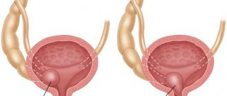
Stenosis can be localized in the initial, middle or terminal part of the renal artery.
Significant, i.e. Having a significant impact on the blood supply to the kidney are narrowings that close the lumen of the vessel by 70% or more, or by 50-70%, provided there is a large difference in pressure before and after stenosis. Pressure is measured using renal ultrasound with Doppler mapping.
In the International Classification of Diseases, 10th revision (ICD-10), renal artery stenosis can be coded as follows :
- I70.1 - atherosclerosis of the renal artery.
- I15.0 – renovascular hypertension.
- I77.1 – narrowing of the arteries.
- I77.3 – muscular and connective tissue dysplasia of arteries.
- N28.0 – ischemia or infarction of the kidney.
- Q27.1 – congenital stenosis of the ostium of the renal artery.
The latter pathology is quite rare. Its cause is congenital malformations in children, fibromuscular dysplasia, and defective structure of the arterial wall.
Bilateral stenoses occur in 20-30% of cases, and such patients have a much greater risk of developing renal failure and early death.
Read our article on how to treat kidney failure at home.
Diagnostic methods
If symptoms appear that indicate kidney problems, you should not delay your visit to the clinic. If the pathology is diagnosed in a timely manner and the doctor’s recommendations are followed, there is a high probability of completely curing the disease or stopping its progression. When going to the hospital, the doctor first asks the patient about the time of occurrence and the nature of the manifestation of the existing symptoms, as well as about the pathologies in the anamnesis of the patient and close relatives.
Then the doctor palpates the abdomen and kidneys, listens to the sounds of the heart and large vessels. To confirm the diagnosis, additional examination is prescribed. If renal stenosis is suspected, diagnosis is carried out using the following diagnostic methods:
- general urinalysis - characterized by an increase in the concentration of leukocytes, erythrocytes, proteins;
- blood biochemistry – increase in creatinine and urea levels;
- MRI (magnetic resonance imaging), CT (computed tomography) - allows you to determine the anatomical and functional state of the kidneys, as well as the presence of tumors;
- Ultrasound of the renal vessels (Dopplerography of the renal vessels, Ultrasound of the renal arteries) - allows you to detect the localization of narrowing and changes in blood flow speed;
- Ultrasound (ultrasound examination) of the kidneys - a decrease in the size of the organ is characteristic;
- duplex scanning of the renal arteries - carried out to assess the degree of narrowing and blood flow rate;
- radioisotope studies (renography, scanning, scintigraphy) - based on the results of the procedure, you can evaluate the correct functioning of each of the kidneys, the patency of the urinary tract, and determine the shape and size of organs;
- excretory urography is a method for diagnosing pathologies of the genitourinary system based on the ability of the kidneys to remove contrast agents. Based on the results of the study, the anatomical and functional state of the organ is assessed;
- angiography of the renal vessels - is carried out to assess the functioning of not only the kidneys, but also individual segments of the urinary system. Based on the results of the study, reliable information is obtained about the degree and location of vascular damage.
In some cases, several diagnostic methods may be required simultaneously. The need arises when the data obtained during the examination is not sufficient to verify the diagnosis. For example, if, according to the results of an ultrasound scan of the renal vessels, a violation of blood flow is detected, then in addition the doctor may prescribe a duplex scan of the renal arteries for a more detailed study of the extent of the damage or other examination methods.
Causes of pathology
The appearance of stenosis is caused by the following pathologies:
- Atherosclerotic lesion (about 80% of cases).
- Fibromuscular dysplasia is a congenital pathology of the structure of the muscular lining of the arteries (15% of cases).
- Other causes (5% of cases): systemic vasculitis, inflammatory diseases of the vascular wall, tumor formations compressing the renal artery from the outside, thrombotic lesions.
Atherosclerosis is a generalized disease in which plaque formation occurs in the arteries of various organs. The vessels most often affected are the heart, brain, legs and kidneys.
Risk factors include: age, smoking, arterial hypertension (AH), increased cholesterol levels, excess body weight, poor diet, and lack of exercise. Vascular damage ranks second after pathology of the coronary arteries, and their combination occurs in 35-55% of patients with cardiac ischemia.
This disease is 5 times more common in women, usually between the ages of 20 and 60 years. Medium-sized arteries are affected: head, liver, intestines, limbs. The renal arteries are narrowed, as a rule, on the left and right sides simultaneously.
Treatment of renal artery stenosis
Drug therapy is auxiliary because it does not eliminate the root causes of arterial hypertension and renal ischemia. Symptomatic antihypertensive drugs and ACE blockers (captopril) are prescribed for old age or systemic arterial disease. Angiographically confirmed stenosis serves as an indication for various types of surgical treatment. The most common type of intervention for fibromuscular dysplasia is endovascular balloon dilation and stenting of the renal arteries.
What are the symptoms?
During ischemia (insufficiency) of the blood supply to the kidney, reflex activation of neurohormonal interactions occurs, the main function of which is to regulate blood pressure.
In the plasma, the concentration of biologically active metabolites increases, causing vasospasm and retaining water molecules. As a result of this, blood pressure increases and renovascular hypertension (Latin vas - vessel, ren - kidney).
Another important consequence of renal artery stenosis and nutritional deficiencies is the gradual loss of viable kidney cells. The kidney tissue is replaced by connective tissue, nephrosclerosis and kidney atrophy appear. With the loss of 70% of nephrons (the structural unit of the kidney), chronic renal failure develops.
The main clinical manifestations of renal artery stenosis:
- Arterial hypertension.
- Decrease in one or both kidneys in size.
- Symptoms of chronic renal failure: thirst, swelling, frequent urination, dry mouth, convulsions, etc. Develop when the glomerular filtration rate decreases to less than 60 ml/min (about half of patients).
- Pain in the lumbar region and blood in the urine develop with a kidney infarction.
It debuts before the age of 30 (in females) or after 50, but in a severe form. There may also be a persistent increase in blood pressure that cannot be treated with conventional medications. The patient complains of headache, dizziness, tinnitus, weakness, and spots before the eyes.
Complications and consequences
The occurrence of this pathology is an extremely dangerous condition due to possible severe complications. So it can cause:
- chronic ischemia;
- renal failure;
- pulmonary edema;
- retinal angiopathy;
- heart attack;
- stroke.
In any case, the longer the patient delays his visit to a specialist and does not pay attention to the symptoms that appear, the greater the likelihood of severe complications.
Diagnostic measures
The examination is based on:
- detailed collection of patient complaints,
- examination by a doctor with auscultation of the heart and renal arteries (a murmur may be heard above them), measurement of blood pressure, palpation of the abdomen,
- general blood tests, urine tests (increased protein content, appearance of cylinders),
- biochemical blood test (increased levels of urea and creatinine).
This algorithm allows one to suspect renal artery stenosis and conduct targeted further examination.
The most informative instrumental methods are:
- Ultrasound. With Doppler mapping they form the basis of screening for renal artery stenosis. The sensitivity of the method approaches 90%.
Ultrasound allows you to see the difference in the size of the kidneys or their bilateral decrease, and color examination allows you to see the difference in pressure before and after stenosis. Signs of significant stenosis according to ultrasound are:
- end-diastolic velocity greater than 0.9 m/sec;
- maximum systolic velocity is more than 1.8 m/sec;
- decrease in resistance index less than 75.
- Magnetic resonance imaging and computed tomography angiography are even more sensitive and allow correct diagnosis in 95% of cases.
- Nephro-photo-scintigraphy (radionuclide angiography and captopril scintigraphy) are radioisotope diagnostic methods based on differences in the blood supply to the renal tissue on both sides.
- Abdominal aortography and selective renal angiography.
With their help, it is possible to more accurately visualize renal artery stenosis.
They are used in complex clinical cases or when it is impossible to use other methods.
The above diagnostic methods are non-invasive, i.e. with them there is no interference in the internal environment of the body.
They are the “gold standard” in detecting renal artery stenosis. They are performed in an angiography room and are carried out under X-ray control.
The doctor makes a puncture on the thigh with a special needle, through which small-diameter catheters (about 2 mm) are inserted into the femoral artery.
The operator then reaches the renal vessels and contrasts them by injecting a special substance directly into the artery of interest. A narrowing of more than 70% is considered significant; 50-70% require additional examination.
In the latter case, the optimal choice is to measure the fractional flow reserve, i.e. pressure difference before and after stenosis. A value greater than 0.90 is considered the threshold, below which stenosis is recognized as the cause of vasorenal hypertension.
Preventive measures
Since the treatment of the pathology is very difficult, surgery is always required, it is better to prevent the development of the problem. People who are at risk should constantly monitor their blood pressure. In addition, it is necessary to monitor weight parameters. Obese people definitely need to lose weight. It is necessary to adjust your diet to keep your cholesterol levels under control. It is important to give up bad habits: smoking, frequent drinking. It is useful to lead an active lifestyle, play sports, or at least do exercises in the morning. Pregnancy is the period when the organs of the future baby are formed. Therefore, mothers during pregnancy should lead a healthy lifestyle and protect themselves from factors that may adversely affect the development of the child’s organs. The first symptoms are a signal to visit a doctor.