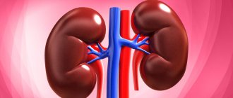
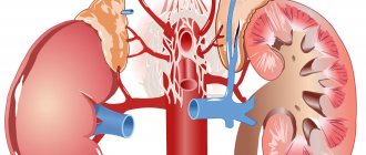
Disease concept
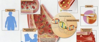
Eclampsia is caused by circulatory disorders due to swelling of brain tissue and a decrease in blood microcirculation in it. In case of serious kidney diseases, acute nephritis, swelling of the brain tissue develops due to water and sodium retention. In pregnant women, the condition is associated with generalized spasm of the cerebral arteries and the formation of intravascular blood clots.
Renal eclampsia in pregnant women is called severe toxicosis, accompanied by a sharp increase in blood pressure. In the absence of competent medical treatment and emergency care, the condition poses a risk to the life of the mother and fetus.
Complications
Thanks to the development of medicine, deaths from this type of eclampsia are rare.
However, death from acute renal or heart failure or from massive cerebral hemorrhage is still possible. The prognosis becomes more difficult when other forms of eclampsia – cerebral, hepatic – are added. In pregnant women, death is possible from disseminated intravascular coagulation. Typically, renal eclampsia does not further complicate the course of acute nephritis - on the contrary, the pathology after attacks often does not become chronic. Complications of renal eclampsia
Causes
The mechanism of development of eclampsia is not fully understood. It is known that the main provoking factor is a strong increase in blood pressure. This causes cerebral vascular spasm and disruption of normal blood circulation. According to the study, it was found that the following factors provoke the disease:
- acute impairment of the kidneys and vascular system;
- diabetes;
- bearing a child at a young age (15 – 18 years) or after 35;
- pregnancy interval is more than 10 years;
- systemic lupus erythematosus;
- genetic predisposition to the disease;
- excess body weight;
- multiple pregnancy;
- attacks of eclampsia in previous pregnancies.
Renal eclampsia develops mainly in pregnant women in the third trimester
Against the background of arterial spasms during pregnancy, women develop renal failure and cerebral ischemia. At the same time, blood flow is significantly reduced, which is accompanied by an increase in blood viscosity. This leads to increased tissue permeability and swelling. The combination of these factors leads to hemorrhages in the tissues of various organs, in particular in the brain.
What is this
Eclampsia is a serious disease and complication of preeclampsia. This is a rare, dangerous condition that causes high blood pressure, leading to seizures, including during pregnancy. Seizures are periods of disrupted brain activity that can cause fatigue, decreased alertness, and seizures.
Eclampsia affects approximately 1 in 200 women with preeclampsia. This pathology can occur even if there were previously no seizures or similar brain disorders.
Symptoms
Eclampsia is always accompanied by a sharp increase in blood pressure. As a result, hemorrhages occur, from which many patients die.
The prodromal period of the disease is accompanied by the following manifestations:
Is proteinuria dangerous in pregnant women?
- severe headache;
- swelling;
- dizziness, nausea;
- retardation of consciousness;
- sometimes delirium, hallucinations;
- feeling of tightness in the chest;
- decreased amount of urine;
- an increase in protein levels in urine.
In terms of vision, many patients note the appearance of a veil before the eyes, flickering spots, blurred vision, and double images.
The prodromal period does not last long or is completely absent. An attack of eclampsia occurs suddenly, sometimes developing against the background of normal health. The duration of the acute course of the disease is 2–3 days, but there are short-term attacks that often go unnoticed.
An attack of eclampsia lasts from 1 to 3 minutes and has several stages:
- The preconvulsive stage lasts about 30–40 seconds, accompanied by twitching of the facial muscles.
- Tonic spasms – spasm of the muscles of the whole body is noted. The patient often loses consciousness, the pupils dilate, and the skin turns blue (10–30 sec).
- Clinical convulsions - the patient has difficulty breathing, often foaming at the mouth. Duration up to 2 minutes.
- The comatose period is a gradual return to consciousness, often accompanied by urinary and fecal incontinence. Sometimes death occurs due to asphyxia, cerebral hemorrhages, and pulmonary edema.
After the attack, the patient’s consciousness is confused, speech is difficult. More often than not, patients do not remember what happened to them. Sometimes the patient becomes anxious after a seizure, nervousness, aggressiveness, attempts to escape, and refusal of medical care.
If signs of renal eclampsia develop, you should consult a doctor as soon as possible
Attacks of eclampsia are rarely isolated; more often the attack is repeated several times during the day. The above-described clinical manifestations are not always observed. Sometimes the attack is short-term in nature, occurring with the patient’s full consciousness or its short-term loss. In this case, renal eclampsia syndrome is accompanied by the following symptoms:
- Strong headache;
- decreased vision;
- lethargy;
- twitching of facial muscles, eyelids;
- decrease in some reflexes.
Important! If an attack occurs, you must call an ambulance as soon as possible. A timely reaction will help avoid many negative consequences.
Renal colic syndrome
Renal colic most often develops with, less often with kinking of the ureter, nephroptosis, or kidney tumor. Pathogenesis: reflex spasm of the ureter due to irritation of its wall with a stone and stretching of the renal pelvis, which is caused by a violation of the outflow of urine from it.
Renal colic has a characteristic clinical picture. Suddenly, usually after walking or jolting, severe, cramping pain appears, localized in the half of the lumbar region where the stone is located. The pain spreads down the ureter, into the groin area, and into the genitals. They are sharp in nature.
During an attack, the patient is extremely restless, screams, moans, and cannot find a place for himself. Renal colic is accompanied by nausea, repeated vomiting that does not bring relief, a feeling of fullness in the abdomen, bloating, and symptoms of functional intestinal obstruction.
Along with the pain, the following phenomena develop: frequent urination, pain and difficulty urinating. The amount of urine excreted is reduced, and at the end of the attack it is increased.
Changes in the urine are detected: single fresh red blood cells, and sometimes severe hematuria. “Urine sand” and small stones can be found in the urine. But during an attack it may not contain pathological impurities if there is a complete blockage of the ureter. The duration of the attack ranges from several minutes to 2-3 or more hours.
When examining the patient, the body temperature is normal, sharp pain is detected when palpating the lumbar region and a positive Pasternatsky sign. The diagnosis of renal colic in urolithiasis is usually confirmed by the typical clinical picture of the disease, changes in urinary sediment and X-ray and ultrasound data.
Renal eclampsia is a relatively rare complication of acute nephritis. It should not be confused with azotemic uremia, a terminal complication of chronic nephritis.
Diagnosis of eclampsia
An attack of eclampsia occurs suddenly and is accompanied by an acute course, convulsions and other characteristic manifestations. The purpose of diagnosis is to identify not eclampsia itself, but its precursors. The following measures are used for this:
- Detection of late toxicosis in pregnant women. Precursors of eclampsia include frequent headaches, blurred vision, nausea, vomiting, irritability, anxiety, or, conversely, apathy. Women often experience skin itching, hot flashes, and difficulty breathing through the nose. However, it is not necessary to have all the above-described signs. The risk of a seizure is often indicated by one or more of these.
- To identify pathology, the patient's medical history must be studied. Provoking factors for eclampsia are hypertension, pathologies of the cardiovascular system, diabetes mellitus, and kidney disease.
- Study of laboratory parameters of blood and urine.
- Blood pressure control. The risk of developing eclampsia is indicated by maintaining high values (140/90 or higher) for 6 or more hours.
During pregnancy, an ultrasound examination of the fetus and placenta is performed to assess their condition. Cardiotocography and Dopplerography are used to determine the presence of fetal hypoxia.
Diagnosis of the disease requires an integrated approach using instrumental research methods
Computer and magnetic resonance imaging allows one to assess the condition of a woman’s brain. To exclude pulmonary edema, a chest x-ray is prescribed.
Renal eclampsia: what it is, clinical picture and treatment methods
Renal eclampsia is a syndrome in which a person with kidney problems loses consciousness, possibly with seizures, due to spasms in the brain.
A similar symptom is rare, but is possible in acute kidney disease, such as nephritis or toxicosis during pregnancy. It manifests itself during high blood pressure with edema. Accompanied by symptoms such as convulsions and fainting.
What is this
Eclampsia is a serious disease and complication of preeclampsia. This is a rare, dangerous condition that causes high blood pressure, leading to seizures, including during pregnancy. Seizures are periods of disrupted brain activity that can cause fatigue, decreased alertness, and seizures.
Eclampsia affects approximately 1 in 200 women with preeclampsia. This pathology can occur even if there were previously no seizures or similar brain disorders.
Causes
Renal eclampsia often follows preeclampsia, which is characterized by high blood pressure after the twentieth week of pregnancy or kidney problems and protein in the urine. If preeclampsia occurs frequently, over time it can progress to a more severe form called renal eclampsia, which severely affects the brain, thereby causing attacks.
Researchers cannot figure out why exactly renal preeclampsia occurs, but there are a number of reasons that can provoke its transition into a complication - eclampsia. Among them:
- High blood pressure . The pressure in the vessels increases so much that they can become damaged and, as a result, limit blood flow. Subsequently, this leads to swelling in the blood vessels of the head, which interferes with the normal functioning of the brain, resulting in seizures.
- Proteinuria . Preeclampsia usually affects kidney function. Protein in the urine is an indicator of this. Normally, the kidneys filter waste from the blood and excrete it in urine. However, the kidneys may try to retain some of the nutrients in the blood, such as protein. If the kidney filters are damaged, protein can leak through them and be excreted in the urine.
- Chronic hypertension.
- Headache.
- Ages up to 20 and after 35.
- Pregnancy with twins.
- Malnutrition.
- Diabetes.
These are the main reasons that can affect the occurrence of such a serious and dangerous disease as renal eclampsia. But there are also risk factors that can affect the occurrence of this disease:
Clinical picture
Preeclampsia and eclampsia have similar symptoms, but their patterns differ in intensity and duration. These symptoms may also be symptoms of other diseases, such as diabetes or kidney disease. The clinical manifestation of eclampsia is:
- Swelling of the face and hands.
- Headache.
- Sudden weight gain.
- Nausea and vomiting.
- Vision problems.
- Difficulty urinating.
These symptoms can appear constantly, but there are also those that come with frequency:
- Cramps.
- Fainting.
- Muscle pain.
- Abdominal pain in the upper quadrant on the right.
Diagnostics
To correctly prescribe treatment, a competent diagnosis of the disease is necessary. The doctor must prescribe a series of studies to confirm the diagnosis and determine the causes of certain symptoms. The main diagnostic measures include examination and questioning, and studies include:
Treatment
The only way to treat renal eclampsia is to treat the underlying disease. If these are kidney problems, then you need to treat them; if the cause is pregnancy, then you just need to give birth.
Drug treatment aims to relieve symptoms, such as lowering blood pressure. If the condition is life-threatening, hospitalization is possible to determine concomitant diseases and the causes of symptoms.
There are also medications to prevent seizures. They are taken together with drugs to lower blood pressure and aspirin in small dosages.
You may have to reconsider your views on nutrition and add more vitamins and minerals, such as calcium, to your food.
Crucial for the treatment of renal eclampsia is the correct implementation of all doctor’s instructions and adherence to the diet. With proper care and control of the disease, it will not cause discomfort in life.
Consequences and complications
When the disease is controlled, complications should not arise, however, if the disease is not treated, it can lead to such serious consequences as disability due to brain damage.
It can also cause swelling of the brain, which is a critical condition that can subsequently lead to death and requires urgent hospitalization.
The main consequences of renal eclampsia include:
If eclampsia occurs during pregnancy, it can cause a number of consequences for the child due to impaired blood flow, which manifests itself in the fetus as asphyxia and subsequently a high risk of morbidity. All these complications can be avoided if you follow your doctor's recommendations and seek medical help at the first sign.
Loading…
Source: https://KardioBit.ru/pochki/lechenie/pochechnaya-eklampsiya-chto-eto-klinicheskaya-kartina-i-metody-lecheniya
Emergency care for eclampsia
If an attack of eclampsia occurs at home, it is important to provide first aid before the ambulance arrives. First of all, the physical safety of the patient should be ensured, since during convulsions the patient often injures himself. The patient should be placed on the left side, with a spoon wrapped in a bandage placed in the mouth to avoid the tongue from sinking. If a decrease in pulse rate and heartbeat is noted, it is necessary to perform an indirect cardiac massage. If there is vomiting, mucus, or foam, it is necessary to clean the patient’s oral cavity. Often the patient’s life depends on how competently and timely emergency care was provided.
The life of a patient with renal eclampsia depends on the correct provision of first aid to a patient.
Features of classification manifestations
The World Health Organization considers pathological disorders in the following sequence:
- Chronic course of arterial hypertension, recorded before conception;
- Hypertension that arose as a reaction of the body to the appearance and development of the fetus during pregnancy;
- Mild stage of preeclampsia;
- Severe stage of preeclampsia;
- Eclampsia.
The development of eclampsia does not always occur according to the scheme considered: it can also occur after a mild degree of preeclampsia.
Domestic obstetrics adheres to a different classification of pathology. Unlike foreign colleagues, Russian doctors assume that preeclampsia lasts a short period of time, followed by eclampsia. In Europe and America, preeclampsia is diagnosed if blood pressure exceeds 140/90 mm. rt. Art., swelling is clearly visible on the woman’s body, and the amount of protein in the daily dose of urine is more than 0.3 g/l.
Russian experts classify these same signs as nephropathy, the severity of which is determined by the severity of symptoms. The stage of preeclampsia is diagnosed if the following symptoms are added to the 3 signs described above:
- Headache;
- Decreased vision clarity;
- Vomiting accompanied by abdominal pain;
- A sharp decrease in the amount of urine excreted.
That is, foreign experts consider nephropathy an emergency condition requiring immediate hospitalization.
The development of nephropathy and the degree of its manifestation are presented in the table.
Severe degree is one of the complicated forms of pathology, when the following symptoms are added to high blood pressure and proteinuria:
- Impaired quality of vision;
- Severe attacks of headache;
- Pain syndrome in the stomach;
- Nausea accompanied by vomiting;
- Readiness for seizures;
- Massive swelling throughout the body;
- A sharp decrease in urine output per day;
- Pain on palpation of the liver;
- Changes in laboratory blood parameters.
The more severe the form of pathological changes, the greater the likelihood that the growing embryo will not withstand drug therapy, and the development of the fetus will be stopped.
There are also forms of the disease, the course of which depends on the time of their occurrence:
- Manifestations during pregnancy. The most common form of pathology. Threatens the life of mother and baby. There is a danger of termination of pregnancy when the fetus cannot withstand the effects of medications used for treatment.
- During the period of childbirth in women. Occurs in 20% of all recorded cases. It poses a danger to the life of the baby and mother. The attack is provoked by childbirth.
- Pathology that develops after the birth of a child. Appears very rarely in the first days after birth.
All forms of eclampsia develop according to the same pattern, therefore, their symptoms and treatment will be identical.
It is worth dwelling on the classification, which is based on dysfunction of any organ. In this case, the clinical picture of the disease will be different.
- Typical form. It manifests itself as severe swelling of the entire body, high blood pressure, intracranial pressure, and proteinuria.
- Atypical shape. Occurs as a result of prolonged labor in women in labor with a weak nervous system. It manifests itself as cerebral edema without pronounced symptoms of swelling of the subcutaneous tissue. In this case, slightly elevated blood pressure and moderate proteinuria are observed.
- The conditions in which renal eclampsia manifests itself differ from the previous 2. Swelling and elevated blood pressure readings are insignificant. It is characterized by a large accumulation of fluid in the peritoneal cavity and amniotic sac.
Treatment of eclampsia in pregnant women
The main method of treating pathology is delivery. In a full-term pregnancy, labor is induced immediately after the mother’s condition returns to normal. For periods less than 37 weeks, the issue of premature birth is decided if eclampsia is accompanied by a significant risk to the health of the mother and child. If the threat is low, therapy is aimed at stabilizing the mother’s well-being, which has a positive effect on the condition of the fetus.
If symptoms are mild, outpatient treatment is allowed with strict implementation of the following measures:
- strict bed rest;
- increasing the amount of fluid consumed;
- reducing salt in the diet;
- blood pressure control;
- examination by a doctor every 2–3 days.
In case of acute symptoms, the woman is hospitalized. During the first few hours of hospital stay, stabilization therapy is carried out, including the following medications:
- calcium gluconate;
- magnesium sulfate.
The dose of magnesium is determined depending on the general condition of the patient, reflexes, blood pressure levels, and the amount of magnesium in the blood serum. Sometimes magnesium sulfate causes fetal lethargy or hypotension, but negative effects are rare.
Treatment of pathology during pregnancy requires a special approach taking into account the condition of the mother and child
If there is no therapeutic effect from the use of magnesium sulfate, patients are prescribed Valium or Hydralazine in the form of droppers.
If delivery is necessary, the method is determined in accordance with the woman’s condition. If no complications arise, labor is induced by administering an Oxytocin solution. If natural delivery is not possible, a caesarean section is performed.
Specifics of definitions
Eclampsia and preeclampsia are conditions of pathological disorders in the body of a pregnant woman. Neither the first nor the second ailment can be called an independent disease, since they are a consequence of insufficient functionality of systems and diseases of internal organs. Moreover, their symptomatic manifestations are always accompanied by disturbances in the functioning of the central nervous system of varying degrees of severity.
Eclampsia and preeclampsia are conditions that occur only in pregnant women, in women during childbirth and in the first days after delivery.
The condition occurs as a consequence of disruptions in the relationship in the mother-placenta-fetus chain during pregnancy. The causes and symptoms of the pathology vary, so in world medical practice there is still no uniform approach to its classification. Thus, in obstetrics in America, Europe and Japan, such syndromes are associated with manifestations of arterial hypertension during pregnancy. Russian doctors believe that such manifestations are gestosis, or rather, their forms complicated by convulsions.
Preeclampsia is a syndrome that develops in the second trimester of pregnancy, with characteristic signs of persistent hypertensive disorders, which are accompanied by edema and the appearance of protein in the urine.
Eclampsia is a clearly manifested symptom of a dysfunction of the brain, the main symptom of which is an attack of convulsions that quickly turn into a coma. Seizures and coma are the result of a malfunction of the central nervous system due to excessive blood pressure.
Prevention
To prevent eclampsia, measures should be taken to prevent late toxicosis in pregnant women. They are carried out in the third trimester of pregnancy and include the following recommendations:
- before pregnancy, it is important to cure chronic diseases;
- regular visits to antenatal clinics during pregnancy;
- blood pressure control;
- periodic urine testing to determine protein levels;
- detection and timely treatment of late gestosis;
- if your blood pressure increases, you need to take medications prescribed by your doctor;
- elimination of stress and emotional tension;
- diet;
- frequent walks in the fresh air;
- quality rest and sleep of at least 8 – 9 hours.
Prevention of renal eclampsia consists of preventing late toxicosis, proper nutrition
Renal eclamptic attacks are a common occurrence among pregnant women and patients suffering from nephritis and other kidney pathologies. A positive prognosis for the patient is possible with timely treatment of eclampsia and its proper prevention. In the absence of medical care, severe complications and death of the patient often occur.
How to treat and can it be avoided?
First, the doctor listens to the patient’s complaints and makes a preliminary diagnosis based on them. Further diagnostic measures are carried out, which include:
- examination using ultrasound and MRI, identifying abnormal areas;
- pressure measurements;
- blood and urine tests.
Eclamptic attacks are not chronic and may appear once, but this does not mean that the disease disappears. There will be no attacks until the disease has passed a certain stage, at which swelling of the brain appears. Therefore, it is important to identify eclampsia at the initial stage in order to avoid possible complications.
In therapy, bloodletting is often used; depending on the patient’s condition, spinal cord puncture is also used to quickly relieve pressure and swelling of the brain. The following medications are used to relieve seizures:
The cause of eclampsia of the kidneys during pregnancy is completely unclear, because of this, the method of stopping convulsions is early delivery. Other methods are also used:
- ensuring peace;
- dehydration therapy;
- eliminating the causes of vascular spasms;
- inhalation of saturated oxygen.
Return to contents
Renal eclampsia causes the following complications:
- damage to the central nervous system;
- psychoses;
- hemorrhage;
- swelling of the brain;
- blood clots;
- partial or complete paralysis.
Return to contents
To prevent the disease, it is necessary to pay attention to the manifestations of late intoxication in pregnant women, as well as carry out preventive measures in children aged 7 to 9 years. Doctors recommend the following preventive measures:
- timely treatment of chronic or acute diseases;
- pressure control;
- maintaining proper nutrition;
- minimizing stress;
- preventive examinations - at least once a year, more often during pregnancy;
- taking vitamin preparations.
If there were primary attacks, it is recommended to reduce the load on the body. Maintain a rest regime, sleep at least 8 hours a day. And also take walks in the fresh air more often. The success of treating the disease depends on therapy and correct methods of prevention; if the disease is left unattended, severe complications and death may occur.
Fortunately, most happy mothers do not know about eclampsia in pregnant women, since this condition occurs only in 0.05% of women. However, among the problems of modern obstetrics, the question of diagnosis and treatment of this disease is more acute than ever and requires further study, because domestic and foreign gynecology interprets it differently.
This dangerous condition is preceded by a variant called preeclampsia. Such pathological changes in the body are recorded in 5-10% of pregnant women. From our article you will learn why these dangerous ailments occur, how to recognize and eliminate them.
Diagnosis
Difficulties may occur when recognizing the disease that caused a coma (an attack of eclampsia). They occur if the patient is under the supervision of a doctor or paramedic not from the onset of kidney disease (acute nephritis), but only during an eclamptic attack. The diagnosis is made easier if the medical history is known and it can be established that the disease began with the characteristic signs of acute nephritis: rapidly developing edema, hypertension and hematuria. These symptoms make it possible to distinguish renal eclampsia from an epileptic seizure.
Symptoms for renal eclampsia and epileptic seizures are similar. In both cases, the attack occurs suddenly. In both cases, tongue biting, involuntary urination, and complete loss of consciousness are noted. Anamnestic data, the presence of arterial hypertension, and edema allow one to diagnose eclampsia in acute nephritis. Epilepsy is characterized by scarring on the tongue, indicating previous seizures.
Sometimes, with renal eclampsia, azotemic uremia is mistakenly diagnosed. Differential diagnosis for these two syndromes is not difficult. The main disease in the first case is acute nephritis, while azotemic uremia develops in a patient with chronic nephritis. With eclampsia, the attack occurs quickly and violently, while uremia is characterized by a long and slow development. A low specific gravity is constantly detected in the urine of a patient with uremia. In eclampsia, the excretory function of the kidneys is not impaired and the urine has a high specific gravity.
In addition, it is not difficult to detect signs of azotemic intoxication in uremia: the smell of urine in the exhaled air, excretory gastritis and colitis, anemia. With eclampsia, these signs are absent. In the blood with uremia, increased residual nitrogen is always detected. With eclampsia, the content of residual blood nitrogen is not increased, and if there is an increase, it is for a short time. Azotemia in renal eclampsia occurs when urination stops (anuria), but this is a short-term phenomenon. With improvement in urine output, azotemia in eclampsia disappears.
Renal eclampsia is observed more often at a young age. With cerebral hemorrhage, patients do not experience seizures. Coma develops acutely within a few seconds; the patient's face is not pale, but, on the contrary, red and hyperemic. The emerging signs of hemiplegia (one-sided paralysis) quickly increase, whereas with renal eclampsia, signs of paralysis rarely appear and soon disappear. Cerebral hemorrhage at a young age is very rare. In the vast majority of cases, it develops in older people.
Specifics of first aid
Considering that the severe condition of gestosis is based on a convulsive syndrome, it is impossible to do without qualified assistance from medical personnel. Treatment methods will also be prescribed by the doctor, and the emergency care algorithm for eclampsia will be as follows:
- Call an emergency medical team, informing the dispatcher about the extremely serious condition of the pregnant woman;
- It is necessary to place the patient on her left side;
- Cover the woman with soft things: blankets, pillows, rugs. This way you can prevent injury during a seizure;
- If necessary, fix the tongue so that it does not stick;
- Between attacks of convulsions, carefully remove accumulated vomit from the mouth.
To eliminate the recurrence of serial seizures, you can administer a magnesium solution intravenously.
The possibilities of transportation are determined by the arriving doctors, and assistance to the expectant mother should be carried out in an emergency vehicle, since artificial ventilation may be necessary to restore breathing. They also carry out emergency measures to reduce blood pressure.
At the initial stage of treatment for pregnant women and women in labor, it is advisable to use drugs that relieve seizures and reduce blood pressure. At the same time, swelling is relieved, which worsens the woman’s general condition.
The use of any one direction of therapy will only worsen the patient’s condition: the use of anticonvulsants without normalizing blood pressure is pointless.
Carrying out sulfate infusion therapy includes the use of such drugs.
- Medicines that relieve seizures:
- Emergency (Droperidol, Magnesia);
- Supporters (Fulsed, Andakin);
- Strengthening the sedative effect (Glycine, Diphenhydramine).
- Drugs that lower blood pressure:
- Emergency (Nifediline);
- Supportive (Methyldopa).
You need to control your blood pressure by taking medications throughout your pregnancy. All medications are used intravenously or intramuscularly.
If the attacks are severe and difficult to treat, emergency delivery is indicated. Indications for its implementation include the following symptoms:
- Bleeding from the birth canal;
- Placental abruption;
- Fetal hypoxia.
At the same time, they begin to stimulate labor after stopping the attack of convulsions, choosing a natural method of delivery, since anesthesia for caesarean section can provoke another attack.
In all other cases, therapy is carried out with magnesia and prescribed medications until the severity of the pathology and the general health of the mother and baby are clarified.
Prevention of dangerous conditions is an important component of maintaining the health of the expectant mother. Preventive measures are used in the following cases:
- The pregnant woman has already had a history of attacks of eclampsia and preeclampsia;
- The attacks occurred in close relatives of the woman: mother or sister.
Preventive measures include the use of Aspirin from the middle of the second trimester (from 75 to 120 mg daily as prescribed by a doctor) and products containing calcium (1 g per day).
How does the syndrome manifest?
The duration of each attack of renal eclampsia is several minutes, but the number of attacks may vary. The development of the syndrome occurs in four stages:
- Precursor stage (preconvulsant). Lasts up to 20-30 seconds, the patient begins to twitch the facial muscles.
- Stage of tonic convulsions (30 seconds). They spread to all large muscles of the body due to muscle tension.
- Stage of clonic convulsions (2 minutes). Foaming at the mouth, activation of convulsions, loss of consciousness, breathing problems.
- Comatose or resolving stage. The patient may come to his senses, or fall into a coma or die (the outcome depends on the assistance provided, the severity of eclampsia, etc.).
Usually the disease develops at the peak of acute glomerulonephritis, less often - after swelling has decreased. At the warning stage, in addition to isolated twitching of the facial muscles, headaches, nausea, and blurred vision may occur. Later, vomiting and increased pain occur, “spots” flash before the eyes, and vision is blurred. A state of stupor may occur, at the same time the pressure increases and the heart rate decreases.
During convulsions, the patient is often unconscious. There is pallor and bluish discoloration of the facial skin, wheezing, dilated pupils, involuntary release of feces and urine, and tongue biting. After returning consciousness or emerging from a coma, a person remains confused in thoughts, speech, memory impairment, aggressiveness, anxiety, and often temporary loss of vision. Later, basic brain functions are restored.
Clinic
An attack of renal eclampsia most often occurs unexpectedly when acute nephritis is at its height. By carefully observing the patient, you can detect a number of signs that precede an attack. In addition to headaches, there is stupor, frequent vomiting, and neck rigidity. Before an attack of eclampsia (the word eclampsia in Greek means convulsions), blood pressure rises sharply, and the protein content in the urine increases. In other cases, attacks coincide with an increase in swelling.
The attack itself begins with sudden tonic convulsions, which are quickly replaced by clonic convulsions of the whole body.
The patient’s skin is pale before the attack (has an appearance characteristic of acute nephritis), and during it becomes cyanotic. Cyanosis extends to the neck. The veins in the neck swell. The eyes are squinted to the side. The teeth are clenched, but foamy saliva, often stained with blood, is released through them, as the patient bites his tongue. During an attack, the patient loses consciousness (is in a coma). Breathing is erratic, sometimes intermittent, sometimes snoring (stertorous), associated with convulsions. The attacks last several minutes and repeat after 1/2-1 hour. Eclamptic attacks in acute nephritis can last for a day or more. In addition to blood pressure, intracranial pressure also increases.
After an attack of eclampsia, the patient, as a rule, plunges into deep sleep, and if he sometimes wakes up, he finds it difficult to navigate his surroundings. Only after the attack is it possible to establish that the patient has suffered complete blindness or partial loss of vision - hemianopsia, which is associated with damage to the visual centers in the brain. With the cessation of attacks of eclampsia in the next 2 days, it goes away.