General information, description of the embryonic organ
Urachus navel - what is it?
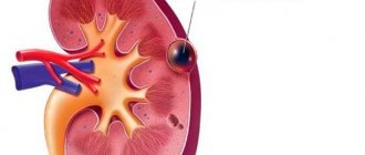
The term in question (medical) is of Greek origin. As you know, it consists of two syllables, which are literally translated as “urine” and “pour out.” Urachus navel - what does this word mean? Another name for this organ is the “urinary duct”. Experts say that the urachus is a tubular formation that gradually develops in the embryo. It connects the anterior bladder and the umbilicus and is located between the peritoneum and the transversus abdominis.
Such an organ is formed in the fetus from the upper part of the allantois. The latter is also an embryonic respiratory organ. This is a kind of embryonic membrane that develops from the ventral wall of the fetal hindgut. By the way, it is the allantois that participates in gas exchange with the environment and the release of waste.
Do you know why the urachus of the navel is needed? What is this? A photo of the formation in question is presented in this article. According to experts, it is through this duct that fetal urine is discharged into the amniotic fluid.
After five months of intrauterine life, obliteration of the duct occurs. It ends by the immediate moment of birth (that is, by 40 weeks of pregnancy). In this case, the median umbilical ligament is formed.
Only a doctor can tell you what a urachus cyst is and how to treat it correctly. The same applies to the possible consequences of this disease.
The main feature of the disease is that it is asymptomatic until the existing fluid becomes infected. The doctor can determine the tumor (cyst) itself after palpation of the abdominal area only if its parameters have reached the size of an adult man’s fist.
The disease has several variants of manifestation and is divided into the following categories:
- urachus cyst without fistula, which appears as a tightly closed capsule with mucous contents inside;
- urachus cyst with umbilical fistula, which is diagnosed as a neoplasm with regular release of internal fluid in the umbilical area and irritation of the skin area;
- urachus cyst with vesico-umbilical fistula, which is accompanied by urine discharge in the umbilical area.
Symptoms
A urachal cyst can appear at any age, even in old age. Basically, the neoplasm does not manifest itself in any way in the early stages, when its size is insignificant. When the cyst reaches a large size, the patient complains of the following symptoms:
- Feeling of pressure on the bladder and bowels.
- Increased gas formation, flatulence, bloating.
- Abnormal bowel movements: constipation or indigestion.
- Painful urination.
When inflammatory processes, suppuration, and infection join the pathology, the following signs appear:
- Swelling of the anterior wall of the peritoneum and pubis.
- Abdominal pain in the abdomen, intestines.
- Redness of the skin on the pubic area and around the navel.
- Increased body temperature.
- Intoxication of the body - weakness, nausea, dizziness.
Causes
The urachus is part of the urinary bladder, so its cystic formation is often called a bladder cyst.
The fetal excretory urinary duct allows all fetal secretions to be discharged into the amniotic fluid. Normal fetal development causes fusion of the urachus by 5 months of pregnancy. In place of the overgrown duct, the middle umbilical ligament appears.
Formed in the embryonic period, the pathology can be detected at any stage of life: a urachal cyst appears in children and adult patients.
The anomaly is divided into the following types:
- Closed type. The pathology of this type is characterized by a closed cystic capsule with fluid, without the formation of a fistula;
- Open (umbilical, vesico-umbilical cyst). This type involves the formation of a fistula, as a result of which embryonic biological fluids entering the walls of the abdomen irritate its surface. Through the fistula, not only embryonic secretions, but also various harmful microbes enter the abdominal cavity, which can lead to the occurrence of a pathological process.
Experts cannot reliably determine the root cause of the pathology. A number of experts are inclined to believe that the cause of urachal cysts in men, women and children is abnormal intrauterine development.
An inflammatory process may occur in the duct due to:
- Umbilical fistula (hole). This pathology is caused by non-closure of the part of the duct closest to the navel. In this case, urine is released into the umbilical area, causing regular irritation. Pathogenic microorganisms can penetrate through the hole, triggering an infectious-inflammatory process;
- Infection of the duct with pathogenic bacteria from the bladder. This situation occurs when the organ is damaged by infections of the genitourinary system;
- The inflammatory process can occur during pregnancy. The growing uterus can put pressure on the bladder, causing inflammation.
According to the observations of specialists, urachal cysts are 3 times less common in women than in the male population. The cystic formation may not manifest itself for a long time and may not increase in size. The hidden dynamics of pathology often makes it possible to detect it in adulthood.
The disease is classified as congenital and appears at 5-6 months of pregnancy. Modern medicine cannot determine the specific reasons that provoke incomplete overgrowth of the urachus. The reason for the development of the disease itself and the formation of a cyst after the birth of a child may be the absence of characteristic suspicions, since the disease is asymptomatic.
Separately, it is worth paying attention to the possibility of the urachus becoming overgrown in two places at the same time, which provokes the formation of a cavity that accumulates the original feces and urine of the fetus. This disorder, as a rule, leads to an increase in the tumor to 10-12 cm in diameter.
This phenomenon is observed when part of the urachus, which is located in the navel area, is not fused. Symptoms of such an anomaly are manifested by weeping of the wound.
What other signs are inherent in umbilical fistula? Urachus of the navel (what it is in adults, we will tell you right now) with non-fusion manifests itself in the form of redness, swelling of the skin around the navel and serous discharge that is observed in the umbilical fossa. In severe cases, they become purulent or bloody. It should also be noted that such an anomaly is characterized by an increase in the patient’s body temperature to 38-39 °C.
How to treat the disease in question? In case of inflammation of the navel, urgent consultation with an experienced surgeon is required. If necessary, bacteriological culture of the discharge is carried out. The method of treating this disease depends on the cause of the inflammation.
Typically, an umbilical fistula is treated through surgery.
This anomaly is characterized by complete non-closure of the urachus. This type is characterized by regular discharge of urine from the wound.
Cystic fistula of the urachus of the umbilicus cannot be treated with a conservative method. To eliminate this pathological phenomenon, surgical intervention is required.
Reasons for education
Since a urachus cyst is formed at the stage of fetal development in the womb, a possible reason for incomplete healing of the urachus duct may be infection of the woman’s body during pregnancy. The causes of this pathology have not been fully studied.
The inflammatory process that can occur in a cystic neoplasm can provoke the activation of pathogenic microorganisms. Infection in the cyst cavity can enter through the fistula tracts or through the bladder.
Bladder diverticulum (signs, treatment)
In what cases does the umbilical urachus develop abnormally? What is this? The reasons for such deviation should only be determined by experienced specialists.
Under certain conditions, the duct in question is not completely blocked. As a result, the following anomalies are formed:
- umbilical fistula;
- vesico-umbilical fistula;
- bladder diverticulum;
- urachus cyst.
What are such deviations and how to treat them? We will tell you about this right now.
This anomaly is characterized by a saccular protrusion of the walls of the bladder. As a rule, congenital diverticula are single. They are located on the posterolateral wall and are connected to the main cavity through a long neck.
As for the acquired disease, as a rule, it is multiple in nature.
How to eliminate the pathology of such a peculiar organ as the urachus of the navel? What is this? Treatment of bladder diverticulum is surgical, especially if it contributes to urinary retention and causes the development of cystitis. The prognosis for the operation is favorable.
It should also be noted that the treatment tactics for acquired diverticulum should include all measures aimed at eliminating the cause of bladder outlet obstruction.
Urachal cyst – in children, women, men, non-union
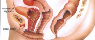
Intrauterine development of the fetus in the first trimester of pregnancy is very important, which is why it is necessary to carefully monitor the degree of fetal formation. Urachus in children is a pathological condition characterized by non-closure of the embryonic urinary duct with the formation of a cystic cavity.
Anomalies of intrauterine development
The excretory canal for the discharge of biomaterial from the embryo into the amniotic waters is called the urachus. During the normal course of pregnancy and the development of the child, it heals by 5-6 months and the median umbilical ligament is formed.
In infants and adults, an obliterated, cavity-free cord is formed at this site, which stretches from the top of the urine reservoir and reaches the navel between the transverse fascia and the abdominal cavity.
A urachal cyst in children is an abnormal condition in which a mass occurs in the bladder duct and appears as an irregular growth with a capsular membrane.
In pathological conditions, non-fusion of the urachus provokes:
- an umbilical fistula forms in the abdominal cavity, causing regular irritation of surrounding tissues with secretions;
- bladder diverticula, which form in the peri-vesical region;
- Vesico-umbilical fistula occurs when the lumen of the passage is completely closed and is characterized by regular discharge of urine into the umbilical region;
- A urachal cyst appears at the site of the duct in the form of a neoplasm with compacted walls, formed as a result of fusion on both sides.
Tumors are filled with mucus, original feces, biomaterial, and serous exudate. Urachal cysts can occur several times more often in women than in men.
The disease grows slowly, which makes itself felt in adulthood and can reach up to 12 cm in length. Infection and inflammation of the formation are dangerous due to complications, death of epithelial cells and abscess conditions.
Tumors of the passage can develop from different layers, epithelial, connective tissue or muscle. A particular danger lies in the formation of bladder cancer.
Based on the TNM classification, all urine storage tumors are divided into large groups:
- cancer without infection in the muscle layer;
- invasive metastatic neoplasm.
Kinds
Abnormal features of the urachus are divided into several classifications:
- Complete umbilical fistula. Characterized by an exposed vitelline passage along its entire length. The lumen of the ileum is revealed upward through the umbilical ring. When there is a significant space of unobliterated vitelline canal, the intestinal viscera are separated through the fistulas, while in the area of the umbilical ring the mucous membrane of the large and small intestines of a bright red color is released.
- Incomplete umbilical fistula. They are formed due to dysfunction of obliteration of the distal region of the duct. A scanty secretion is produced, and when infected, they become purulent. When examining the wound between the scanty epidermis, a pinpoint fistula opening can be identified, from which a small amount of liquid content is released.
- Ileal diverticulum. Pathology in which the proximal part of the vitelline duct is kept unobliterated. It is located on the platform of the ileum opposite the mesentery and looks like a vermiform appendix.
- Vitelline duct cyst. When both ends of the ureters and the remaining unobliterated middle part are obliterated, a closed cavity appears, which gradually stretches, fills with secretions of the mucous membrane, and a cyst is formed.
- Urethral duct cyst. For a long period of time, the cyst is small in size and does not manifest itself in any way. When it suddenly expands, it presses against the bladder, causing the urge to urinate. It has the ability to suppurate and is characterized by an increase in temperature, pain, redness and swelling of the tissues around the navel, and tension in the abdominal cavity.
Causes
The urachus cyst is formed in the initial stages of embryonic development. The etiology of the disease is still not fully understood. The main reasons for the production of pathology are the infected organism of the mother.
In pregnant women, it is affected due to displacement of internal instruments as a result of the growth of the uterus and newborn, which puts pressure on the formation and causes pain in the lower abdomen.
The urachus of the navel in an adult makes itself felt during the inflammatory process due to the activation of pathogenic microflora. The infection manifests itself as a result of damage to the bladder, kidneys, and weakened immunity.
Symptoms
Formations of small sizes do not cause manifestations. A visual physiological examination does not make it possible to determine the presence of an anomaly; the disease is detected when pathogenic microorganisms enter.
The main features are:
- prolonged healing of the umbilical wound in newborns, while blood is constantly released and can fester;
- a large tumor begins to compress the urine reservoir, resulting in difficulties with emptying, frequent urge to urinate, a feeling of incomplete emptying of the bladder, pain in the lower part of the abdominal cavity during emptying;
- bloating, flatulence, problems with bowel movements occur;
- after the cyst combines with a urine accumulator, purulent-blood secretion occurs in the biomaterial;
- in the presence of a fistula, purulent exudate will separate from the navel area, and when pressing or straining the abdominal muscles, streaks of blood will also emerge;
- the only and most complete non-closure of the tract is characterized by the constant separation of urine from the navel;
- women experience nagging pain as during menstruation, cystitis, intestinal spasms;
- the surrounding epithelium may become inflamed and develop a rash;
- the presence of an umbilical fistula after the appendix falls off indicates non-closure of the upper part of the meatus;
- severe variants of the malaise provoke acute abdominal syndrome, in which it becomes swollen, tense, painful and increased in size several times;
- The baby does not sleep well, constantly burps, and is developmentally delayed.
A cyst of the passage in childhood is a congenital anomaly, and it can become inflamed and become very dangerous for the child’s health.
Diagnostics
To determine and establish the degree of deviation, it is necessary to conduct a complete examination of the patient:
- Examination of the abdominal cavity, palpation.
- Cystography. X-ray method uses dyes. The picture shows the structure, shape, volume and abnormal formations of the internal organs of the pelvis.
- Ultrasound examination (ultrasound). The exact location of tumors, the urethra, the presence of stones, and inflammatory processes are determined.
- Computed tomography (CT). Shows in more detail the tendency of neoplasms, structure, contents, relationship with the bladder and abdominal cavity.
- Fistulography. X-ray irradiation is used in conjunction with contrast elements. Determines the length of the fistula, directions, branching.
- General analysis of blood and urine (OAM, UAC)
- Cystoscopy. The bladder is examined, the presence of structures, and pathologies of the formation of the internal organs of the small pelvis. To do this, anesthesia is used and a cystoscope is inserted into the girl’s urethral canal.
- Magnetic resonance imaging (MRI). It is used to determine the presence of abnormal changes, as well as a three-dimensional image of neoplasms.
- Biopsy with histological verification. It studies not only malignant, but also benign tumors; histology of cysts is needed to determine its type and develop a plan for further examination and therapy.
If acute pain and bloated abdomen occur, the laparoscopic method is used.
Treatment
The pathological disease cannot be treated at home or in traditional medicine. The only and effective method is surgical manipulation in a hospital.
In childhood and adolescence, excision surgery is performed faster. If the canal is partially closed, removal will not be required; it is possible to treat it with medication. In newborns, to prevent infection, treat the umbilical wound daily with antiseptic solutions.
When a fistula is formed, the surgical technique involves excision of it along with the navel; with the intravesical type, the surgeon performs manipulation in case of tissue damage. You can only be treated for the presence of cysts if they are removed.
Drug therapy is prescribed after the operation. Is to use:
- antibacterial agents;
- anti-inflammatory drugs;
- ointments, creams, gels for local anesthesia and healing;
- antispasmodic medications;
- vitamin complexes.
The operation begins with the help of anesthetic agents, after consultation with a urologist, obstetrician-gynecologist and a complete diagnosis of the body’s condition.
The function is to cut tissue to gain access and open the tumor. After the cysts are removed, stitches are applied and covered with a bandage. The duration of the postoperative period is at least 20 days.
Consequences and prevention
If you do not go to the clinic in a timely manner to diagnose the pathology, the cyst may burst and peritonitis may occur. In such cases, the principle of cure is urgent surgery.
In the postoperative period it is necessary:
- follow the doctor's recommendations;
- take medications in a timely manner;
- systematically handle sutures and visit dressing rooms;
- Do not open the wound or bandage yourself;
- undergo regular preventive examinations with a doctor;
- do not change treatment tactics;
- exclude the use of folk remedies;
- cure infectious diseases.
Removal methods
In newborn children, if no inflammatory processes are detected, surgery is not performed; up to 1 year of age, the urachus heals on its own.
To remove tumors the following are used:
- laparoscopic method. If the capsule is intact and does not contain pus, an endoscopic apparatus with video is used. Surgical methods are not used; special instruments are used to puncture the abdominal cavity;
- laparotomy. Abdominal surgery under general anesthesia.
Source: https://FlintMan.ru/kista-uraxusa/
Diagnostics
A large urachus cyst can be palpated through the anterior abdominal wall in the form of a tumor-like formation located between the pubis and the navel.
In the diagnostic process, it is important to distinguish a urachal cyst from a bladder diverticulum, an umbilical cyst, or a hernia of the anterior abdominal wall. For this purpose, the diagnostic algorithm includes cystography, cystoscopy, ultrasound of the bladder, MRI or CT. To find out whether the open urinary duct communicates with the bladder, fistulography is performed after the inflammatory phenomena have subsided.
In the case of suppuration or perforation of a cruchus cyst, patients are often admitted to a hospital with an acute abdomen clinic, and the correct diagnosis can only be established during diagnostic laparoscopy or laparotomy.
If a large urachus cyst can be detected after palpation and defined as a tumor between the pubis and the navel, then in other cases diagnosis requires an ultrasound examination (ultrasound). The study itself must be carried out carefully and always under the supervision of a doctor. This is the only way to distinguish this disease from bladder diverticulum or hernia of the anterior abdominal wall, which have similar symptoms.
The entire list of diagnostic procedures includes:
- cystography (x-ray examination);
- cystoscopy (using an endoscope to examine the inner surface of the bladder);
- Ultrasound of the bladder;
- Fistulography, which allows you to determine the presence of a connection between the open urinary duct and the bladder;
- MRI (magnetic resonance imaging);
- CT (computed tomography).
If the process of suppuration has already begun in the body or perforation of the cyst (violation of the integrity of the tumor), patients are admitted to hospital treatment with symptoms of an acute abdomen. The correct diagnosis can be made by a specialist after laparotomy or laparoscopy.
As for medical specialists, the patient can be examined by a surgeon, urologist or andrologist. The first suspicion of a cyst is given by a palpable tumor located between the navel and pubis. Most often it occurs in men and is dangerous primarily by the risk of violating the integrity of the cyst and the entry of infected mucous fluid onto the surfaces of other internal organs.
When examined by a urologist, large cystic formations are determined by palpating the anterior abdominal wall.
To confirm the diagnosis of “urachal cyst” code according to ICD 10, the doctor prescribes laboratory and instrumental examination methods:
- MRI, CT, allowing to determine the communication of the cystic capsule with other organs;
- Ultrasonography. A urachus cyst is clearly visible on ultrasound; the location and size of the formation can be determined;
- Cystographic examination, which determines the condition of the bladder, the presence of a cystic formation in its cavity, and structural changes in the organ;
- Cystoscopy. The procedure helps to examine in detail the mucous membrane of the bladder, allows you to diagnose inflammatory processes, structural abnormalities, and damage to the walls of the organ. New growths are determined qualitatively.
After eliminating inflammation of the cystic cavity, fistulography can be performed. The procedure is carried out in the presence of fistulas and allows you to track their progress. In cases where the patient is admitted with an “acute abdomen” (swollen and hardened abdominal cavity), laparotomy and laparoscopy are performed.
When the diagnosis is confirmed, surgery for a urachal cyst is scheduled for the very near future. This is necessary to prevent serious complications and consequences.
Umbilical fistula on ultrasound
Non-closure of the urachus in the area of the umbilical ring manifests itself in the first days after birth - a weeping navel, maceration of the skin. An umbilical fistula at a later date indicates drainage of an infected urachal cyst.
It is important to distinguish between patent urachus, umbilical vein and vitelline duct. When the umbilical vein is not closed, the discharge is bloody. When the vitelline duct is not closed in the navel, there is a bright red tumor, the skin is severely irritated by alkaline discharge.
Drawing. A man complains of purulent discharge from the navel. On ultrasound, just below the navel there is a heterogeneous rounded lesion, which communicates with the umbilical fossa due to a hypoechoic tubular structure; connection with the bladder is not determined; blood flow in the surrounding tissues is slightly increased. Diagnosis: Infected urachal cyst, umbilical fistula.
Treatment of urahust cyst
The pathology is not eliminated by conservative therapy; surgical intervention and removal of the cystic formation are necessary.
In children, surgery to remove the urachus is performed when the duct is completely blocked, when the urachus is completely excised. The procedure is performed in the first days of a newborn’s life.
When the lumen of the canal is partially closed, surgery is not performed, since the anomaly may self-heal. To prevent infection, the navel area is lubricated daily with a solution of brilliant green. The procedure is carried out after bathing the baby.
The classical treatment direction is used before surgery to eliminate the inflammatory process and normalize the patient’s condition.
Preoperative treatment of cysts in the bladder in men, women and small patients includes:
- Anti-inflammatory non-steroidal drugs (NSAIDs);
- Products with antibacterial characteristics;
- Local preparations (gels, ointments).
At the end of classical therapy, examinations are carried out to confirm the absence of inflammatory processes. An operation is prescribed, refusal of which can lead to serious complications (abscesses, omphalitis).
The surgery is performed under local anesthesia. The surgeon opens the cavity, removes its contents, and drains it. When the inflammatory process spreads to nearby tissues, they are also removed. If a fistula has formed, it is removed along with the navel. After excision of the cystic cavity, the wound bed is sutured, and a bandage is applied to the suture.
The postoperative period lasts about 3 weeks. During recovery, it is necessary to take healing, anti-inflammatory drugs to improve postoperative tissue regeneration.
After removal of a urachus cyst, the patient should avoid physical activity and should not lift heavy objects. During the entire rehabilitation period, the patient must adhere to bed rest, rest as much as possible, and move minimally.
Treatment
The surgical operation performed is the only method of successful treatment of the disease. A urachal cyst must be removed to prevent the risk of infection and further development.
The most effective treatment methods are determined by the doctor individually and depend entirely on the results of the research. If it is necessary to treat a cyst with sterile contents, then, as a rule, surgical intervention is performed by extraperitoneal excision of the urachal cyst. In this case, an abscessed cyst with spreading purulent contents requires other treatment methods - mandatory and immediate opening and removal of pus from the affected cavity (drainage of the abscess cavity). Only after some time (during the cold period) is it possible to perform a standard operation to excise the urachal cyst.
If the presence of a urachus cyst in an adult man has caused the formation of an umbilical fistula and the development of omphalitis, then before surgery a course of conservative therapy with the use of antibiotics, dressings and ultraviolet irradiation is mandatory. This is necessary to reduce the activation of the inflammatory process. Only after such treatment can surgery be prescribed.
Taking medications can only be conditioned by symptomatic treatment of the disease, reducing the severity of pain. Since the disease itself is dangerous due to the entry of pus into the abdominal cavity, at the first suspicion, a thorough examination of the body and strict adherence to the instructions of the attending physician is necessary.
Prevention of urachus cysts
To prevent postoperative complications, after surgery for urachal cysts, tests are performed: CT scan, cystoscopy, urine cytology. The patient is under the supervision of a doctor throughout the recovery period and must strictly follow all medical recommendations.
If the root cause of the urachus cyst is unclear, prevention is difficult. General recommendations include:
- Strengthening the immune system (adequate physical activity, proper rest, compliance with nutritional rules, intimate hygiene, taking vitamin complexes);
- Prevention of infectious diseases of the genitourinary system.
Prevention
When a person is diagnosed with a urachal cyst, they should undergo a comprehensive examination and receive timely treatment to prevent serious complications.
During the rehabilitation period, the patient needs to adhere to medical recommendations: rest more often, do not overexert yourself physically and emotionally. It is worth protecting your stomach from lifting weights.
Urachal cyst: its signs and treatment methods - Pediatric Urology
Recovery period
As mentioned above, abnormal development of the umbilical urachus is not treated with conservative methods. This deviation can be congenital or acquired. In the acute period, surgical intervention is contraindicated. Usually the operation is performed after the inflammatory process has subsided.
Only an experienced surgeon should perform procedures for excision of cysts, diverticula, etc. In most cases, such operations are well tolerated by patients. The outcome is almost always favorable.
After surgery, the doctor is required to instruct the patient about the recovery period. The patient is prohibited from engaging in physical labor and lifting weights. He should spend most of his time in bed, lying on his back. The patient is also prescribed a strict diet.
If you follow all the doctor’s recommendations, the excision site should heal successfully in the shortest possible time. If the patient feels pain and discomfort in the abdomen, then you should immediately contact the surgeon who performed the operation. The same applies to cases of discharge from the navel (blood, pus, etc.).
For a speedy recovery of the patient, various vitamin and mineral complexes can be prescribed.