Cystoscopy is performed with a catheterization cystoscope, and the bladder is examined. The mouth of the ureter is located, which must be catheterized. A disposable ureteral catheter No. 5–6 with a mandrel is inserted into the channel of the working element of the cystoscope, observing asepsis, and advanced into the bladder. Using an Albaran fork, the end of the catheter is directed into the opening of the ureter and carefully moved along the ureter to the obstruction, trying to bypass it. If this is successful, then the catheter is advanced to a height of 20–25 cm. The guideline for determining the location of the distal end of the ureteral catheter is the centimeter scale on its surface. The wire mandrin is removed from the catheter. After this, if the catheter is in the pyelocaliceal system, urine begins to flow through it in a stream or in frequent drops and the acute pain syndrome immediately passes. Now the cystoscope is turned with its beak up, the Albaran fork is lowered and the catheter is advanced into the body of the cystoscope up to the rubber cap. Release the washing liquid and carefully remove the cystoscope. After the beak of the cystoscope appears from the external opening of the urethra, the catheter is grabbed with the fingers of the left hand and held in this position, and the cystoscope is “removed” from the catheter with the right hand. The ureteral catheter is left in the renal pelvis for 24–48 hours (no more than 72 hours). After this time, if the PC was caused by a small (less than 5 mm) stone, 2-3 ml of glycerin and 2-3 ml of 1% novocaine solution are injected through the catheter into the renal pelvis or ureter, and the catheter is removed. After this, the stone may pass on its own.
If the catheter cannot be passed above the stone, and the stone is X-ray negative, then the catheter is left in this position and DLT of the stone is performed (guidance along the distal end of the catheter). If the catheter manages to move the stone into the pelvis, the catheter is left in the pelvis and DLT of the stone is performed.
To facilitate the passage of the catheter above the stone, it is advisable to use ureteral catheters with a specially modeled distal end (bayonet-shaped, hook-shaped, etc.).
The procedure of uncontrolled catheterization of the renal pelvis for ureteral stones is fraught with the risk of developing a specific complication in the form of perforation of the ureteral wall, renal pelvis or renal parenchyma. The danger of this complication increases when trying to overcome an obstacle with a catheter with a mandrel. This complication can be suspected by the absence of urine discharge from the catheter after passing it to a height corresponding to the position of the renal pelvis. You can confirm that the catheter has gone beyond the wall of the ureter or pelvis by injecting a contrast agent through the catheter and taking an x-ray (see the topic on ureteral injuries).
Therefore, catheterization of the renal pelvis should be performed in the X-ray office, which will allow monitoring the position of the catheter at any time.
In addition to perforation of the ureteral wall during catheterization of the pelvis, acute pyelonephritis and even septic shock may develop after a few hours or days, even after successfully performed catheterization. Therefore, as Yu. A. Pytel and I. I. Zolotarev (1985) rightly point out, cystoscopy and catheterization of the ureter cannot be considered as indifferent manipulations for the patient, since they are fraught with the possible development of severe complications, therefore “. Catheterization of the pelvis should be used only after all other less severe treatment measures have no effect.”
If renal colic is caused by a ureteral stone whose diameter is more than 5 mm and its spontaneous passage is doubtful, then treatment should be started immediately with ELT of the stone or with URS with contact lithotripsy. If it is impossible to use these treatment methods, catheterization of the renal pelvis is performed or a self-retaining catheter-stent is installed (see Chapter 4).
Date added: 2016-02-02; ; ORDER A WORK WRITING
Catheterization is the introduction of a special tube (catheter), which can have a different shape and length, diameter and material of manufacture, into certain cavity structures or channels of the body for the purpose of treatment or diagnosis.
Why is a catheter placed?
In general, catheterization is indicated in the following cases:
To determine ureteral patency and the degree of obstruction in it; In order to obtain separate urine for the diagnosis of leukocyturia and determine its origin; To eliminate urinary stasis; Bougienage; Reduction of stones; To carry out the retrograde pyeloureterography procedure; To restore urinary outflow in acute forms of pyelonephritis or ureteral occlusion by a stone.
Indications for the procedure may vary depending on the gender and age of the patient.
During pregnancy
In pregnant women, the risk of developing kidney pathologies increases significantly, which is associated with changes in the genitourinary organs during pregnancy. Kidney damage negatively affects pregnancy and fetal health.
Catheterization may be prescribed for pregnant women in the following cases:
Renal hydronephrosis; Chronic or gestational pyelonephritis.
Catheterization in pregnant women with pyelonephritis performs a serious therapeutic task - it relieves the patient of renal blockade.
The catheterization procedure in men is technologically a more complex procedure, since the length of the male urethra is longer than that of the female. Indications for catheterization in male patients are:
Urinary outflow disorders; Inflammatory processes in the urinary system (for the purpose of rinsing); For chronic or acute urinary retention; To obtain urine from the pelvis for the purpose of studying it in more detail; For therapeutic purposes, the technique is used to administer medications.
Usually a soft catheter is used, but if it is not possible to insert it, then a rigid tube is used, for example, with prostate adenoma or with strictures of the urethra.
In women, catheterization of the kidney and ureter, in addition to diagnostic purposes and some of the above diseases, can also be performed for acute nephritis and urolithiasis. When to do this is decided by the attending physician.
If there are stones in the kidneys, a catheter helps restore urinary flow, which is blocked by the stone.
Catheterization is often performed in women with acute inflammation of the kidneys, such as pyelonephritis. Here the catheter can be used for drug flushing and as a diagnostic method.
How is catheterization performed?
Renal catheterization is the insertion of a catheter into the ureter or pelvis. A similar procedure is performed using a special urethrocystoscope through which a ureteral catheter is advanced. The procedure requires strict adherence to antiseptic and aseptic rules. Sometimes catheterization is prescribed for the purpose of sounding or drainage.
There are fundamental differences when installing a catheter for people of different sexes. This is due to differences in the body structure of men and women. In any case, regardless of gender, the catheter is generously lubricated with a special antibiotic ointment. It is very important to install the catheter without damaging the genital organs, for which sterile Vaseline is used.
A container is placed between the patient's legs to collect urine. The patient should not feel pain when the catheter is installed correctly, and there should be no bleeding. In order for the outflow of urine to occur as easily as possible, the catheter must be checked in advance for leaks and air bubbles removed from it.
- Wash hands with soap and brushes.
- Treats them with alcohol and iodine.
- Treats the patient's external genitalia.
The patient must first thoroughly wash the genitals.
The catheter is inserted into the bladder using tweezers, a certain amount of urine appears - this indicates that the catheter is inserted into the canal in the right way. In men, catheterization is somewhat more difficult than in women. The catheter must be inserted so that it does not have kinks.
In order to protect the mucous membrane of the urethra and not cause unnecessary injury to it, before removing the device, a furatsilin solution must be very carefully injected into the bladder with a syringe, then the previously installed catheter is removed.
After use, clean the tool in warm water and soap. Catheters must be sterile, for which they are boiled at high temperature and stored in a solution of boric acid (sometimes carbolic acid). The containers in which tools are stored must be hermetically sealed. If a disposable device was used, it is disposed of after use.
Renal catheterization is a procedure during which a doctor inserts a catheter into the pelvis or ureter. It is performed using a special device - a urethrocystoscope, through which a ureteral catheter is advanced.
Renal catheterization is performed for the following purposes:
- determining the patency of the ureter and the presence of obstacles in it;
- eliminating stagnation of urine;
- bougienage;
- performing retrograde pyeloureterography;
- restoration of urine outflow in case of ureteral occlusion by a stone or acute form of pyelonephritis.
If kidney catheterization is performed for diagnostic purposes, then after obtaining the necessary biological material, the doctor removes the catheter back. If there are indications for therapeutic treatment, the catheter in the kidney is left indefinitely. It can be used to inject medications into the kidney.
Contact the Yusupov Hospital if you need a nephrostomy. The cost of the operation can be clarified by calling the clinic.
Executing the procedure
Most often, during the catheterization process, rubber catheters of different lengths and diameters are installed. The procedure is carried out using a cystoscope, at the end of which there is specialized equipment for the most accurate installation of the tube - an Albarran lift.
The microcamera on the device visualizes the advancement of the catheter:
Before the catheter is inserted, a cystoscope is inserted into the bladder; if the procedure is performed on men, then a solution of local anesthesia is first injected. Before insertion, the urethral opening must be treated with an antiseptic. The cystoscope is thoroughly disinfected before insertion and treated with vaseline or glycerin oil. For women, this procedure is performed in a supine position with hips apart and knees bent. If necessary, the bladder cavity is first washed to remove bloody or urinary debris and filled with saline solution. Then they look for an outlet into the ureter by rotating the microcamera of the cystoscope. When the ureteral orifice is located, the microcamera is brought to it so that it enlarges and is located directly in the center of the visual field. Then the catheter is carefully inserted. Having reached the desired place, it is fixed. How long the procedure lasts depends on your goals.
As for the period for which a catheter is placed, it all depends on the purpose. For diagnostic purposes, the doctor obtains the necessary biomaterial and removes the device back. If the procedure is carried out for therapeutic purposes, then the catheter can be left indefinitely, for example, to drain urine from the pelvis. Then a special tube is used, which has loops at the end for better fixation.
If there are problems with urination, catheterization is carried out 6 times a day (every 4 hours). When wearing a catheter for a long time, an infectious process may occur, so periodically the doctor removes it, washes it and puts it back. Please note that only an experienced specialist can flush the catheter.
Diagram of catheter insertion into the kidney
Ureteral catheterization is performed for diagnostic or therapeutic purposes.
Indications for use
Installation of a catheter is a rather painful procedure for the patient, but sometimes it is the only option for recovery. The need for this medical procedure often arises in patients who contact urologists with certain complaints and symptoms. To differentiate the diagnosis and prescribe adequate therapy, the doctor prescribes a series of diagnostic studies, and subsequently uses catheterization as a method of diagnosis or treatment.
A special tube made of flexible and elastic material is used for this purpose. Using this method, it is possible to perform an X-ray contrast examination of the organs of the genitourinary tract, surgery to remove a benign or malignant neoplasm, stent the ureter or eliminate scar narrowing of its canal. Often this procedure is used in combination with catheterization of the renal pelvis. A catheter in the kidney is a necessary manipulation when removing stones, tumors, contrast or plastic surgical correction of tissue.
Therapeutic objectives
Ureteral urological catheters are used as a conduit for delivering pharmaceuticals directly to the affected organ. Also, using this tool, you can quickly normalize the outflow of urine in cases where the disorder is caused by malignant neoplasms of various locations or hyperplastic changes in the prostate gland in men.
The main indications for using a ureteral catheter for therapeutic purposes:
- Impaired patency of the ureter as a consequence of complications of urolithiasis;
- Secondary strictures caused by infectious and inflammatory processes, adhesions and radiation therapy;
- Omond's disease is localized in the fatty tissues of the retroperitoneum and leads to mechanical compression of the urinary tract;
- Growth of neoplasms into the canal cavity, deformation of the walls by a tumor or modified enlarged lymph nodes;
- Drainage of the renal pelvis in case of obstruction by a blood clot, stone or salts;
- The need to install a terminal ureterocutaneostomy when it is impossible for the patient to provide urine outflow in any other way.
Diagnostic purposes
The installation of ureteral catheters can be an additional diagnostic tool for a differentiated diagnosis when receiving uninformative urine tests. Catheterization helps obtain urine in an unchanged form, without introducing the microflora of the patient’s genital organs. As a result, the specialist receives a clean laboratory sample.
The catheter is used to obtain an isolated sample from the right or left kidney separately. This allows you to accurately determine the location of the infectious process. Using this technique, it is possible to accurately assess the amount of fluid remaining in the patient after urination, which is an important diagnostic criterion for identifying many urological pathologies.
Insertion of a catheter is effective for detecting the level of leukocytes, microorganisms, tubercle bacilli and salts in the urine. Using this manipulation, it is possible to determine the patency of the ureter along its entire length, without the use of additional medical equipment.
Structure of the urinary system
In the functional cells of the kidneys, nephrons, blood is filtered from metabolic products.
The resulting urine flows through the nephron tubules into the storage system of the kidneys - into the calyces and pelvis, and from there through the ureters it enters the bladder.
There it accumulates and, when a certain volume is reached, is excreted through the urethra.
Normally, in an adult, the length of the ureter is 25–30 cm. Its diameter is uneven, it narrows at the exit from the pelvis, at the connection with the bladder and at the intersection with the iliac vessels.
In men, the ureter intersects with the vas deferens, and in women it is located behind the ovaries.
The walls of the ureter consist of three membranes. On the outside, it is covered with a layer of connective tissue, which forms a kind of protective case. The inside is lined with a mucous membrane of transitional stratified epithelium.
The most developed is the muscle layer, which consists of longitudinal and circular muscle fibers. Their contractions ensure the flow of urine from the renal pelvis to the bladder.
Catheter into the bladder
This procedure is carried out for treatment, probing, drainage of the urinary tract, as well as for the purpose of making an accurate diagnosis. The main factors for conducting medical catheterization include the following indications:
- Help in establishing the patency of the ureters, as well as in removing obstacles in them in the form of stones.
- Bougienage procedure.
- Removal of stones in case of stone disease of the kidneys or bladder. Carrying out retrograde pyelourethrography.
- Establishing normal urine outflow during exacerbation of pyelonephritis.
- Obtaining qualitative analyzes from each individual kidney.
- Detection or absence of pathological processes in the bladder and kidneys.
- For better administration of drugs into the genitourinary organs.
In some cases, catheterization may be performed on patients diagnosed with urinary incontinence. In this case, the duration of his stay in the organ will be determined by the attending physician.
Reasons for installing a catheter
Ureteral catheterization is performed for retrograde pyelography. In this case, 2 - 3 ml of contrast agent is injected into the kidney through a catheter, which is excreted in the urine, then a series of photographs are taken.
This method allows you to evaluate the excretory function of the urinary tract, the presence, shape and location of stones, and violations of the internal structure of the kidney.
The rhythm of urination during catheterization also plays an important role. Normally, urine is released in 4-5 drops at intervals of 30-40 seconds.
Continuous urine production in large portions indicates hydronephrosis. With this disease, the size of the calyces and pelvis increases, which often leads to atrophy of the kidney tissue.
In addition, using catheterization, you can take urine for analysis directly from the renal pelvis. The concentration of leukocytes, urea and chlorides, and the presence of Mycobacterium tuberculosis are checked.
You can take urine samples from different kidneys and compare the test results.
Catheterization allows you to determine the exact cause of the disturbance in the outflow of urine from the kidney. This may be caused by blockage of the lumen of the ureter with a stone or compression of its walls by a tumor of nearby organs.
If, at the moment of overcoming the obstacle, blood comes out of the ureter, and then clear urine, then this sign is called Chevassu’s symptom. He talks about the presence of a tumor in the ureter.
When the ureter is obstructed, stagnation of urine develops in the kidneys. This may be accompanied by severe pain. In addition, increased pressure inside the kidney can lead to hydronephrosis.
Catheterization restores the flow of urine. This alleviates the patient’s condition and prevents irreversible changes in the structure of the kidney. In many cases, catheterization can remove the stone from the ureter.
Causes of stones getting stuck
The location of stones in places of narrowing of the ureter is possible if their diameter is more than 2 mm.
There are 3 jam segments:
- The lumen connecting the renal pelvis and the urinary canal is 2 mm in size.
- Narrowing of the area of intersection with the iliac vessels equal to 4 mm.
- More than 70% of stones are retained in the vesicoureteral segment, the diameter of which is no more than 1 mm.
Ureterolithiasis occurs as a result of an imbalance in the colloid composition of urine. A deficiency of protective organic substances that maintain phosphates, uric acid and calcium oxalate in a dissolved state leads to their sedimentation on the protein components of urine.
Among the exogenous factors causing imbalance are:
- hot climate promoting dehydration;
- the presence of a large amount of protein foods in the diet;
- deficiency of vitamins A and D, which negatively affects the absorption of calcium;
- hard drinking water, oversaturated with lime and calcium salts;
- insufficient physical activity.
There are a number of diseases that cause the formation of salt accumulations in the ureter:
- pathologies of the endocrine system;
- anomalies of the anatomical structure of the ureters;
- infectious diseases of the urinary system;
- injuries associated with violation of bone integrity, diseases of the skeletal system;
- heredity.
As a result of these reasons, the outflow of urine is disrupted and the kidneys secrete substances that contribute to the development of oxaluria, uraturia, and phosphaturia.
Technique of the procedure.
Catheterization is carried out using rubber catheters 50–60 cm long and 1–3 mm in diameter. They are made of plastic or silk fabric.
Sometimes they are additionally coated with bismuth salts, this helps to obtain a clearer image on an x-ray.
Catheterization of the ureters is carried out using a cystoscope specially designed for this purpose. It has one or two channels for inserting catheters.
At the end of this cystoscope there is a special device for accurately guiding the catheter to the mouth of the ureter. This device is called an Albarran lift.
In addition, the cystoscope has a special eyepiece that displays an image of the internal surface of the organ on the screen.
First, the cystoscope is inserted into the bladder. If the manipulation is performed on a man, then a local anesthetic solution is first injected into the urethra.
The cystoscope is then lubricated with sterile glycerin or petroleum jelly and inserted into the bladder. This procedure is performed for the woman while lying on her back; she is asked to bend her knees and spread her hips.
The area of the external opening of the urethra is pre-treated with an antiseptic.
If necessary, the bladder is washed to remove any remaining urine or blood, then filled with saline solution. The eyepiece at the end of the cystoscope is rotated until the orifice of the ureter is found.
Then the eyepiece is moved so that its image is magnified and is in the center of the field of view. The catheter is then inserted into the bladder through a channel in the cystoscope.
When its end is visible on the screen, it is lifted using the Albarran elevator, fixed at the desired angle, and catheterization of the right or left ureter is performed.
The installed catheter may be left in place for several days to drain urine from the pelvis.
In this case, a so-called self-fixing catheter is used. Its ends are bent in the form of a loop, this allows it to be fixed in the kidney.
In addition to conventional catheterization for diagnostic purposes, a urethropyeloscope is inserted into the ureter, which has an eyepiece and an irrigation system.
The eyepiece allows you to display an image of the inner wall of the ureter, and a solution is supplied through the irrigation system, which expands its lumen.
In order to prevent bacterial complications after catheterization, uroseptic and antibacterial drugs are prescribed.
The passages of the nephrons carry urine into the calyces and pelvis. And only then - along the ureters to the bladder. After it is filled, the liquid exits through the urethra.
In males, the ureter intersects with the vas deferens, and in females it is located behind the ovaries.
The ureter is surrounded on the outside by connective tissue; it performs protective functions. Inside there is a mucous membrane consisting of epithelial tissue.
The most developed are the rows of longitudinal and circular muscles. By contracting, they ensure the discharge of urine from the renal pelvis into the bladder.
Structure of the human urinary tract
Kidney drainage technique
The installation of a nephrostomy is controlled by radiography or ultrasound monitoring. The catheter is inserted into the kidney tissue through the abdominal wall. Its edge should be in the kidney itself. Using the installed drainage, urine is discharged into a specially installed sterile container - a urinal, which is changed regularly.
Oncologists at the Yusupov Hospital prefer puncture nephrostomy. Reviews about this operation are positive. Percutaneous nephrostomy is easier to tolerate for patients diagnosed with cancer than open surgery. The nephrostomy kit includes a puncture needle made of stainless steel, an adapter for nephrostomy, a spiral guide stylet, an auxiliary stylet made of stainless steel for the catheter, a catheter for nephrostomy device, a plastic adapter with a connecting connector and a funnel for connecting to a urinal.
Nephrostomy is a preliminary step before kidney surgery. It allows chemotherapy, as well as some specialized examinations of the urinary tract and kidney tissue for tissue inflammation and metastases. Before the procedure, doctors at the oncology clinic examine the patient:
- general urine and blood tests;
- detailed and biochemical blood test;
- ultrasound examination of the kidneys;
- urography;
- computer and magnetic resonance imaging.
Doctors at the Yusupov Hospital perform all studies using equipment from leading global manufacturers. Modern techniques are used for laboratory analyses. Patients can undergo all complex examinations at partner clinics.
How long do people stay in the hospital with a nephrostomy? Nephrostomy is not considered a particularly dangerous or complex procedure. The overwhelming number of patients leave the oncology clinic a few hours after the operation. If they follow simple recommendations, they feel comfortable and no complications arise. Doctors do not recommend doing physical exercise, lifting weights, or overexerting after installing a nephrostomy.
Indications for catheter installation
A remedy for improving kidney function, which relieves pain and normalizes urination
A catheter is placed in the ureter to examine the condition of the kidneys. In medical terminology, this method is called retrograde pyelography - two to three milliliters of a special substance are injected into the kidney through a catheter, which is released along with urine, after which a series of images are taken.
What do the results show?
This method makes it possible to evaluate the functioning of the urinary canals, the presence, nature and location of stones, and the failure of the internal kidney system.
The rhythm of urine output during catheterization is of no small importance. It is considered normal to release urine in an amount of four to five drops in an interval of thirty to forty seconds.
The continuous outflow of urine in large quantities indicates an increase in the size of the calyces and pelvis, which entails atrophy of the renal tissue. This disease is called hydronephrosis.
Also, using catheterization, it is possible to collect urine for examination directly from the renal pelvis. Check the content of leukocytes, urea and chlorides, the presence of tuberculosis bacillus.
It is possible to collect urine samples from each kidney and compare the results.
Ureteral rubber catheters
What is determined
With the help of a catheter, it is possible to specifically detect the factors of failure of the outflow of urine from the kidneys. This may be due to the presence of stones in the ureter or its strangulation as a result of swelling of adjacent organs. The case when, when overcoming the obstacle, blood came out of the catheter, followed by light urine, indicates the presence of a tumor in the cavity of the ureter. This indicator is called the Chevassu symptom. If the patency of the ureter is impaired, urine retention occurs in the kidneys. In such a set of circumstances, a person is bothered by acute pain syndromes, and increased intrarenal pressure can provoke hydronephrosis. With the help of catheterization, the outflow of urine is regenerated, which helps to alleviate the patient’s well-being and removes irreparable changes in the structure of the kidneys. In most cases, with the participation of catheterization, it is possible to exclude stones from the ureteral cavity. Note! User recommendation!
For the prevention of diseases and treatment of kidneys, our readers recommend the Monastic Collection of Father George. It consists of 16 beneficial medicinal herbs that are extremely effective in cleansing the kidneys, treating kidney diseases, urinary tract diseases, and cleansing the body as a whole.
Get rid of kidney pain..."
Nephrostomy replacement
Replacement of the nephrostomy or repeated nephrostomy is performed by oncologists at the Yusupov Hospital in case of displacement, loss of the drainage tube, or its blockage with an excessive amount of salts present in the urine. Complete removal or replacement of the nephrostomy occurs during a careful operation. Drainage can be removed from the kidney if there is no need for its further use, after the outflow of urine has been restored naturally.
In order to check whether the urinary system is functioning correctly, the drainage tube is filled with a special solution. It allows you to clearly view all the urinary organs on x-ray. A permanent nephrostomy is installed in those patients who, due to the physiological characteristics of the body or the late stages of a malignant neoplasm, are contraindicated for radical surgery.
Patients often ask: “If a kidney nephrostomy is installed, how long do they live after surgery?” Life expectancy depends on the disease for which the operation was performed. The prognosis is individual in each case. A patient can live with a nephrostomy for many years if it is properly cared for.
Operation progress
The catheterization session is carried out in a disinfected room using sterile gloves. The doctor carries out this process together with the nurse. Initially, a cystoscope is introduced, the bladder is washed, filled with a special solution, and only then a catheter is inserted.
Catheterization process - catheter installation
During a catheterization session for men, the patient lies on his back, bends and spreads his legs to the sides. The external opening of the urethra is cleaned with the composition of furatsilin, the genital organ is wrapped in a sterile napkin, and the foreskin is pushed back to the beginning of the organ. The catheter is generously lubricated with a sterile solution of glycerin or petroleum jelly and inserted into the urethral canal, threading the phallus onto the catheter tube. Once sample collection is complete, the catheter is carefully removed.
The sequence of actions during catheterization for women: the patient also lies on her back, bends and spreads her legs to the sides. The urethra and perineum are subject to hygienic cleansing. The entrance to the vagina is covered with sterile napkins. A sufficiently lubricated catheter is inserted into the urethra approximately ten centimeters in length, until the urine protrudes. When an insurmountable barrier is formed, the manipulation is terminated.
Installation of a catheter for a man and a woman
To cause the least damage to the mucous membrane of the urethra, before removing the catheter into the bladder with a syringe, inject a solution of furatsilin.
The reservoir for urine waste should be located below the patient's pelvis; it is necessary that there are no creases or folds on the surface of the catheter. After catheterization, the urinary bowel movement takes three to four minutes. If urination occurs over a longer period of time, the device may be the wrong size for the patient. When catheterization is carried out for a long time, the area around the catheter must be cleaned twice a day with warm water and soap. This serves as a preventive measure against infections entering the urinary tract. If sepsis develops, the urine acquires a pungent odor and a cloudy color.
Patients with difficulty urinating require catheterization every three to four hours. The interval between procedures is reduced if the patient takes a large volume of liquid. When there is damage to the spinal cord, the signals sent by the walls of the bladder do not reach the brain. As a result, the person does not feel the urge to urinate. Such patients will complain of headaches, decreased heart rate, increased sweating, and high blood pressure. If there are such complaints, the person needs emergency catheterization.
“Doctors are hiding the truth!”
Even “neglected” kidney stones can be quickly eliminated. Just don't forget to drink once a day...
>
Nephrostomy - catheter installation
Upon completion of the catheterization procedures, it is recommended to take baths for several days in a row; they help eliminate inflammatory processes. The most commonly used solution is a weak manganese solution; you need to fill a basin and sit in it for a while. Be sure to ensure that all the crystals melt in the water. No less useful are baths made from a herbal infusion of chamomile, sage, and St. John's wort. The procedures should be performed repeatedly throughout the day.
In men
Due to the structural features of the male genital organ, the procedure itself is considered more complex. For its implementation, soft catheters are mainly used, and if insertion is impossible, catheters of a rigid structure. In addition to the above diseases, in the stronger sex, catheterization is used for prostate adenoma and urethral strictures.
Regardless of gender and age, catheter placement should be carried out with extreme caution so as not to damage the internal surface of the organs. To do this, the device is treated with a special ointment, that is, sterile Vaseline. A correctly placed catheter does not cause pain or bleeding.
Before removing the device, furatsilin is injected into the bladder using a syringe to protect the mucous membrane from damage, and only after that it is carefully removed from the cavity of the urethra. All manipulations must be carried out by a specialist in his field.
And a little about secrets...
Have you ever suffered from problems due to kidney pain? Judging by the fact that you are reading this article, victory was not on your side. And of course you know firsthand what it is:
Discomfort and pain in the lower back Morning swelling of the face and eyelids does not at all add to your self-confidence... It’s somehow even embarrassing, especially if you suffer from frequent urination... In addition, constant weakness and ailments have already become a firm part of your life...
Now answer the question: are you satisfied with this? Can problems be tolerated? How much money have you already wasted on ineffective treatment? That's right - it's time to end this! Do you agree? That is why we decided to share an exclusive method that reveals the secret to combating kidney pain. Read the article >>>
Leave a comment 3,305
To diagnose and treat a number of diseases of the urinary system, a ureteral catheter is used, which is significantly different from the urethral one. With its help, an X-ray examination of the kidneys is carried out, stones and concretions are removed. Inserting a catheter into the ureter is a complex procedure. It is performed by highly qualified doctors only in a hospital setting.
Catheterization of the ureters is used in the diagnosis and treatment of pathologies of the urinary system.
Comparative characteristics, which operation is better
The advantages of lithotripsy are:
- simple conditions for the rehabilitation period;
- low cost of the procedure;
- with contact lithotripsy, the doctor can completely visually control the progress of the operation;
- shorter duration compared to other methods of stone removal;
- minimal risk of injury;
- no need for a large surgical incision.
The only disadvantage of lithotripsy is the need for natural release of crushed pieces, which leads to irritation of the urinary system and the development of renal colic.
- minimally invasive approach to treatment;
- possibility of a one-time procedure;
- hospital stay no more than 3-4 days;
- short rehabilitation period.
Compared to lithotripsy, this type has a greater number of contraindications and complications.
Turning to a treatment method such as abdominal surgery is recommended as a last resort. Surgery has a number of negative consequences:
- long rehabilitation period;
- possible dangerous consequences;
- high injury rate.
Surgery is prescribed if the diameter of the stone exceeds 1 cm and other treatment methods are ineffective. Compared to surgery, endoscopic ureterolithotomy has a lower risk of infectious and other complications, low blood loss and a shorter rehabilitation period.
The disadvantage is the impossibility of using medical equipment with the narrow structure of the ureter.
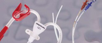
Ureteral catheters
For catheterization of the ureters, sterile disposable catheters are used, made of flexible plastic, equipped with a rigid wire mandrel (conductor) and a centimeter scale. To prevent catheters from bending, they are packaged in durable covers or cases. To avoid damage to soft tissue during installation, there is a spherical profile at the end of the instrument. Thanks to the ring marks, the insertion depth of the device is adjusted, and the holes on the sides https://www.youtube.com/watch?v=ZtuHcvAlM5M allow urine to be removed and medications to be administered.
In essence, a ureteral catheter is a tube 710 mm long. To carry out catheterization of the right ureter, a catheter with red markings is used, and for the procedure above the left - blue. Instruments are marked with numbers from 3 to 8 to indicate tube diameters from 1 mm to 2.66 mm, respectively. Catheters are made of non-toxic, hypoallergenic materials and undergo radiation sterilization. The shelf life of the devices is 3 years.
Duration of use
Depending on the purpose, ureteral catheters are installed for different times. The timing of installation depends on the diagnosis, the severity of the clinical symptoms of the pathology and the general condition of the patient.
The service life is also affected by the characteristics of the material used in the manufacture of this medical product:
- Silicone devices can be used for up to 30 days. This is due to the fact that bacteria do not multiply on the surface of medical silicone; such catheters do not tend to grow into organ tissue.
- Latex catheters can be used for up to 7 days. Often such products are used for diagnostic purposes or in the early postoperative period.
- models with silver coating are indicated for long-term use, without the need for replacement more than once every 3 months. These are the safest, but most expensive designs.
Purposes of ureteral catheterization
Ureteral catheterization is comparable to surgery. It is carried out only when clearly necessary. This procedure involves retrograde pyelography, checking the patency of the ureter, and collecting urine directly from the kidney. Catheterization is used in the treatment of a number of diseases; it allows medications to be introduced into the body and urine to be removed, for which the catheter is sometimes left in the ureter for several days.
Catheterization as part of treatment
During the treatment of pathologies of the urinary system, installation of a catheter in the ureter allows:
- Normalize the flow of urine from the affected kidney.
- Remove stones and concretions from the ureters.
- Remove pus from the kidney if the pelvis is infected (comparable to opening an abscess).
- Rinse the renal pelvis if there is a purulent process in the organ.
- Ensure drainage and rapid recovery after surgery.
Return to contents
Diagnostic purposes
As part of diagnostics, catheterization allows:
- Obtain an accurate urine test when directly collected from the kidney.
- Determine the volume of residual urine.
- Conduct a urodynamic examination.
- Identify the composition of urea, determine the number of leukocytes, chlorides, and tuberculosis bacteria contained in it.
- Take a urine test from a specific kidney, which is not possible with regular test collection.
- Determine the presence of a stricture (narrowing) of the ureter and its exact location.
The ureteral catheter is installed using a cystoscope.
Types of operations to remove stones
In medical practice, there are various types of operations to remove stones from the ureter:
- Remote and contact lithotripsy.
- Ureterolithoextraction.
- Nephrolithotomy
- Endoscopic ureterolithotomy.
- Surgical abdominal operation.
Only a doctor can select a specific procedure for stone removal based on the research performed.
Preparation for ureteral catheterization
Before the procedure, the patient is examined and anamnesis is collected to promptly identify contraindications. Catheterization is performed under general or local anesthesia. If local anesthesia is used, the patient may be prescribed antispasmodic drugs. Depending on the purpose of catheterization and the characteristics of the disease, the patient may need to take antibacterial agents. Sterile instruments are prepared for catheterization. The doctor conducts a conversation with the patient, explaining the essence and purpose of the planned procedure. If the catheter is to provide drainage, then its installation is the final phase of the operation and preparation for this procedure is not required.
Possible contraindications
Before inserting the catheter, the doctor takes a medical history and reviews laboratory results.
During catheterization, one should take into account the patient's possible contraindications to the procedure.
The main contraindications are:
- Lack of urine in the lumen of the bladder, which may be a consequence of the development of anuria. The presence of a pathological condition is confirmed by ultrasound examination.
- The occurrence of obstruction of the urethra, which is provoked by sphincter spasm. Catheterization in such a situation is carried out after the introduction of antispasmodics into the patient's body.
It is not recommended to carry out the procedure if the patient has laboratory-confirmed infectious processes localized in the organs and tissues of the urinary tract. A medical device in such a situation can provoke the spread of the inflammatory process.
Carrying out the procedure
Ureteral catheterization has been practiced since 1899. This is the most effective way to deal with stones. To install the catheter, a special device is used - a cystoscope. Passing the catheter is possible only through this device after it is installed in the bladder. Due to the peculiarities of the physiological structure, catheterization in men and women has some differences.
Catheterization in men
To ensure the catheter reaches the ureter, a cystoscope is inserted into the bladder at the beginning of the procedure. This instrument is a tube equipped with a light system, a camera that transmits an image to a monitor, channels https://www.youtube.com/watch?v=ZtuHcvAlM5M through which catheters will be passed and a special device that brings them to the ureter (Albarran lift) . The procedure is performed under general anesthesia or local anesthesia, which will require prior injection of an anesthetic into the urethra.
The patient lies on the table with his legs spread apart. Sterile instruments and containers are prepared for collecting tests, removing urine, etc. The external opening of the urethra is treated with an antiseptic solution, the penis is wrapped in a sterile napkin and pulled back. The foreskin is pushed back towards the base of the penis. The cystoscope, lubricated with glycerin oil, is carefully inserted into the bladder. If necessary, before installing catheters, the bladder is washed and then the organ is filled with saline solution. Using a camera on a cystoscope, the doctor finds the entrance to the desired ureter and inserts a ureteral catheter into the bladder through a special channel. When it appears on the monitor, it is lifted using the Albarran lift, set at the required angle and advanced into the ureter.
Carrying out catheterization of the ureters is equivalent to surgical intervention and requires compliance with all sanitation rules.
Carrying out the procedure in women
It is much easier for women to insert a cystoscope into the bladder, since the female urethra is shorter and wider than that of men. To carry out the procedure, the patient lies on the table, bending her knees and spreading them apart. The external genitalia and the opening of the urethra are disinfected with an antiseptic. When treating the perineum, movements are made from top to bottom. The vagina is covered with a sterile napkin. After inserting the cystoscope into the bladder, further steps for installing the catheter are the same as for the procedure for men. To avoid infection of the urinary system after the procedure, the patient (regardless of gender) is prescribed uroseptic drugs and antibiotics.
What to do after removing the instrument
Catheters are installed for different periods. It all depends on the diagnosis and condition of the patient. Instruments made of silicone can be used for a month, latex ones for about 6-7 days; if the catheter is coated with a layer of silver, the tube in the ureter can remain in the body for up to 3 months. How long and how the catheter will be used is decided only by the attending physician.
To avoid inflammation, after the procedure of removing a catheter from the urinary tract, it is worth taking baths with a light solution of potassium permanganate or chamomile infusion. Regularly clean your genitals with a sterile swab soaked in this solution or infusion. Such procedures are performed over the next few days.
To avoid infection entering the organs and the occurrence of urethral fever, you must carefully follow all the rules of asepsis and antisepsis when performing this procedure.
Possible complications
Catheterization of the ureter can lead to the following complications:
- Perforation of the urethral wall. During the insertion of the necessary instruments into the bladder, the urethra can be damaged, up to the complete destruction of the integrity of its wall with the formation of a false passage. This occurs when using very rigid instruments or when force is used if the advancement of the device along the urethra is difficult (for example, with prostate adenoma or stricture). This complication is accompanied by pain at the site of injury, bleeding, and lack of urination. Catheterization is canceled until the urethra is completely restored.
- Reaction to urinary emptying. Occurs in elderly https://www.youtube.com/watch?v=ZtuHcvAlM5Milty, weak patients suffering from pathologies of the cardiovascular system and disorders of the kidneys. If a full, distended bladder is quickly emptied, a serious malfunction of the kidneys can occur, including uremia or anuria. To prevent this from happening, during the procedure the urine present in the bladder is released gradually in small portions.
- Epididymitis. If sanitation rules are violated, men may develop an inflammatory process in the epididymis. Suppuration and septicemia occur - blood poisoning with pyogenic bacteria.
- Urethral fever. It occurs some time after the procedure due to the penetration of pathogenic microorganisms into the blood through the damaged urethra. Manifested by profuse sweating, chills, fever, and cardiac dysfunction. To prevent this pathology, the patient is prescribed antibiotics.
- Violation of the integrity of the ureteral wall. As a result, retroperitoneal phlegmon (suppuration) develops. High probability of death.
Perforation of the urethra or ureter requires immediate surgical intervention. No one is immune from complications. Definitely, before agreeing to carry out this procedure, the patient must make sure that experienced doctors will work with him, and the hospital has all the necessary equipment.
During pregnancy
Since during pregnancy in women the genitourinary system undergoes significant changes, kidney pathologies often develop, which is dangerous both for the woman in labor and for the child. If a patient is diagnosed with renal hydronephrosis or chronic pyelonephritis, then catheterization in this case is simply necessary to remove the renal blockade.
In women outside pregnancy, the insertion of a catheter may be prescribed for the diagnosis of diseases and the development of certain pathologies.
- For acute nephritis.
- For urolithiasis.
- In inflammatory processes of the urinary system.
- For acute urine retention and impaired urine outflow.
- For administering medications and obtaining qualitative analysis directly from the renal pelvis.
Stoma removal
Indicators of successful treatment, after which the nephrostomy can be removed, are:
- formation of a fistula tract to ensure the natural outflow of urine (in patients with severe pathologies);
- restoration of normal urine drainage through natural urinary drainage channels.
Typically, drainage is carried out for 10-15 days. Pregnant women are not recommended to use nephrostomy for more than 4 days due to the high risk of developing disorders. In any case, removal of the nephrostomy is prescribed by the attending physician based on the results of control tests and assessment of the degree of restoration of urinary diversion function. An equally important indicator is the absence of irreversible diffuse damage to the renal parenchyma (fiber).
If the operation was performed for the purpose of performing medical procedures (chemotherapy, stenting of kidney vessels), the nephrostomy is removed after restoration of the physiological outflow of urine.
Sometimes a drainage tube is installed indefinitely. Most often with severe cancer. In this case, the nephrostomy tube needs to be replaced periodically. It must be performed in an operating room. Unlike the initial surgery, tissue puncture is not required as a fistula is created.
Nephrostomy: self-care at home
Basically, after the operation, the patient is sent home on the same day with detailed instructions from the attending physician. The patient should not engage in sports or physical activity.
Proper care of the nephrostomy and carrying out preventive measures to protect against possible inflammation in the kidney are also important. A salt-free diet is required so that the flow of urine is not delayed.
After abdominal surgery, standard care is required. The tube is removed after a fistula tract appears to allow fluid to drain away.
It is unacceptable for the tube to fall out before a fistula tract appears. This can cause complications because it is difficult to put it back. But if the catheter does fall out, the doctor will replace it within 24 hours.
Regardless of the timing of catheter installation, it requires careful care:
- To avoid urinary infection, it is advisable to regularly flush the drainage with saline solution.
- Also, the area of the puncture wound should be clean, it should be washed with antiseptic solutions (furacillin or chlorhexidine), applying a sterile bandage.
- Periodic cleaning of the urine bag. The urinal has a sealed clasp and a special mark indicating the fluid level at which the device needs to be changed. If the bag is not changed in a timely manner, backflow of urine into the renal pelvis is possible. Because of this, kidney infection, suture divergence and increased blood pressure are possible.
- Constant flushing of the kidney. Active drainage should be used. To do this, 2 stomas are placed in the pelvis. By applying an antiseptic to one stoma, a washing liquid with stagnant urine and sand residues comes out of the second.
After nephrostomy, the patient goes home with detailed instructions from the doctor on how to care for the drainage and prevent inflammation. During the entire period of wearing the catheter, physical activity is prohibited, otherwise the nephrostomy may fall out; a salt-free diet is observed. To prevent urinary infection, regularly wash the wound and drainage with sterile saline solution.
If the catheter is installed for a long-term or lifelong period (for life), the nephrostomy is periodically replaced. In particular, renephrostomy is required when the drainage tube is blocked by salts contained in urine. The same manipulation is required if the catheter falls out, which cannot be allowed until a natural fistula tract has formed for the outflow of urine due to the risk of infection and problems. Replacement is made within 24 hours.
You need to care for your stoma through the following procedures:
- Rinse the nephrostomy with saline solution (20 ml of 0.9% sodium chloride). You can wash it at home if you follow your doctor’s recommendations. If you need to change the device. For this purpose, special kits are sold that contain a removable adapter and the tubular catheter itself.
- Wound care by rinsing with an antiseptic. You need to rinse the inlet with an antiseptic (“Furacillin”, “Chlorhexidine”), followed by applying a sterile, dry bandage. If a gauze dressing is used, it is changed daily. When using a sterile transparent dressing, change it every 3 days.
- Emptying the urinal after reaching the level marked on the device. If the replacement is untimely, there is an increased risk of biofluid reflux into the drainage and kidney, excess pressure in the renal pelvis with suture divergence and loss of drainage.
- Active kidney lavage. The technique is used when a paired organ is infected. To do this, two stomas are inserted: through one, a washing solution is supplied, through the second, stagnant urine with traces of sand is removed.
- Keeping dry. The patient needs water treatments (bath, swimming are prohibited), but it is important to keep the area around the wound dry for at least 14 days.
- Providing protection. If a patient is undergoing chemotherapy through a stoma, it is important to provide protection in the form of sterile gloves when emptying the urine collection container.
- Providing assistance. A patient with a nephrostomy needs the assistance of at least two people to change dressings and empty the urine bag, especially with double drainage.
Since drainage is a foreign body for the body, it can become infected at any time. Every patient who has a nephrostomy is warned about this. The catheter should be maintained regularly. Methods for preventing the infectious process include:
- Rinsing the drainage tube with saline solution and antiseptics. For this purpose, the drug “Chlorhexidine” or “Furacilin” is used.
- Applying a sterile dressing to the area of the postoperative wound.
- Timely emptying of the urine bag.
- Check with a doctor every 2 weeks.
Patients who have a nephrostomy should not exercise or lift heavy objects. Kinking the drainage tube can lead to complications, so the catheter should be in one position.