Features of use
A catheter is a flexible tube that is inserted into a blood vessel. The purpose of using the device is to suction fluid and administer medications into the vein. The device performs 2 functions: blood carrier and hemodialysis. The long-term internal catheter consists of two tubes (lumens).
The arterial lumen functions to collect blood from the body and transport it to the catheter. The venous lumen returns blood from the apparatus to the body. The device allows you to control the speed of blood flow. The main parameters range from 200 to 500 ml per minute.
By what criteria to select
The pharmacy offers a wide range of short- and long-acting venous and arterial catheters. There are several criteria by which a device is selected.
The amount of time the device will be used must be determined. It is necessary to select the exact diameter of the needle so that there are no difficulties during insertion. If the visibility of the veins is poor, a tourniquet is used. Medical devices from each manufacturer have a certain functionality and cost, the material is made with a special coating that prevents rapid resistance.
When choosing a catheter, there is an important condition: a smaller diameter ensures long-term blood patency. Devices with a large diameter can close venous areas and damage the vein lining.
Only a doctor can select the correct device. Carrying and installing on your own increases the risk of infection.
A long-term hemodialysis device ensures maximum renal function. Promote cleansing of poisons and unhindered blood transport in the body.
The article was approved by the editors Link to main publication
Select a category or subcategory
B. Brown in Russia Dialysis catheters Haemocat Signo / Hemocat Signo
Main types
Long-term hemodialysis catheters come in the following varieties:
- Double lumen.
- Tunnel.
- Permanent.
- Triple lumen.
- Hemodialysis kit.
The first type provides a short-term effect of cleansing the blood of harmful substances. Used as access to vessels during hemodialysis, hemofiltration, plasmapheresis, hemoperfusion. The main feature is that two lumens are located in one vessel. The necessary blood flow is ensured, which reduces the risk of blood clots.
The tunnel type acts for a long time, for several weeks or months. Inserted through a special hole in the chest.
A permanent catheter for hemodialysis is installed in the cervical, femoral artery, and subclavian vein. Used for immediate blood pumping. Installs easily.
The triple-lumen catheter is made of polyurethane. Designed for infusion of large volumes of fluid and hemodialysis.
We recommend reading
- Droppers to improve cerebral circulation
- Fistula for hemodialysis: pros and cons of the technique
- How to clean the blood vessels of the heart in the hospital and at home
The kit includes the following components: one catheter, a needle-introducer, a graduated guide, two injection plugs, a dilator, a scalpel, sterile wipes.
What information is missing from the article?
- Detailed review of medications
- More practical treatments
- Innovative developments in this area
- Qualified expert opinion
Types of catheters
According to their configuration, catheters are divided into single-, double- and triple-lumen. There are also models with a large number of gaps, but they are used in rare cases.
According to the possible duration of wearing this instrument, they are divided as follows:
- short-term, or acute;
- long-term (chronic);
- permanent catheter for hemodialysis.
Since hemodialysis must be performed regularly, up to several times a week, catheterization of the central vein with a long-term or permanent instrument is advisable.
Installation and removal rules
Catheters with two lumens are installed using a special Duo-Chez introducer - a thin-walled needle that ensures correct insertion into the vessel. Thanks to the device, 2 tubes are connected into one. Installed using the Seldinger technique in the subclavian and internal jugular vein. A prerequisite is that the patient must take a lying position at an angle of 15 degrees. In this state, the veins swell as much as possible, which eliminates the occurrence of air embolism. After catheterization, it is important to close the device to prevent air from entering the vein.
Installation of a medical device requires strict adherence to sterility.
The introduction process includes the following steps:
- Determination of the insertion site, anesthesia with 0.5 ml of lidocaine. The medicine is administered intradermally.
- Carrying out catheterization of the vessel (unimpeded collection of blood into the syringe indicates correct installation).
- The needle is inserted through the string conductor until resistance occurs. If the barrier appears earlier, the manipulation is repeated again.
- Then the needle is removed.
- Then they take a scalpel and make an incision near the conductor.
- Then a dilator with a built-in catheter is inserted along the guide wire.
- Now you should grab the end of the string protruding from the catheter and rotate it to pass through the skin.
- Carefully remove the guidewire and dilator. Make sure that there is free pumping.
- Then the tube is connected to the catheter.
- Be sure to secure the insertion site with a suture and apply a sterile bandage.
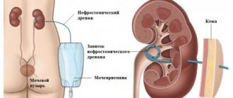
A tunneled catheter is placed in a large vein in the neck. And it is fixed at the site of scar tissue formation. Then it moves deep into the vessel and is released through the skin (approximately 6 inches from the injection site).
It is important to ensure safe handling and care of the blood pumping device to ensure long-term use.
Instructions for using catheters:
- The injection site must be kept clean.
- Make sure that the two ports are pinched.
- It is strictly forbidden to lie in a bathtub, jacuzzi, or sauna with the device installed.
- While taking a shower, the bandage is covered with polyethylene film and secured with an adhesive plaster. If the compress gets wet, it should be changed immediately.
- It happens that catheters become unstuck and bleeding occurs. In this case, it is necessary to pinch the bruise and immediately seek medical help.
- Touching the installed device should not be painful. In ideal condition there is no swelling, redness, or moisture.
It is important to ensure that there is no damage; both caps from the sides are compressed.
The bandage is changed every 3 days, or when it falls off or gets wet. Apply with clean hands and treat the installation site with a special solution. The compress is fixed with a transparent sticker.
If the procedure for inserting a catheter into a vein is incorrect, blood poisoning may develop. If you experience chills, high body temperature, burning sensation, increased sensitivity, swelling and pain, you should immediately call a doctor.
Vascular access
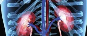
To effectively perform hemodialysis, it is necessary to create a vascular access through which blood will move into the dialysis machine, undergo purification in the dialyzer and return safely back to the patient's body. Before starting the first hemodialysis procedure, it is necessary to prepare access to the bloodstream. This vascular access allows blood to be easily withdrawn from a vein, sent through lines to the dialyzer and then back into the body. This access is created surgically on an arm, leg, or near the collarbone. There are three main types of vascular access for hemodialysis: AV fistula, vascular graft, and central venous catheter.
Pay close attention to symptoms that may indicate an infection: redness, swelling, pain, high heart rate, and fever. If you experience these signs, contact your dialysis center physician immediately.
Hemodynamics after AVA
After the formation of AVA, the conditions of local and central hemodynamics begin to change.
In the artery that feeds the fistula, the volumetric blood flow velocity (VVV) increases tenfold.
The diameter of the brachial artery gradually expands within a year after the formation of the fistula.
The diameter of the radial artery increases in the first month and then remains virtually unchanged.
AVA “robs” the peripheral bed, because the flow is directed directly into the venous system.
Distally the AVA, the blood flow initially decreases sharply, but is then filled with an influx of collaterals.
OSC in the fistula and the distal segment of the tributary artery mainly depends on the diameter of the anastomosis.
The wider the anastomosis, the higher the CSC in the fistula and the lower in the distal segment of the afferent artery.
As long as the diameter of the anastomosis is not wider than the distal part of the artery, the flow to the periphery is antegrade.
Equality has been achieved - the volume of blood to the periphery in systole is equal to the reverse wave of diastole.
When the anastomosis continues to grow, the reverse diastole flow may exceed the antegrade systole flow.
Retrograde blood flow in the distal segment of the artery confirms the steal syndrome.
Systolic pressure index is the ratio of PSV distal to the AVA and PSV on the opposite side.
With subclinical ischemia, ISD ranges from 0.6 to 0.8; in case of critical ischemia, ISD is up to 0.4.
Ischemia in the periphery of the limb is more likely to occur with a proximal than with a distal AVF.
Ischemia is aggravated by diseases with damage to peripheral vessels - diabetes, atherosclerosis, etc.
An indication of access-associated ischemia is restoration of perfusion when the fistula is compressed.
Significant access-associated limb ischemia may require removal of the fistula.
Through the AVA, arterial blood quickly returns to the heart, and the volume load on the cardiovascular system increases.
The risk of heart failure is high when the AVF/SV ratio becomes greater than 0.3.
When CSC at the anastomosis is more than 3000 ml/min, chronic heart failure develops.
Hemodialysis: Frequently Asked Questions
How is hemodialysis performed?
During hemodialysis, blood is removed from your body, filtered through the dialyzer, and returned to the body. Dialysis is also called extracorporeal blood purification, since purification is carried out outside the human body using a special “artificial kidney” apparatus. In a dialyzer, blood flows through hundreds of small tubes. These tubes are made of special membranes with thousands of small holes that allow certain toxins and chemicals to pass through and be removed from your blood. These chemicals and toxins are then removed by a dialysis machine. At the other end of the tubes is specially treated water called dialysate. This is very pure water that has undergone a special purification process in the dialysis center, that is, removing from it all chemicals that could harm your body during the procedure. After purification, this water is mixed according to a special recipe with the dialysate concentrate in the dialysis machine and then passes through the dialyzer.
How does the dialyzer clean my blood?
A dialyzer has two main parts: one part is for blood and the second part is for dialysate. Both of these parts are separated from each other by the dialyzer membrane. Blood and dialysate never mix and always remain isolated from each other. Red blood cells, proteins, and other essential components remain in the blood because they are too large to pass through the membrane. Smaller waste products such as urea, sodium and potassium, as well as excess fluid, pass through the membrane and are removed. Changes can be made to the dialysate fluid to suit your specific needs. These changes are determined by your dialysis appointment.