Pain in the kidney area may indicate the development of Ormond's disease
Ormond's disease, also known as retroperitoneal fibrosis, retroperitoneal fibrosis, is a serious condition during which the fatty tissue of the retroperitoneum becomes inflamed. As a result of such inflammation, compression and disruption of the patency of tubular structures in a given anatomical area gradually occurs; it becomes fibrous. Most often, fibrous pathology affects the ureters.
Fortunately, Ormond syndrome is not very common, and the incidence among the male population is twice as common as among females. The lethality of the disease is directly dependent on how active the obstructive process is and the severity of complications from it.
Among the causes of the disease, experts identify the following.
- Inflammatory diseases of the female genital organs
- Cholangitis and cholecystitis
- Ileitis
- Pancreatitis
- Lymphangitis
- Vasculitis
- Retroperitoneal hematomas resulting from trauma
Also, doctors do not exclude the immunoallergic development of retroperitoneal fibrosis.
CT results
As a rule, the onset of this fibrotic disease occurs in the retroperitoneal tissue, which surrounds the iliac vessels, in the places where they intersect with the ureters. Next, fibrosis spreads to the sacral promontory and to the hilum of the kidneys. According to statistics, 30% of the disease occurs on both sides. White fibrous tissue, which closely resembles plaque, is located along the periaortic lymph nodes. The tissues that surround the lymph nodes are also involved in fibrotic processes; they become tightened. However, damage to the retroperitoneal structures does not occur. But the vessels and ureters are so involved in these processes that it is sometimes difficult to determine the boundaries between adventitia and fibrous tissues.
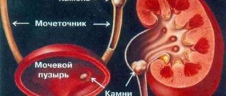
First of all, the results of the disease are displayed on the ureters - they are compressed. Further compression is observed in the area of the inferior vena cava, the aorta and its main arteries. Due to the fact that the passage of urine through the ureter is disrupted, hydronephrosis, pyelonephritis develops and, as a result, chronic renal failure and kidney shrinkage. With retroperitoneal fibrosis, doctors sometimes encounter complications such as intestinal obstruction, obstruction of venous vessels, arterial and hepatic obstructions.
Symptoms
Patients with retroperitoneal fibrosis can present a wide variety of complaints, which depend mainly on the stage of the disease, as well as on the degree of activity of pathological processes. There are three main stages in the development of the disease: in the first, the disease makes its debut (just beginning to emerge), in the second, it actively develops, and in the third, it reduces its progression.
Causes of the disease
In the debut period, patients feel pain in the lower back. They are whiny. Often the onset of these pains occurs on one side, but over time they spread to the opposite side. They often radiate to the back, stomach, and external genitalia. During this period, patients may notice minor changes in blood pressure (it increases), as well as asthenic symptoms (headache, fatigue, sweating). Also, Ormond's pathology in this period can manifest itself as a tumor-like formation located in the pelvis or abdomen.
The next two stages are characterized by the progression of symptoms. True, the process of progression is slow. The pain increases somewhat, but the amount of urine produced decreases. Anuria often develops because it becomes difficult for the ureters to perform their functions. With a unilateral course of the disease, the functions of the diseased kidney are compensated by the healthy organ. The situation is much more complicated with bilateral fibrosis. In this case, the products of protein metabolism are not able to be excreted in urine and enter the blood. A septic condition occurs - uremia. In this case, you cannot do without an artificial kidney apparatus; the patient must be connected to it as soon as possible, otherwise there is a risk, and not a small one, of death.
Along with renal symptoms, symptoms of arterial hypertension progress in the second and third stages. It develops due to excessive pressure on the renal artery. Increased pressure is manifested by a headache that “pulsates” in the back of the head.
With retroperitoneal fibrosis, venous hypertension also develops. Its cause lies in compression of the inferior vena cava. It is not surprising that patients suffer from varicose veins in the lower extremities. Men often notice varicocele. As a complication of this condition, the development of thrombosis of the inferior vena cava cannot be excluded. But this complication is quite rare.
IMPORTANT! If you notice at least one of the signs of the disease in yourself, consult a specialist. Remember, the golden rule for retroperitoneal fibrosis is that early treatment will yield better results.
Prevalence
Ormond's disease is, fortunately, extremely rare: only one case per two hundred thousand people. As a rule, it is detected in men from thirty to sixty years old. Women of the same age category get sick half as often. Mortality is low, and in most reported cases was caused by complications rather than by the disease itself.
Most often, the ureters, which are well covered with subcutaneous fat, come under compression. They find themselves, as it were, in a tight, narrow case and, due to its rigidity, cease to perform their function.
Diagnostic measures
The early stages of the disease can only be determined by laboratory blood tests. When conducting tests, the rate at which red blood cells settle is determined - it is increased.
At subsequent stages, it is possible to use excretory urography. The images show dilated pelvises and altered contours of the ureters. An attentive and experienced doctor will notice that a deformed ureter is the result of external compression.
CT and ultrasound are also actively used for diagnostic purposes in Ormond's disease. With their help, compacted fiber is determined.
The most reliable results regarding Ormond's disease are obtained by a biopsy of the lumbar region - it determines fibrin fibers in the tissue.
If there are clear manifestations of the disease in the heart, blood vessels or digestive organs, irigography and contrast aortography may be informative and useful.
Classification
Clinicians distinguish between primary and secondary Ormond's disease. Primary, or idiopathic fibrosis, occurs on its own for unknown reasons. Scientists have several explanations for this:
- breakdown of genes responsible for the structure of connective tissues;
- specific manifestation of autoimmune aggression;
- inflammatory changes.
Secondary retroperitoneal fibrosis is associated with changes in tissue characteristics due to a previous long-term disease. This could be a chronic infection (or carriage of a pathogen), connective tissue diseases, and others.
Therapeutic measures
Treatment is selected and prescribed taking into account the reasons that led to fibrous changes in the retroperitoneal space. So, if the cause of fibrotic pathologies lies in taking certain drugs, their immediate discontinuation is required. Often this is enough to cure.
Tomography results for Ormond's disease
When the cause of retroperitoneal fibrosis lies in malignant neoplasms, therapy is selected taking into account the cell types of the tumor.
These fibrotic pathologies respond well to treatment with glucocorticoids. However, surgery is often required to free the ureters, especially if retroperitoneal fibrosis is widespread.
In any case, the main goal of therapeutic measures aimed at patients with fibrotic tissue changes is aimed at restoring and maintaining the patency of the ureters, and preventing any complications of this pathology.
Drug therapy can be used only if the passage of urine is maintained and with moderate degrees of reduced ureteral tone. As noted, glucocorticosteroids are used for these purposes. They are combined with immunosuppressants and absorbable drugs. This treatment is especially effective in the initial stages of the disease, when ureteral obstruction is controllable. If necessary, additional drugs may be prescribed that relieve inflammation, have an antibacterial and detoxifying effect.
Non-surgical treatment can improve the well-being of patients due to the fact that it restores the patency of the ureters. True, among the “disadvantages” of such treatment are frequent possible relapses, which have even more pronounced symptoms and an aggravated course with possible changes.
Surgical treatment is carried out in advanced stages or when disorders increase. The most common indications for surgery are:
- pronounced unilaterally or bilaterally dilated ureters
- grossly deformed ureters
- stricture
- constant exacerbations of pyelonephritis
- increasing renal failure
- nephrogenic arterial hypertension
Symptoms
Contrast-enhancing fibrosis covering retroperitoneal structures causing obstruction and displacement of the ureter and vessels.
Displacement of the aorta and IVC anterior to the vertebral bodies suggests a malignant etiology. Invasion and destruction of bone and/or soft tissue structures suggests an aggressive process of infection or malignancy. Multiple subcentimetric lymph nodes are often seen in non-malignant RPF, likely reactive, secondary to the disease process, and should not be confused with malignancy.
Usually occurs in adults, rarely in children. Patients have pain in the lower abdomen, side or lower back.
Other symptoms may include:
- Fever.
- Peripheral edema.
- Phlebitis.
- Deep vein thrombosis.
- Weight loss.
- Nausea and vomiting.
- Fatigue.
- Renal dysfunction.
One third of cases are drug related (especially methylsergide, adrenergic blocking agents and methyldopa). Can happen with:
- Trauma.
- Immune-mediated connective tissue disease.
- Malignant neoplasms (lymphoma, sarcoma or carcinoma).
May be part of multiple fibrosclerosis (systemic idiopathic fibrosis).
A poorly defined fibrous mass, usually centered on the 4th and 5th lumbar vertebrae and often involving one or both ureters (hydronephrosis) or the vasculature (Panel A).
Traditional treatment
For retroperitoneal fibrosis, traditional medicine can be used. As a rule, they come down to the use of all kinds of herbal compresses and lotions.
You should not place high hopes on herbal medicine. Often, such therapy provides only a temporary improvement in the condition and slightly stops the development of fibrosis.
Medical specialists do not always recommend the use of such remedies, because sometimes some complications may develop after them. Therefore, before using any prescription, consult your doctor, this will give some guarantee that you will not harm your own health.
You should not get carried away with such treatment for another very common reason. The fact is that after changes for the better appear or when the progression of the disease stops, patients often ignore traditional medicine and willfully refuse drug treatment. As a result, retroperitoneal fibrosis begins to progress and ends very badly. Fibrosis progresses to more complex stages, which are much more difficult to treat. And in this case it is unlikely that it will be possible to do without surgery. Therefore, many urologists strongly recommend using compresses and lotions in combination, along with medications. This will bring much better results, although it does not guarantee a complete cure.
Diagnostics
Ormond's disease is characterized by a number of laboratory and instrumental signs that help distinguish it from other diseases of this group.
In a clinical blood test, you can see an acceleration of the sedimentation time of red blood cells, a biochemical analysis indicates an increased amount of alpha globulins and C-reactive protein, and an increase in the content of urea and creatinine (signs of chronic renal failure).
From instrumental studies, radiography is performed to look at the contours of the kidneys and muscles around them. Then a contrast agent is injected into the blood and observed how quickly it filters out. In this case, you can see the contours of the cups and pelvis of the kidneys, the shape, location of the ureters and their deviation to the central plane of the body. In addition, scintigraphy with radioactive atoms can be performed in order to assess the functional state of the urinary system.
Do not forget about such a method as ultrasound. It visualizes not only hollow structures, but also blood flow in important vessels of the retroperitoneal space. Sometimes CT diagnostics of retroperitoneal fibrosis is used as an additional method to clarify the topography of individual organs before surgery. If necessary, you can resort to venocavagraphy. This method allows you to visualize the inferior vena cava, its branches and collaterals, see their location and patency along the entire length of the vessel.
Not only targeted diagnosis is important, but also the search for the etiological factor that gave impetus to the development of the disease. Therefore, it is recommended to conduct an examination of the pelvic and abdominal organs for hidden inflammatory diseases.
Dietary regime
Patients are recommended to follow a strict diet, in which fried, salty, sour and smoked foods are excluded from the diet. It is especially important to adhere to such canons in nutrition in the early stages of the disease, when the main goal of doctors is to prevent the progression of pathology.
The main directions of therapy for the disease
If you have symptoms of a narrowed ureter, it is recommended to follow a drinking regime - drink as much liquid as possible during the day in order to increase the circulation of urine, prevent its stagnation and progression of the disease.
Differential diagnosis
Retroperitoneal fibrosis (Ormond's disease or RPF) must be distinguished by its clinical manifestations from other pathological narrowings of the ureters (strictures and achalasia), as well as from bilateral hydronephrosis. The latter is rare, since a violation of the outflow of urine on both sides at once will lead to acute renal failure and require emergency treatment, in contrast to slowly progressive fibrosis.
To clarify the diagnosis and differentiate it from oncological pathology, several puncture biopsies can be done, followed by histological examination. In rare cases, it is possible to establish the fact of the disease only after diagnostic laparoscopy with collection of material for pathological examination.
In addition, an important difference between Ormond's disease is that the ureters are compressed at the level of their intersection with the iliac arteries, and not in an arbitrary location. Sometimes it becomes necessary to distinguish RPF from atypically located pancreatic cysts, neoplasms of the digestive tract, tuberculosis of the kidneys and ureters.
In addition to all of the above, the patient may need to consult a pulmonologist, phthisiatrician, oncologist and cardiologist.
Possible complications and prevention
Perhaps the most dangerous complication of Ormond's disease is anuria and renal failure. These conditions develop due to stagnation of urine.
The next complication is arterial hypertension. It appears as a result of a narrowed renal artery. You should not lose sight of such dangerous complications as varicose veins, chronic intestinal obstruction and intestinal intoxication.
To prevent Ormond's disease, it is important to eliminate factors that may cause its development. Be sure to treat all diseases, especially chronic ones.
You should also be attentive to your own health, undergo timely examinations in order to identify retroperitoneal fibrosis at its earliest stages, which will allow you to cure it using conservative methods and avoid surgical intervention.
Causes of retroperitoneal fibrosis
There are two theories about the origin of Ormond's disease. The first is based on inflammatory, and the second on immune reactions. Also, in some medical sources, Ormond's disease belongs to the group of true collagenoses, that is, systemic connective tissue diseases.
There are many factors that, according to experts, can lead to the occurrence of retroperitoneal fibrosis. These include malignant neoplasms, chronic pancreatitis, chronic hepatitis, tuberculosis of the spine, radiation exposure, large hematomas of the lumbar region, exposure to toxic drugs and chemicals. Despite such a large list of risk factors, in many patients with Ormond's disease none can be identified. Then we have to talk about idiopathic retroperitoneal fibrosis.
The mechanism of development of the pathology is associated with the fact that under the influence of these factors, sclerosis of the fatty tissue of the retroperitoneal space occurs, as a result of which the vessels located there and the ureter are mechanically compressed. This leads to hydronephrosis or pyelonephritis, and ultimately to shrinkage of the kidney. In addition, a clinical picture of intestinal obstruction may occur due to compression of fatty tissue on the colon.
Forecast
The development of Ormond's disease in each individual patient depends on the stage at which the pathology was detected and on the rate of progression of fibrosis. It is also important to take into account the conditions of the urinary system, the presence of complications and birth defects. In most cases, conservative therapy brings temporary positive effects. The most successful method of surgical treatment was considered to be plastic surgery of the ureter and its relocation. After surgery, it is recommended to take long-term steroid medications to improve the prognosis of survival and quality of life. Relapses are possible, but they are significantly delayed compared to isolated drug therapy.
The main cause of death is chronic renal failure. Therefore, the prognosis remains unfavorable. In some cases, when the disease is detected at a late stage, the probability of death is more than sixty percent. Therefore, the sooner the pathology is detected, the more successful the treatment will be.
Additional facts
Adhesive disease is a pathological condition caused by the formation of connective tissue adhesions (adhesions) in the abdominal cavity. The human body is uniquely structured; at a certain point in time it turns on protective mechanisms that can prevent the development of severe complications, but this is reflected in the general condition. In order to protect healthy organs from damaged structures, connective tissue is formed around the pathological focus. Of course, it cannot fill the lost function, but it allows you to fill the void and protect the surrounding tissues from pathology. At first, this tissue is loose, then it becomes denser and sometimes ossifies. This is how adhesions are formed. Adhesions in the abdominal cavity are connective tissue cords that connect the peritoneum and internal organs. Adhesions tighten organs and limit their mobility, create conditions for disruption of their functions, and therefore often become the cause of serious diseases, for example, intestinal obstruction or female infertility.
Adhesive disease