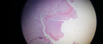
Papillary necrosis is also known in medicine as necrotizing papillitis. This disease is characterized by destructive processes that affect the renal medulla and predominantly affect the renal papillae. Such destructive processes cause dysfunction of the kidney and morphological changes in it. For a long time, luminaries of science and medicine believed that papillary necrotizing disease was a fairly rare phenomenon and did not occur very often. Moreover, it was first mentioned only in 1887. And only in the late 60s of the last century, scientists came to the conclusion that about 1% of patients with urological diseases, and more than 3% of patients with diagnosed pyelonephritis, are susceptible to papillary necrosis. By the way, it is noteworthy that female representatives are twice as susceptible to this disease.
Necrotization of renal papillae
Causes of renal papillary necrosis
Conditions contributing to the development of renal papillary necrosis:
• chronic and acute pyelonephritis; • obstruction of the urinary tract; • sickle cell hemoglobinopathy; • urogenital tuberculosis; • liver cirrhosis, chronic alcoholism; • irrational use of painkillers; • kidney transplant rejection; • radiation therapy; • diabetes; • systemic vasculitis.
More than 50% of patients with renal papillary necrosis have two or more of the listed causative factors, i.e., the pathological process in most people has a multifactorial origin. Nephrologists identify several main causes of papillary destruction:
• Impaired blood supply to the medulla.
Systemic vasculitis, vascular changes due to diabetes mellitus, cholesterol deposits due to atherosclerosis cause a decrease in blood flow with a deficiency of nutrients and oxygen in the papillary apparatus. Thrombosis of the renal vessels with ischemic phenomena can be caused by sickle cell anemia, disseminated intravascular coagulation syndrome and other conditions associated with hypercoagulation.
• Increased intrapelvic pressure. Urodynamic disorders initiate stagnation of urine in the collecting system, which contributes to the development of pyelorenal reflux. Urine is a breeding ground for pathogenic and opportunistic microorganisms, so in the future the situation is aggravated by the addition of an acute inflammatory process. Impaired ureter outflow and pelvic hypertension can be caused by a tumor, calculus, iatrogenic ligation of the ureter during surgery, etc.
• Purulent destructive process in the kidneys.
Sometimes inflammation of the apices of the renal pyramids is secondary and is considered as a complication of severe purulent-destructive processes: apostematous pyelonephritis, renal abscess, carbuncle, purulent inflammation. Microbes in the process of life produce proteolytic exotoxins that melt the parenchyma of the organ. The process involves the papillae, pyramids, and other structures of the kidney.
• Drug-induced nephropathy.
One of the most common but preventable etiological factors is the use of analgesics, in particular phenacetin and its highly toxic metabolite p-phenetidine. Therefore, the recent growing popularity of nonsteroidal anti-inflammatory drugs (NSAIDs), especially those that inhibit cyclooxygenases (COX-1, COX-2), has led to a relatively high incidence of adverse events in patients at risk of developing renal papillary necrosis.
Symptoms of necrosis of the renal papillae
Necrotizing papillitis has no pathognomonic symptoms, so a number of urological and nephrological pathologies that occur with impaired urodynamics are initially excluded
Renal papillary necrosis has a variable clinical course, which ranges from a chronic, protracted and relapsing form to an acute, rapidly progressive form. The latter is rare, but its consequences are devastating, leading to death from sepsis and renal failure. Patients with the chronic form may be asymptomatic until the diagnosis is made incidentally by radiological studies or by detection of mucous papillae in the urine. The symptomatic form manifests itself as episodes of pyelonephritis and hydronephrosis, imitating the symptoms of nephrolithiasis.
The most common signs include: • fever with chills; • pain in the lumbar region and/or abdomen; • the appearance of blood in the urine (hematuria).
Acute renal failure with decreased urine production (oliguria) or complete absence of diuresis (anuria) is rare; the appearance of these symptoms may indicate a fulminant form of renal papillary necrosis, which requires dialysis and has an increased risk of mortality.
Nephrologists consider the diagnosis of papillary necrosis even in an asymptomatic patient with a burdened urological or nephrological history, if tests show evidence of deterioration in renal function. An acute violation of the patency of the ureter against the background of blockage of dead papillae by necrotic masses always provokes renal colic with the release of bloody urine. Pyonephrosis or secondary acute/recurrent pyelonephritis aggravates the course of the disease due to chills, confusion and sepsis.
Differential diagnosis is carried out with infection, tumor, congenital anomalies of the structure of the urogenital tract, metabolic disorders (nephrolithiasis), hemotamponade of the ureter, tubulointerstitial nephritis, glomerulonephritis, renal hemorrhage, perinephritic abscess, etc.
Symptoms
The clinical picture will differ depending on the affected segment.
The following symptoms are characteristic of tuberculous papillitis:
- minor discomfort;
- rapid fatigue and decreased performance;
- subfebrile temperature values;
- progressive loss of body weight;
- the appearance of painless hematuria, which is caused by erosions and ulceration of the renal papillae;
- internal hemorrhages.
Papillitis of the stomach, intestines and pancreas in its clinical picture has the following symptoms:
- pain in the epigastric region;
- belching and heartburn;
- violation of the act of defecation;
- attacks of nausea and vomiting;
- bloating;
- the appearance of a characteristic rumbling sound;
- pale skin;
- severe headaches;
- increased gas formation;
- fragility of hair and nail plates;
- heart rate fluctuations;
- heaviness in the stomach;
- feeling of oversaturation or incomplete emptying;
- fast saturation.
In cases of development of rectal papillitis, the symptoms will be:
- constant or periodic pain in the anus;
- sensation of a foreign object in the anus;
- anal bleeding;
- swelling of the affected tissues;
- itching and burning;
- leakage of intestinal contents from the anus - because of this, maceration of the skin of the perianal area appears.
Symptoms of ocular papillitis are presented:
- decreased visual acuity;
- blurry or double images before the eyes;
- photophobia;
- increased lacrimation;
- swelling of the retina;
- dilation of blood vessels around the disc;
- hemorrhages.
Catarrhal, i.e., superficial papillitis of the tongue or with localization on the palate is accompanied by:
- swelling and pain;
- change in the shade of the mucous membrane - it becomes redder;
- drooling;
- unpleasant sensations while eating food;
- bleeding gums;
- unpleasant taste in the mouth.
Symptoms of tongue papillitis
Diagnostic measures
Diagnosis of necrotizing papillitis relies on imaging studies and laboratory tests. Kidney CT is one of the highly informative methods in urology and nephrology
The diagnosis of “necrosis of the renal papillae” is based on laboratory and instrumental research methods and is determined after excluding other nosologies with a similar clinical picture. If acute ureteral obstruction is manifested by hematuria, renal colic, a series of visual tests are performed to exclude nephrolithiasis and blocking stones. Approximately 15% of all stones are radiopaque (especially uric acid stones), so in addition to radiography, computed tomography is performed. CT scanning is the most informative way to visualize all stones (with the exception of rare stones consisting of indinavir crystals in patients with HIV infection while taking certain drugs). Computer tomograms can also show urothelial tumors; if a neoplasm is suspected, cytological examination and endoscopic diagnostic procedures are additionally performed. Tomograms performed even without contrast show the entire anatomy of the renal collecting system, which makes it possible to diagnose inflammation, hydronephrosis, abscess, etc.
Ultrasound is informative only in the later stages of the disease; early reversible conditions may not be visible. However, ultrasound scanning is performed as a first-line study to diagnose potential urological problems: urolithiasis, pyelonephritis, renal abnormalities, hydronephrotic transformation, etc.
Retrograde pyelography is an invasive test that involves endoscopic access; the images visualize a “clogged” calyx or a filling defect in the ureter. Retrograde (ascending) contrast administration increases intrarenal pressure and may result in pyelovenous drainage of infectious material, thereby increasing the likelihood of sepsis. This risk is less when antibacterial drugs are prescribed. Bilateral retrograde pyelography is preferred for assessing the upper urinary system in patients with contraindications to intravenous enhancement.
Excretory urography always requires the use of an iodinated contrast agent, but a contraindication to it is azotemia (increased creatinine levels in the blood), which can occur in patients with necrosis of the renal papillae.
Other restrictions:
• diabetes; • dehydration (dehydration); • allergies; • asthma; • thyrotoxicosis; • plasmacytoma.
Signs of papillary necrosis on urograms:
• shrinkage and unevenness of the papillae with expansion of the calyx arch in the form of hooks; • ring shadow; • calix without papilla; • partially calcified filling defect of the renal pelvis; • contrasting cavities the size of grains of rice in the papillae (pathognomonic for the medullary form of renal papillary necrosis).
In these cases, a CT scan is preferred and demonstrates intracavitary necrotization of the papillae, allowing for diagnosis.
Laboratory diagnostics include:
• urine microscopy; • general blood analysis; • blood biochemistry (creatinine, urea, sugar, ALT, AST, bilirubin, etc.) • coagulogram (prothrombin + thromboplastin).
If the patient has a hyperthermic reaction, bacterial culture of the biomaterial is performed for microflora and sensitivity to antimicrobial drugs is determined. Patients with hematuria require a full-scale diagnosis, since papillary necrosis does not exclude, for example, a bladder tumor. When assessing the patient's medical history and the clinical picture as a whole, further examination may be required - for example, tests for tuberculosis, analgesic abuse (look at the levels of salicylates and acetaminophen in the blood and urine), diabetes mellitus, etc.
Diagnostics
If one or more of the above symptoms occur, you should consult a therapist, who, if necessary, will refer the patient to other specialists for consultation.
The most significant diagnostic methods are instrumental examination methods, which are preceded by the following primary diagnostic measures:
- study of life history and medical history - to establish the most characteristic physiological or pathological cause of inflammation of the papillae;
- a thorough physical examination of the problem area. If anal papillitis develops, a digital examination of the rectum will be required. Optic nerve damage cannot be diagnosed without an ophthalmological examination;
- a detailed interview with the patient to determine the severity of symptoms.
In the diagnosis of papillitis, laboratory tests of blood, urine and feces are often not performed, but general tests are prescribed if necessary.
Instrumental diagnostics may include:
- anoscopy and sigmoidoscopy;
- radiography with contrast;
- Ultrasound of the abdominal cavity;
- CT and MRI of the head.
After establishing the etiological factor, the patient may be referred for consultation to a gastroenterologist, ophthalmologist, nephrologist and dentist. Depending on who the patient goes to, he will need to undergo a number of specific laboratory and instrumental diagnostic measures.
Treatment
Treatment of renal papillary necrosis requires hospitalization
Therapy for renal papillary necrosis is aimed at:
• reduction of ischemia; • restoration of normal acid-base balance; • increased hydration; • treatment of the root cause of the pathological process, for example, maintaining normal blood glucose levels; • suppression of microbial flora by prescribing antibacterial drugs taking into account sensitivity.
Drug therapy
Conservative treatment of necrotizing renal papillitis is possible with normal urodynamics.
Since ischemia is a fundamental factor in the development of renal papillary necrosis, the patient in intensive care needs to eliminate hypoxia. In case of acute inflammatory process, broad-spectrum antibiotics are prescribed intravenously, a sufficient amount of solutions is infused, and drugs that alkalize the urine are used. Stopping analgesics may stabilize renal function. Patients with hematuria may require blood transfusion (exchange for sickle cell anemia), and those with diabetes may require insulin therapy.
Surgery
Patients with renal papillary necrosis undergo diagnostic or therapeutic urological intervention according to indications. The urologist evaluates any obstruction with urodynamic abnormalities, hematuria, infection, neoplasm, as well as possible complications.
Acute obstruction with concomitant urinary tract infection requires emergency percutaneous nephrostomy to drain urine, installation of a ureteral stent, endoscopic removal of necrotic masses, followed by antibiotic therapy.
Sometimes the final diagnosis is made during a search operation
In severe cases, for example, with emphysematous pyelonephritis that is resistant to drug action, organ-sapping surgery—nephrectomy—can be life-saving. With a bilateral process and, accordingly, bilateral nephrectomy, hemodialysis will be required.
Surgical treatment is used to eliminate anatomical abnormalities that predispose to urinary stagnation. These include:
• vesicoureteral reflux; • narrowing of the ureteropelvic segment; • ureteral strictures; • ureterocele.
If the cause of renal papillary necrosis is nephrolithiasis, one of the operations is performed to help restore urodynamics.
Prognosis and prevention
The prognosis of necrotizing papillitis is individual in each case. The prognosis of renal papillary necrosis depends on the etiology of ischemia, the number of associated pathological factors, the affected area, the involvement of both kidneys, and the general health of the patient. In weakened people with age, the prognosis is serious, as well as in people with developed sepsis and multiple concomitant diseases. Patients with diabetes who are noncompliant and prone to severe episodes of hyperglycemia are at greatest risk of developing complications.
Given the synergistic nature of factors predisposing to papillary necrosis, it is important to control chronic diseases - sickle cell anemia, diabetes and cirrhosis. For patients with these nosologies, analgesics and NSAIDs should be prescribed exclusively by a doctor after taking into account all the pros and cons, with mandatory monitoring of laboratory parameters during treatment. If a urinary tract infection is suspected, therapy and observation by a nephrologist or urologist is indicated. In persons with symptoms of obstruction of the lower parts of the urogenital tract, prolonged catheterization should be avoided and rational antibiotic therapy should be carried out.
Publication date 08/31/2019
Types of pathology
In practice, doctors distinguish 5 types of pathological process.
- Necrosis affecting the renal papillae - necrotizing papillitis - can be acute or chronic.
- Canalicular appearance - in this case, the epithelium of the renal canals is damaged.
- Cortical view - in this case, the tissues and cells of the surface of the organ are damaged.
- Caseous appearance - rather acts not as an independent pathology, but as a consequence of a previous disease.
- Focal view - marked by point lesions of the glomeruli of the organ, while the kidneys themselves function normally.


