What is being studied?
There are two types of kidney ultrasound:
- Ultrasonography is a method for studying kidney tissue.
- Doppler ultrasound (USDG) is a method for studying blood vessels. In some cases, a modified method of three-dimensional vascular echography is used - duplex ultrasound scanning (USDS).
Ultrasonography is performed with the patient lying, on his side, sitting or standing. The kidneys are examined in longitudinal, transverse and oblique sections. This method allows you to determine the number and location of the kidneys, their shape and size, evaluate the clarity of the contours, the structure of the renal sinuses, parenchyma, and the collecting system. The mobility of the kidneys is determined by assessing their position during inhalation and exhalation. In addition to the kidneys, the sonographer will usually scan the bladder, ureters and adrenal glands.
Echography allows you to verify echo-positive (uroliths, tumor nodes) or echo-negative formations (cysts), as well as signs of inflammation.
To assess hemodynamics and determine blood flow parameters, an ultrasound doppler (or ultrasonography) is performed, in which the patient can be in a sitting, lateral or lying position.
Ultrasound of the bladder and ureters
The bladder is a hollow organ consisting of smooth muscle muscles. It stores urine until it is “evacuated” at the request of the body.
The most common reason for a bladder ultrasound is to check whether the bladder is emptying. The urine that remains in the bladder after urination (“post-void”) is measured.
If it stagnates in the bladder for a long time, problems may arise, for example:
- enlarged prostate (prostate gland in men);
- urethral stricture (narrowing of the urethra);
- organ dysfunction.
Bladder ultrasound can also provide information about:
- walls (their thickness, contours, structure);
- diverticula (sacs) of the bladder;
- prostate size;
- stones (uroliths) in the cavity;
- large and small neoplasms (tumors).
During a bladder ultrasound, the ovaries, uterus, or vagina are not examined.
Preparation for an ultrasound of the kidneys and bladder includes a fasting diet (about 10 hours) and normal bowel movements.
If you do not check for residual urine after urination, a full bladder is necessary. You may be asked to drink a lot of water an hour before the test.
The ultrasound probe is placed between your belly button and your pubic bone. The image is viewed on the monitor and read on the spot. To check your bladder drainage, you will be asked to go out and empty your bladder. When you return, exploration will resume.
To keep your bladder full, you will need to drink at least 1 liter of fluid 1 hour before your appointment. Avoid milk, carbonated drinks and alcohol.
If you have a permanent urinary (urethral) catheter installed, you need to check with your specialist about the preliminary preparation before scanning.
Norm
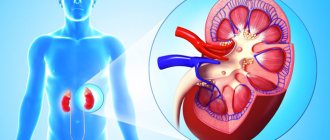
The kidneys are located in the retroperitoneal space on both sides of the spine at the level of the 12th thoracic – 2nd lumbar vertebrae. Surrounded by adipose tissue. The left one is slightly higher than the right one due to the pressure exerted on it by the liver. The kidneys are relatively mobile; displacement of up to 1.5-3 cm is allowed in the vertical direction. According to statistics, in 90% of people the size of the left kidney exceeds the size of the right. In this case, the difference in size should not exceed 3 cm.
The shape of the buds resembles beans. They have smooth, clear contours. The parameters are as follows: length – 11 cm ±1 cm, width – 5.5 cm ±0.5 cm, thickness – 4.5 cm ±0.5 cm. The difference in the parameters of the left and right kidneys normally does not exceed 1 cm.
The structure of the kidney tissue is homogeneous. The thickness of their parenchyma ranges from 1.5-2.5 cm, gradually decreasing with age to 1.1 cm.
The parenchymal-pelvic index (PPI), which assesses the functional capabilities of the kidneys and is determined by the ratio of the thickness of the anterior and posterior parenchyma to the thickness of the central echogenic complex (CEC), is of diagnostic importance. The value of PLI gradually decreases with age: from 1.6 - up to 30 years to 1.1 - over 60 years.
During a renal echo scan, the sonographer examines the adrenal glands and ureters, which are normally virtually invisible, as well as the inferior vena cava, located to the right of the spine, which is tube-shaped and less than 2.5 cm in diameter.
The essence of the procedure
Have you been trying to heal your JOINTS for many years?
Head of the Institute for the Treatment of Joints: “You will be amazed at how easy it is to cure your joints by taking the product every day for 147 rubles ...
Read more "
For contrast diagnostics using radiography, computed tomography or magnetic resonance imaging, preparations containing iodine or radioisotopes are used - substances that are “caught” by the beam scanner. The sonographic method (ultrasound) differs in principle in that for ultrasonic waves the chemical composition or radioactivity does not matter, but only the density of tissues, their ability to reflect or absorb sound waves.
The least dense gases are ordinary air and any gas; they do not reflect waves, but absorb them, and black areas resembling holes form on the screen. This is why gases are the best contrast for ultrasound. It is dangerous to introduce air itself into cavities, and even more so into vessels. Therefore, air suspensions were invented from gas bubbles with a diameter of 2 microns (0.002 mm), which easily penetrate the capillaries, through their walls and are removed from the body without causing air embolism (blockage of blood vessels).
Features of blood flow research
Renal blood flow ultrasound begins with scanning the abdominal aorta for aneurysms, atherosclerotic lesions, and stenoses. His research is conventionally divided into external and internal. With an external scan, the sonographer examines the proximal, middle and distal parts of the renal artery; with an internal scan, the sonographer evaluates the internal renal blood flow in the arcuate vessels in the upper, middle and lower poles.
The main indicators that the doctor focuses on are:
- resistance index (RI);
- systole-diastolic ratio (SDR);
- pulsation index (PI).
With their help, the degree of change in blood flow is assessed. Elevated numbers indicate a possible narrowing of the lumen of the arteries or a decrease in the speed of blood flow.
Ultrasound scanning also makes it possible to establish the location of blood clots, assess the condition of vessel walls and verify signs of atherosclerosis, and “see” varicose veins. The method provides significant assistance in monitoring the regeneration of arteries and veins in the postoperative period.
Who is it prescribed to?
There are several ways to perform an x-ray. Some of them involve the introduction of the contrast agent Urografin or Omnipaque into a vein or through a urinary catheter. It is necessary to consider which radiographic techniques are used.
The contrast agent contains iodine. The drug is intended for administration into the cavity and blood vessels. When introduced into the bloodstream, it enhances visualization of the vascular bed.
Survey radiography. This x-ray examination of the kidneys is performed without the introduction of contrast. The area of the entire urinary system is projected onto a film on which the following information will be available to the specialist:
- stones in the renal pelvis and urinary canal;
- position of the kidneys (prolapse or displacement);
- kidney development (doubling or underdevelopment);
- bladder condition;
- passages of the urinary canal;
- condition of the intestinal walls, which is indicated by increased gas formation (perforation of the intestinal walls).
A survey radiography of the kidneys will allow the doctor to decide whether surgical intervention is necessary to remove kidney stones or treat the patient conservatively.
Cavity system of the organ
The cavitary system is also called the collecting system (PSS), renal sinus, or central echogenic complex, which is normally characterized by reduced echogenicity. The main function of the CLS is the accumulation and removal of secondary urine. The most common changes in the pelvis are:
- Hydronephrosis is a pathology of CLS of various origins, caused by impaired urinary diversion. Most often of an obstructive (urolitis) or compression (tumor) nature.
- Uroliths that provoke severe pain.
How to prepare for a kidney ultrasound
Preparation begins three days before the ultrasound date.
How to properly prepare yourself for an examination:
- Foods that stimulate the formation of intestinal gases are removed from the menu: potatoes, milk (fresh), raw fruits and vegetables, cabbage, sweets, black bread, etc.;
- the patient is recommended to start taking enterosorbents (activated carbon, fennel and the like) to absorb existing gases and reduce the formation of new ones;
- in the evening of the day before the appointed date of the study, you should not eat a heavy dinner - only a light meal of easy-to-digest foods is acceptable;
- Eating on the day of the ultrasound depends on the type of examination being performed. If only a kidney examination is being done, there are no dietary restrictions; the same patients who are scheduled for an abdominal examination should refrain from eating;
- If an ultrasound scan of the kidneys covers a bladder, you should not urinate before the diagnostic procedure. To fill the bladder, you can drink water an hour before, but it is also important not to overfill. When you feel a strong urge to go to the toilet, you can empty your bladder a little. Sometimes the doctor himself may tell the patient to do this;
- It’s worth taking a bottle of water, a clean towel or wet wipes. After the ultrasound, they wipe off the conductive gel applied to the skin. In addition, you should not wear “ceremonial” clothes, since it is quite difficult to completely wipe off this gel even with wet wipes, and it does not wash well;
It is advisable to have some money with you if the examination is paid. It is difficult to say how much a kidney ultrasound costs; the cost of a kidney ultrasound depends on the pricing policy of the clinic.
Parenchyma echogenicity
Parenchyma is the kidney tissue that performs the functions of filtration and excretion. Parenchyma comes in three varieties:
- The cortical or outer layer, which has medium echogenicity, in which urine is formed.
- The medulla, represented by 12-18 pyramids, which are well defined in a healthy kidney and have reduced echogenicity. The purpose of the medulla is to transport urine from the cortex to the pelvis.
- Cortical tissue located between the pyramids. This type of parenchyma is called columns or “pillars” of Bertinni.
Renal emphysema and xanthulognulomatous pyelonephritis
The formation of gas in the calyces and pelvis indicates renal emphysema. It appears in the following cases:
- Infection with anaerobic bacteria.
- Diabetes mellitus, in which impaired carbohydrate metabolism causes the level of carbon dioxide release.
- Renal-intestinal fistula, which occurs due to purulent and inflammatory diseases of the kidneys or perinephric tissue.
If not diagnosed and treated in a timely manner, renal emphysema can be fatal for the patient. During an ultrasound examination it can be detected by the presence of:
- stones caused by bacteria;
- calcification, when a distal acoustic shadow is obtained on ultrasound or the kidneys cannot be detected, since they are completely filled with gas and resemble a loop of intestine.
Xanthogranulomatous pyelonephritis is also a rare form of kidney disease, as is renal emphysema. And in the same way it is very dangerous. Inflammation appears due to the accumulation of xanthoma cells and macrophages around the renal calyces and parenchyma, then completely engulfs the entire organ and replaces healthy cells with inflammatory and “foamed” cells.
Kidney pyelonephritis
During sonographic examination it manifests itself in:
- increasing the size of the organ;
- a decrease in the level of echogenicity or have hypoechoic and echogenic foci;
- the presence of stones that are almost impossible to detect.
Characteristics of pathologies
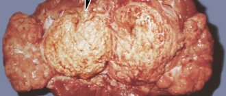
Ultrasound scanning of the kidneys can identify: neoplasms of a malignant and benign nature, uroliths, foci of inflammation, abscesses and cysts, transplant rejection, fluid accumulation inside the organ or in the perinephric tissue, the presence of air in the renal pelvis system, dystrophic changes and congenital developmental anomalies. Doppler ultrasound allows you to assess the state of renal blood flow and possible vascular abnormalities.
The most common deviations include:
- Unilateral kidney aplasia, kidney duplication or loss of pairing as a result of removal of one of them. Duplication can be complete or partial, in which two pelvises are visible on the echogram, from which a “Y”-shaped ureter arises.
- “Horseshoe kidney” is the fusion (fusion) of the lower poles of the kidneys in front of the aorta.
- Renal hypoplasia (healthy kidney length less than 7 cm). Usually the process is one-way.
- Nephroptosis up to dystopia (atypical arrangement of organs in the pelvis). Ectopia can be simple (the kidneys are located on opposite sides of the spine) or cross (the kidneys are located on one side).
- An increase in the thickness of the parenchyma as a result of inflammation or edema, or a decrease in thickness as a result of pathological or age-related involution.
- Vesicoureteral reflux is the inability of the ureters to keep urine from flowing back into the urinary tract.
- Polycystic kidney disease, congenital or acquired.
- The appearance of foci characterized by anechoicity (space-occupying formations containing air) or hyperechogenicity (foci of amyloidosis, benign neoplasms, oncology, glomerulonephritis or diabetic nephropathy).
- The appearance of hyperechoic inclusions in the pyelocaliceal system (uroliths with a diameter of up to 5 mm).
- Traumatic kidney damage.
Hypertension can lead to narrowing of the renal arteries, trophic disorders of the renal glomeruli and their subsequent destruction.
Traumatic injuries
Kidney injuries are usually caused by blows from blunt objects, falls from a height, gunshot wounds, and wounds caused by piercing objects. Kidney injuries are divided into closed and open:
- The group of closed injuries includes: bruise, contusion and crushing of the kidney, subcapsular rupture, capsular rupture with damage to the mandibular joint, rupture of the ureter, damage to the vascular sinus.
- Open injuries include: a puncture or cut wound, a bullet or shrapnel wound.
The diagnosis is primarily based on severe pain, medical history, hematoma in the lumbar region, as well as micro- or macrohematuria.
Features of kidney ultrasound for pyelonephritis
Pyelonephritis is an acute or chronic inflammation of the kidneys of an infectious nature, affecting the renal pelvis system, the tubular system, interstitial tissue and, in some cases, blood vessels. The causative agents are usually viruses or bacteria. Women and girls get sick 6 times more often than boys and men due to the structural features of their genitourinary system. Pyelonephritis is accompanied by pain in the lumbar region, increased temperature, frequent and painful micturition, as well as leukocyturia. According to morphological data, three types of pyelonephritis are distinguished: acute, chronic, chronic with exacerbations.
At the onset of the disease in the phase of serous inflammation, ultrasound may not reveal pathological changes. As it progresses, the echographic picture is characterized by uneven contours, limited mobility of the kidneys due to edema of the fatty membrane, as well as inflammation and an increase in the volume of the organs themselves. A decrease in renal mobility may also be due to the expansion of the renal joint due to obstructive changes. Pyelonephritis is supported by deformation of the cups and pelvis, stricture or atony of the ureters. At later stages, areas of increased echogenicity (areas of scarring) are identified, and a decrease in the size of the kidneys is observed due to their sclerosis.
Complications after urography with contrast agent
Complications after this diagnostic measure, in most cases, depend on the number of x-ray examinations performed over a long period of time.
It is important! A special place is occupied by nephrotoxic effects and allergic reactions. A huge number of modern X-ray contrast agents contain iodine atoms, and intravenous urography is contraindicated if you are allergic to iodine.
The risk group includes patients with bronchial asthma, previous allergic reactions to contrast agents and other severe allergic reactions.
Source