Kidney hemangioma is a benign formation, which is a pathological growth of the organ parenchyma; it includes changed cells. Often the formation is diagnosed in elderly patients. Women are more likely than men to encounter this problem. Among newborns, renal hemangioma is observed in 3% of the total mass of babies.
A benign tumor can very often develop into a malignant tumor. Before starting therapy, the doctor must carry out diagnostic procedures and identify possible dangers to the patient’s life. You can avoid sharply negative consequences by contacting a specialist in time and starting hemangioma therapy.
Causes
The exact reasons for the formation of vascular formation in the kidney have not been studied, but scientists agree that heredity plays an important role in this matter. Hemangioma begins to develop in the prenatal period, when intensive division of vascular cells and their proliferation occurs.
The following factors can cause hemangioma in a child:
- Drinking alcoholic beverages and the effects of nicotine on the body of a pregnant woman,
- Diseases of viral and infectious etiology in the first three months of gestation,
- Radiation exposure to a woman during pregnancy.
The risk of developing a neoplasm increases in children who, while still in the womb, encountered various kidney diseases, for example, renal eclampsia. The peculiarity of hemangioma is that, having appeared even before the baby is born, it begins to grow rapidly during the period of active growth of the child. These moments occur between the ages of six and eight years, as well as puberty. In adult women, tumor growth is observed during pregnancy, which may be due to an increase in blood circulation in her body and hormonal changes. In approximately seven percent of children, the hemangioma resolves spontaneously before the age of five.
Possible complications and prognosis
The disease poses a danger to human health because it can cause the following pathologies:
- internal bleeding (due to rupture);
- neoplasia can give rise to carcinogenesis;
- there is a risk of developing renal failure;
- Arterial hypertension is formed (when the renal vessels are compressed).
In some cases, necrosis of the hepatocyte tissue of the liver is possible if the hemangioma puts pressure on the organ.
Forecast:
- if there is a tumor up to 3 cm and its stability, nothing threatens life and health;
- tumor growth can lead to its rupture and bleeding, but with timely surgical assistance, as a rule, everything ends well;
- when cancer develops: if treatment is started at stage 1 or 2, then the five-year survival prognosis is favorable, but in the presence of metastases, the chances are much lower.
As a rule, renal hemangioma can be successfully corrected and treated.
Classification
Hemangiomas according to their structure are divided into:
- Capillary - consist of small capillaries and in most cases affect the human skin.
- Cavernous (cavernous) - neoplasms from large vessels with cavities filled with blood. It is this type of hemangiomas that develops in the kidney tissue.
- Mixed - include both capillaries and vessels.
Hemangioma, in addition to vascular tissue, may also include adipose or muscle tissue. In this case, the pathology will be called renal hemangiolipoma or hemangiomyolipoma. Cavernous kidney tumors are the most dangerous, since the tumor tissue is constantly growing, new caves are formed, which increases the risk of bleeding and other equally dangerous complications.
Preventive measures
Since this disease is more genetic in nature, no means of prevention have been found. All measures include proper lifestyle and nutrition, regular testing and ultrasound examination of the kidneys.
Treatment most often involves drug therapy or surgical excision of the tumor with partial or complete removal of the kidney. For small formations, the patient is monitored dynamically.
- 5 minutes to read
All neoplasms in medicine, depending on the nature of the course, are divided into malignant and benign. Kidney hemangioma belongs to the second type of tumor. The disease develops against the background of accelerated growth of organ tissue. The neoplasm consists of intertwined blood vessels, which are covered with elastic soft tissue. Often the tumor does not affect the patient’s quality of life and health status.
Content
According to statistics, the disease is more often diagnosed in women than in men. Elderly patients are at risk.
Symptoms and stages of the course
At the initial stage of this disease, there are no manifestations. The nature of the tumor is such that it can “sleep” for decades, and a person will not even be aware of its presence until complications begin. As the hemangioma grows, symptoms begin to appear, in most cases, this happens very suddenly. Once the diameter of the tumor becomes more than five centimeters, the following clinical picture develops:
- pain begins in the lower back, radiating to the groin,
- body temperature rises,
- colic is felt in the kidney area,
- hematuria (blood in the urine) develops
- the general health and condition of the patient worsens,
- appetite begins to disappear
- having trouble sleeping,
- the person becomes weak and gets tired quickly.
After the stage of proliferation (active growth), in some children the stage of independent resorption of hemangioma begins. In other cases, it grows throughout life and if left untreated, the tumor may rupture. This complication is accompanied by a sharp drop in blood pressure, dizziness and fainting, severe weakness and acute abdominal syndrome.
In ninety percent of cases, the rupture of a renal hemangioma ends in the death of the patient, therefore, having learned your diagnosis, you should not hope for “what if it passes” and delay treatment. The last stages of hemangioma growth lead to kidney failure, severe pain when going to the toilet and swelling.
Characteristic signs
Hematuria is one of the signs of the presence of a tumor
The early stages are hidden until the tumor grows to a large size, when pressure is exerted on the parenchyma and neighboring organs, the functioning of the kidneys is disrupted, as a rule, the first sign will be arterial hypertension, which is caused by compression of the renal artery. Hypertensive crises are difficult to stop, and after a short-term remission the situation worsens again.
Other signs of pathology are listed below:
- constant fatigue syndrome;
- decline in working capacity;
- pain, especially often at night, sensations can radiate to the lower back and less often to the groin area;
- heat;
- phlebeurysm;
- blood in urine.
Problems with urination and pain may be a consequence of compression of the ureter or due to the presence of blood clots. Hematuria is evidence of a violation of the integrity of the tissue structure of an organ or efferent ducts. The tumor can easily be injured and rupture. The appearance of such symptoms is a significant reason to visit a urologist in the very near future.
Large neoplasias are dangerous because there is a high risk of their rupture. Internal hemorrhage is quite difficult to stop; the resulting clots block the ureters.
Pay attention to the signs of ruptured renal hemangioma:
- severe weakness;
- renal colic or acute pain in the lower back on the side where the formation is located;
- loss of consciousness or fainting states are not excluded;
- lowering blood pressure.
Important. Rupture of a kidney angioma is extremely life-threatening, so the patient must be immediately taken to a medical facility.
Diagnostics
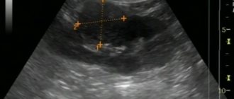
Since a vascular tumor can occur without symptoms for a long time, it is often detected during a preventive ultrasound or during examination for some other reason.
The main diagnostic methods that doctors can resort to when making a diagnosis are as follows:
- X-ray,
- Ultrasound examination (ultrasound) of the kidneys,
- Computed tomography (CT),
- Magnetic resonance imaging (MRI).
If the data obtained to determine the type of tumor is not enough, additional studies are carried out - biopsy, radioisotope study. Urine and blood tests are also taken. Many people visit clinics abroad to make a diagnosis, for example, studies in Germany and other European Union countries make it possible to diagnose pathology in the shortest possible time and carry out therapy there.
Determination of kidney hemangioma, causes and treatment of pathology in an adult
Renal hemangioma is a benign neoplasm consisting of a large number of normal and abnormal vessels on the skin or internal organs. Neoplasia is 3 times more common in women than in men.
In the International Classification of Diseases, 10th revision (ICD-10), renal hemangioma is designated by code D18. It may contain arterial, venous and capillary vessels. Treatment methods depend on the extent of the pathology and the patient’s health status.
In this article we will look at why hemangiomas appear.
Description of the disease
When you hear the combination of words “renal hemangioma,” not everyone will understand what it is. This is the name for benign tumors of blood vessels, which can occur in all organs.
New growths lying in the skin are usually very noticeable as purple or red spots. They contain arterial, venous or capillary elements. If the tumor consists primarily of supporting cells of the vascular wall (“glomus cells”), it is called a glomangioma. Lymphatic vessels can also form angiomas: lymphangiomas.
In the past, different terms were used interchangeably. Currently, the concept of “hemangioma” is used to refer to acquired proliferation of blood vessels.
Deviations - arteriovenous fistulas - are already present at birth and are grouped under the name “vascular malformations”. Acquired defects in which normal blood vessels become thicker than normal (varicose veins and aneurysms) are not called hemangiomas.
Mechanism and stages of development
Capillary hemangiomas in the kidney in adults are thought to be hamartomatous proliferations of vascular endothelial cells.
Neoplasms usually have 2 growth phases: proliferative and involutional. The proliferative phase of rapid growth occurs after 8-18 months.
Pathologically, it is characterized by an increase in the number of endothelial and mast cells, the latter being a stimulus for vascular growth.
The involutional phase is characterized by slow regression of hemangiomas. During this stage, the number of mast cells decreases to normal levels and the activity of endothelial cells decreases.
Characteristic manifestations
Hemangioma of the right kidney causes nonspecific symptoms: muscle pain near the spine, bleeding, dizziness, fatigue, or urinary problems. If the tumor is very large and easily visible, it may limit kidney function.
Hemangioma may be sensitive to pressure. In rare cases, internal bleeding, pain and inflammation of the affected skin may occur. If the pathology continues to grow, signs of renal failure occur. Especially newborns and the elderly need rapid treatment.
Varieties
There are congenital and infantile hemangiomas. Infantile ones are much more common than congenital ones.
Infantile hemangiomas appear as a small red dot or spot in the first four weeks of life.
In the subsequent period, rapid growth is observed, which slows down increasingly at 6 months after birth. After about six months, vascular growth stops completely.
Subsequently, natural regression of neoplasia begins. This phase takes years; sometimes complete disappearance of the hemangioma is observed.
Infantile hemangiomas are mostly limited to a specific area, but can affect an entire body segment. Congenital forms usually mature at birth. If more than 10 hemangiomas appear, so-called hemangiomatosis occurs.
Attention! A clinical trial conducted by scientists showed that the presence of already 3 neoplasms increases the likelihood of additional hemangiomas in the liver or brain. For this reason, ultrasound examinations of the abdomen and skull of affected children are performed in collaboration with local radiologists.
Potential Complications
If the disorder is left untreated, multiple hemangiomas can form, which pose a risk to the baby or woman during pregnancy. Some patients develop urogenital or anal malformations.
Large hemangiomas (>3-5% body surface area) increase the risk of heart failure, hypothyroidism, and thrombocytopenia. In extremely rare situations, patients die from internal bleeding.
Patient examination
First, a physical examination is performed and an anamnesis (medical history) is taken. In laboratory studies, immunohistochemical staining is positive for factor VIII. Imaging tests help confirm the diagnosis.
CT scans poorly detect renal hemangioma without bone erosion. The lesion usually enhances with intravenous contrast. Magnetic resonance imaging (MRI) detects pathological conditions less effectively than ultrasound (ultrasound).
Angiography can reveal large and well-perfused hemangiomas. Cardiac echocardiography and great vessel imaging are useful in diagnosing PHACES syndrome.
In histological examination of the microspecimen, proliferation (growth) of one layer of endothelial cells and pericytes is observed. Endothelial cells are characterized by a significant amount of endoplasmic reticulum (a branched network of membranes that penetrate the cytoplasm of the cell).
Treatment
With renal hemangioma, early diagnosis and timely initiation of treatment help save the patient’s life. For small neoplasms of non-critical localization, expectant therapy is usually sufficient.
In most children, hemangiomas disappear on their own. Therefore, treatment is usually not required. If minor skin changes remain, they can be corrected with surgery or laser therapy.
It is important to know! For life-threatening hemangiomas, deterioration of renal, cardiac, or vascular function must be prevented. It is recommended to use medications - non-selective beta blockers.
They lead to rapid narrowing of the affected vessels, reduce the size of the tumor and improve the patient’s condition.
Surgical measures are also recommended for the treatment of pathology. Laser correction is only conditionally applicable due to insufficient penetration depth.
It is prohibited to use folk or untested remedies (Vishnevsky ointment) for hemangiomatosis on the left or right side of the kidney.
Traditional medicine
Since 2008, patients with hemangiomas have been prescribed non-selective beta blocker therapy. Propranolol is a drug used primarily to treat cardiovascular diseases.
For critical localized hemangiomas that cannot be treated with cold, Propranolol is used in a low dose. By December 2013, 120,000 children had been successfully treated with the drug. It is still not officially approved for the treatment of hemangioma.
Parents are informed about therapeutic alternatives, benefits, risks and side effects of the medication.
Since Propranolol affects the cardiovascular system, an ECG and echocardiography are performed before starting therapy to rule out congenital heart disease. The medicine is prescribed during a two-day hospital stay. It should be taken three times a day.
Drug treatment lasts 6 months. When using Propranolol, tumor growth slows down and improves the condition of patients. After completion of therapy, in rare cases, further development of hemangioma may occur.
Then the drug is again prescribed to sick children in a course that lasts 2-3 months.
Often the use of beta blockers is supplemented with Cortisone or interstitial laser treatment. Only in isolated cases is open surgery required.
Alternative Methods
Fast-growing hemangiomas in cosmetically unfavorable areas can be treated with cryotherapy (cold treatment). It has a penetration depth of only about 2 mm. As an alternative, laser therapy using dye can be performed. The laser penetrates 0.7 mm. Such methods are not used to treat renal hemangioma.
Prevention of pathology and prognosis
There are no absolute or relative methods for preventing the disease. A healthy lifestyle or other measures do not prevent the onset of pathology. According to recent studies, cannabinoids (substances found in cannabis and some other plants) increase the risk of benign vascular neoplasms.
The growth phase lasts 9 months. Then comes the expansion phase (onset: 4-8 weeks after birth) - an unpredictable and rapid growth of vascular tissue that continues until 1 year of age. Growing neoplasias appear as warm, swollen, bright or dark red nodules.
Larger tumors lose about 10% of their volume annually. After 5 years, 50% of the tumor completely disappears, after 7 years - 60%, and after 9 years - 90%. For infantile hemangiomas, the course is favorable even in the absence of treatment; for congenital hemangiomas, the course is conditionally good.
Treatment options
The dynamics of the disease, symptoms and treatment are closely interrelated. In cases where a renal hemangioma has no symptoms and the neoplasm is less than 3 cm in diameter, doctors choose observational tactics and do not prescribe therapeutic measures to the patient. If the size of the hemangioma is more than three centimeters, doctors carry out drug therapy or surgically remove the vascular tumor. The method of therapy may also depend on the patient's age, concomitant diseases, the presence or absence of complications and the general condition of the patient.
Treatment with medications
Scientists in the field of hematology and vascular diseases have found that antihypertensive medications can lead to a reduction in the size of hemangiomas. But this method has not yet been fully studied and doctors are in no hurry to resort to it. Most often, to combat vascular tumors of the kidneys, doctors tend to use hormonal drugs and cytostatics. The most commonly prescribed hormonal drugs include corticosteroids such as Diprospan and Prednisolone.
Hormonal medications have side effects, so they are taken strictly as prescribed and according to medical recommendations.
If a hemangioma is diagnosed in a child, he may be prescribed Vincristine or Timolol. How long it is necessary to take medications, as well as what the dose will be, is decided by the attending physician individually in each specific case.
Removal methods
If the tumor grows too rapidly, surgical intervention is prescribed. Doctors often resort to laparoscopy, because this method of removal has a number of advantages:
- no large incisions,
- less pronounced pain syndrome in the postoperative period,
- virtually no trauma to nearby tissues and organs,
- very low risk of blood loss,
- absence of rough scars after wound healing,
- reducing the risk of infection of the surgical field,
- faster and easier recovery period.
After the operation, doctors send part of the tumor for histological examination. If malignant cells are found in the tissues of the hemangioma, the patient is prescribed radiation and chemical therapy.
If hemangiomas are too large or if complications occur, the operation is performed through an open approach (laparotomy). During surgery, the entire tumor and adjacent tissue are removed. Removal of the organ (nephrectomy) is necessary if the hemangioma occupies its entire area. After the operation, the patient is prescribed antibacterial drugs and analgesics. If, some time after surgery, a relapse develops or metastatic vascular neoplasms appear, chemical and radiation therapy is necessary.
ethnoscience
Folk remedies can be used only for small hemangiomas (up to three centimeters in diameter).
Any actions using alternative medicine must be agreed with the doctor who made the diagnosis, since some recipes can only provoke the growth of hemangioma and lead to serious complications.
To combat vascular tumors of the kidneys, fans of traditional medicine use the following methods:
- Make an infusion or decoction of walnut shells and membranes, and drink twice a day for a month.
- Squeeze juice from burdock, drink 1 tsp. half an hour before meals one month.
- Prepare a balm with rose hips, chaga, yarrow, wormwood, honey and cognac. This product must be used for three months.
Therapy with folk remedies does not always help everyone, so doctors recommend using it only as an auxiliary treatment.
What is a hemangioma?
Kidney hemangioma is a neoplasm that forms on a plexus of blood vessels. It is usually benign in nature. But under the influence of external or internal factors it can acquire a malignant course.
On this topic
- Digestive system
Differences between sigmoidoscopy and colonoscopy
- Natalya Gennadievna Butsyk
- December 9, 2020
The tumor has a number of features, including:
- Innate character. A predisposition to the development of pathology develops during the period of intrauterine development. After the birth of a child, when rapid growth is observed, the neoplasm can also grow rapidly. Pregnancy can also trigger the formation.
- older or elderly patients
- No symptoms for a long time. It is the asymptomatic course of the disease that complicates the diagnosis of a neoplasm at the initial stage of its development. The diagnosis usually occurs during diagnostics for other pathologies, when an ultrasound or MRI is required.
- Localization. A neoplasm is formed in the walls of the pelvis. In rare cases, it is found in the cortex.
Hemangioma is diagnosed in rare cases and is not considered a common pathology. Due to the fact that its presence is established only in the later stages, when the tumor acquires significant dimensions, treatment is necessary. This is because it can cause serious consequences.
In medicine, it is customary to divide all benign neoplasms into the following types:
- Simple. Formed from capillaries.
- Cavernous. The basis of its occurrence is the choroid plexuses.
- Combined. Combines both types.
Depending on the location, they are divided into internal and external. Hemangioma belongs to the cavernous type and has an internal localization. Since it does not affect the functioning of systems and organs, it is benign.
But a tumor can pose a threat to human health in cases when it reaches a significant size and is injured. The growth of the tumor leads to compression of neighboring organs, blood vessels and nerve endings. In severe cases, the damaged kidney is removed.
In addition, there is a high risk of hemangioma degenerating into a malignant tumor. That is why, when diagnosing the disease, specialist supervision and, if necessary, surgical intervention are required.
Vascular tumor treatment methods
I would like to note once again that renal hemangioma is a benign neoplasm and, accordingly, the prognosis for such a diagnosis is generally favorable. In some cases, when this vascular tumor is diagnosed, treatment is not even prescribed. This occurs when the size of the tumor does not exceed three centimeters. But, despite the lack of treatment in such cases, patients are recommended to undergo systematic examination. This is necessary so that you can monitor the process of tumor development and, if necessary, begin timely treatment.
By controlling the size of the tumor, specialists, if it begins to develop, first of all prescribe the necessary drugs that prevent its increase, and in some cases even lead to its reduction. These are mainly hormonal drugs, drugs to lower blood pressure and increase immunity.
But, unfortunately, situations sometimes occur in which drug treatment does not stop the growth of the tumor. In this case, the doctor prescribes a planned operation. If a patient's hemangioma ruptures, surgery is performed as an emergency.
The doctor will prescribe the necessary treatment
Such surgical intervention is not new, but has been performed quite often for many years. In addition, modern medical equipment significantly increases the chance of a positive outcome of the operation, even though in such cases doctors never undertake to predict the outcome. This is primarily due to the fact that the entire tumor is composed of blood vessels. That is why biopsy material is never taken before surgery, and all studies are carried out directly during the operation, as a result of which the final decision on the course of the surgical intervention is made. Only after all the studies have been carried out can the surgeon decide to remove the kidney completely or just the hemangioma. This decision is influenced primarily by the fact whether the hemangioma has entered the malignant stage or not. If the results of the biopsy indicate a malignant process, the kidney is removed completely; if not, then only the vascular tumor is removed.
After the operation, each patient must undergo systematic examinations. They are necessary not only for the correct course of the rehabilitation period, but also for preventing relapse. In addition, you should strictly follow all the recommendations of your doctor and adhere to a certain diet during the rehabilitation process.
Treatment of hemangioma with folk remedies
When diagnosing a hemangioma, if it is small in size and does not develop, you can try to cope with this disease using traditional methods. But before you do this, you should definitely consult with your doctor. This is necessary so as not to provoke its development. It is also worth considering the fact that any traditional methods are not able to rid you of a vascular tumor completely. They are only effective in combination with medications.
As practice shows, the following means lead to a positive result:
- decoction of crushed walnut shells;
- juice from burdock leaves;
- decoction of oat seeds.
In order to select the necessary course of treatment and dosage of the decoction in combination with medications, discuss this method of treatment with a urologist, who, based on the individual characteristics of your body, will select the best and most effective remedy.
Forecast
Seeing a doctor on time and receiving appropriate treatment creates a favorable prognosis. Kidney hemangioma is not prone to recurrence, but the patient is still required to regularly check the kidneys and take tests. A low-salt diet is recommended.
Tumors up to three centimeters in size remain under medical supervision. Under certain circumstances, the tumor does not change in size throughout the patient's life. There are no preventive measures to prevent the disease. Regular visits to the doctor and tests, a healthy lifestyle and giving up bad habits will minimize the unfavorable process of growth and development of the tumor.