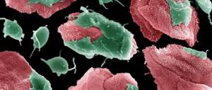
The urinary system under the influence of unfavorable factors is at risk of developing tumors
. Pathology occurs more often in men than in women; if we exclude cancer, then pathology accounts for about 3% of patients.
Tumors of the genitourinary system
can be divided into benign and malignant.
It is worth noting that a tumor is formed due to the uncontrolled growth of organ cells. Such growth is provoked both by improper functioning of the organ and by external unfavorable factors.
Benign formations:
such formations do not pose a danger if diagnosed and treated in a timely manner. Benign tumors do not create metastases, do not affect adjacent tissues and do not create relapses. Most often, formations appear in the kidneys and prostate gland and can lead to the development of adenoma, lipoma, cyst and angiomyolipoma. As a rule, papillomas form in the bladder, which can develop into malignant tumors.
Malignant formations:
such formations pose a critical risk to human life. Malignant cells can be formed in the blood or lymphatic system, and lead to the formation of metastases in other organs, as well as to relapses. Malignant tumors include: prostate adenoma or cancer of the kidneys, ureter or reproductive system.
Causes of tumors of the urinary system
The occurrence of tumors, both benign and malignant, can be influenced by the following factors:
- The presence of papilloma virus (HPV) in the body or on the human body
- Smoking is the main provocateur of tumors. The risk of cancer in smokers is doubled.
- Untreated inflammatory processes in the body (Prostatitis, Cystitis, STIs, Pyelonephritis, Inflammatory diseases of the female genital organs, etc.)
- Effect of industrial carcinogens on the body
- Ionizing radiation.
Classification
Of all cancer cases, bladder cancer accounts for 5%. This is the 5th most common cancer among cancer pathologies. Most patients are elderly men. The incidence rate among them is 4 times higher than among women. The average age of the patient is about 60 years, but it becomes lower from year to year.
This type of cancer is dangerous because it has no symptoms at the initial stage. And later stages of bladder cancer are more difficult to treat, take longer and are less successful. For bladder cancer, the prognosis depends on how early the patient seeks help. If this occurs at the stage of metastasis, then a cure is unlikely.
Bladder cancer affects the walls of the organ, where foci of transition of normal cells into tumor cells appear. Transformation begins with epithelial cells forming the inner surface of the organ. With the help of modern diagnostic methods, a tumor can be detected before it begins to change the muscle layer. But if cancer cells leave the bladder and form metastases in other organs, the disease is almost impossible to stop.
Bladder cancer affects different types of cells. Depending on this, there are several types of disease:
- transitional cell carcinoma (accounts for up to 90% of cases);
- squamous cell tumor (the second most common type, most often develops due to chronic cystitis);
- adenocarcinoma;
- poorly differentiated carcinoma;
- lymphoma and other rare but common forms.
Symptoms of tumors of the urinary system
Symptoms of tumors include:
In men, neoplasms in the genitourinary system
It occurs more often in old age, and is accompanied by the formation of a tumor in the prostate gland (as a rule). Symptoms of the disease include:
Women have an increased risk of developing tumors
in the bladder due to treatment of cervical or ovarian cancer using radiation.
Diagnostics
There are several ways to diagnose a tumor of the genitourinary system. By visual examination, as well as by palpation, large tumors can be identified. If the size of the formation is small, the following methods are used:
- Examine the bladder cavity using an endoscope. This method produces biomaterial for biopsy.
- Ultrasound is used. In addition to the fact that using ultrasound you can find out about the size and presence of a tumor, this method determines the depth of metastases.
- X-ray examinations using a contrast agent are prescribed.
- A clinical urine test is used to check for the presence of atypical cells in the urine.
- Computer and magnetic resonance imaging methods are used for a more detailed analysis and compilation of a complete picture of the disease.
Cost of consultation?
| Name of service | Price, rub.) |
| Primary appointment with a urologist-andrologist | 1500 rub. |
| Repeated appointment with a urologist-andrologist | 1000 rub. |
| Primary appointment with an obstetrician-gynecologist | 1500 rub. |
| Repeated appointment with the obstetrician-gynecologist | 1000 rub. |
| Primary appointment with an obstetrician-gynecologist (candidate of medical sciences; doctor of medical sciences) | 1700 rub. |
| Repeated appointment with an obstetrician-gynecologist (candidate of medical sciences; doctor of medical sciences) | 1200 rub. |
| Prescription of treatment (drawing up an individual treatment regimen) | 700 - 2500 rub. |
All our services and prices
Treatment of tumors of the urinary system
Treatment of tumors is divided into two methods: conservative treatment and surgical treatment.
Conservative method
is based on treatment with medications, as well as chemotherapy and radiation. Often resort to stimulation of the immune system using immunotherapy.
Toward the surgical method
can be attributed to direct surgical intervention in the human body to remove a tumor, in various ways.
It is important to remember that anyone can reduce the risk of pathological neoplasms. To do this, you need to follow a number of preventive methods:
Sign up for an examination for tumors of the urinary system
Make an appointment
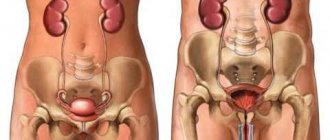
Causes, types, symptoms and treatment of kidney cysts in women
A kidney cyst is a benign neoplasm that consists of connective tissue filled with fluid (primary urine), pus or blood. They are the consequence and cause of various pathologies of the urinary system. Benign neoplasms are diagnosed more often in women over 40 years of age. Consultation and treatment with a urologist and nephrologist is required.
Why do cysts develop?
A kidney cyst in women can be a congenital or acquired disease. The cause of the first may be a difficult pregnancy, during which pathologies and anomalies in the structure of internal organs develop. Congenital cysts are diagnosed in infants due to genetic diseases.
The process of formation of kidneys and their tubules is negatively affected by bad habits of the mother and infectious diseases during pregnancy. Congenital pathology can be hereditary. The risk of developing a neoplasm increases if close relatives have been diagnosed with cases of a similar disease.
The acquired disease develops against the background of chronic inflammatory and infectious processes in the kidneys. Women with high blood and kidney pressure, as well as patients with tuberculosis, which leads to the proliferation of fibrous tissue, are at risk.
The risk of cyst formation increases with urolithiasis, which disrupts the normal process of urination. Stagnation of urine leads to its accumulation in the kidneys, which gradually fills the cavities of the connective tissue. Damage to the connective tissue of the organ as a result of injury also leads to disruption of the process of accumulation and discharge of urine and the formation of a cyst.
Classification
Kidney cysts in women can be simple or complex. Simple (solitary) neoplasms are small in size and can be located in any part of the organ. Complex or multi-chambered ones consist of several simple vesicles, which are wrapped in one connective tissue. This type is diagnosed quite rarely.
In polycystic disease, formations cover the entire surface of the kidneys. Polycystic disease develops as a result of genetic failures and is a hereditary pathology. Spongy kidney is a type of congenital hereditary pathology in which the organ has the appearance of a sponge with multiple brushes. Cysts of this type are the most dangerous.
Depending on the type of tissue on which the pathology is localized, cysts can be:
- parenchymal;
- sinus;
- subcapsular;
- parapelvical;
- multilocular.
Parenchymal disease directly affects the internal tissues of the organ and occurs without obvious clinical signs. Subcapsular are located under the capsule of the organ and rarely lead to complications. Multilocular ones affect various parts and are the most dangerous; surgical intervention is necessary.
The neoplasms located in the sinus of the organ do not affect the renal pelvis and ureters, and therefore do not lead to impaired diuresis and stagnation of urine. Less common is the parapelvical type, which provokes the growth of fibrous tissue in the cavity of the pelvis. Diagnosed in women over 50 years of age.
Based on the contents of the connective tissue, cysts can be filled with:
- Serous fluid is the most commonly diagnosed type.
- Pus due to infection.
- Blood that can leak out when the integrity of the kidney vessels is disrupted.
- Other hard and soft tissues, hair, fat. It is rare; genetic diseases lead to this appearance.
Classification of cysts according to Bosniak depending on the possibility of malignancy:
The 1st type is easily determined during ultrasound examination, is a benign neoplasm and has a simple structure. It can go away on its own, so surgery is rarely required.
Type 2 - multiple cysts with a large number of septa. Requires drug treatment, rarely surgery. It has a subtype that requires urological control, as it is located in large areas of the organ and contains multiple septa. There is a high probability of their growth into a malignant tumor.
The 3rd type is dangerous. Cysts have compacted walls, and folds form in some places. Surgery is necessary, as the likelihood of malignancy is high.
Type 4 – cancerous tumors. Treatment by an oncologist is required.
Symptoms
The first symptoms occur when the cyst reaches a significant size, when it puts pressure on the kidney or surrounding organs. As a result of pressure, urination is impaired: pain and burning appear, the single volume of urine decreases, and blood clots may accumulate.
Painful symptoms occur that intensify with movement and physical activity. The pain is localized in the area of the affected organ and radiates to the lumbar and groin area. Due to a violation of the outflow of urine, intrarenal pressure increases, which entails an increase in blood pressure. In the presence of other diseases that led to the formation of cysts, symptoms of the underlying disease appear (urolithiasis, pyelonephritis and others).
Cysts in the kidneys lead to decreased immunity and the development of infection, which increases the risk of inflammatory diseases of the kidneys and other organs of the urinary system. When an infection occurs, signs of general intoxication of the body appear, the urine becomes cloudy, and the pain becomes severe.
When a cystic formation ruptures, acute symptoms appear:
- pain in the abdomen and lower back;
- increased body temperature;
- uremia;
- constant thirst;
- urine volume increases;
- the color of the skin changes;
- chills and tissue swelling appear.
If the fibrous membrane ruptures, urgent surgical intervention is required.
Diagnostics
Symptoms of cyst formation are often confused with other diseases of the genitourinary system, so cysts are often discovered by chance. Among diagnostic measures, clinical studies are mandatory. A general blood test allows you to assess the patient’s condition and the likelihood of infection. A general clinical urine test allows you to assess the condition of the kidneys and other organs of the urinary system. During the examination of urine, the presence of proteinuria, uremia, and leukocyturia is determined.
Among instrumental diagnostic methods, ultrasound examination of the kidneys is widely used. In the ultrasound image you can clearly see the size of the cyst, its appearance and location. X-ray diagnostics allows you to obtain more accurate data when ultrasound images are insufficient. An X-ray with a contrast agent helps determine the exact size.
Computed tomography and magnetic resonance therapy make it possible to study the physical properties of the formation, as well as the general condition of the organ tissues and the size of the pyelocaliceal system. In order to exclude oncology, a cytological examination of the fibrous contents is necessary.
Treatment
Depending on the characteristics of the kidney cyst in women, treatment is carried out individually. A general recommendation from your doctor would be to follow a diet that will help reduce the load on your kidneys. Restrictions include animal proteins and salt.
For small benign tumors that do not have a negative effect on the flow of urine, surgical intervention is not used. A woman needs to undergo a follow-up ultrasound examination regularly, twice a year. When other diseases occur, they require urgent medical treatment to help avoid complications.
For cystic formations that impede the outflow of urine, the most commonly used minimally invasive procedure is puncture. It is performed under local anesthesia using a special syringe with a benign tumor diameter of no more than 50 mm. Using an ultrasound machine, the source of the disease and the accuracy of needle insertion are determined. The contents of the fibrous membrane must be removed; instead, a special substance is introduced to promote gluing of the walls of the cyst.
If the kidney cyst in women is more than 50 mm, laparoscopic surgery is required. With this type of surgery, three minor punctures are made through which an endoscope is inserted and removal is performed. The risk of complications and relapses is minimal.
If the neoplasm compresses neighboring organs, leads to an increase in pressure, interferes with the flow of urine, if an infection is attached, the membranes are ruptured or are malignant, an open operation is required. Surgery with open access allows you to completely remove the cyst and prevent complications.
Phytotherapy
If the size of the benign neoplasm is small and the symptoms do not cause discomfort, after consultation with a doctor, you can use traditional medicine recipes. A decoction of cedar shells will help get rid of the cyst: 30 g of shells per 200 ml of water, brew and leave. Take a decoction of 50 ml three times a day before meals.
An effective remedy is a gruel made from burdock leaves. To prepare, simply grind the leaves in a blender or pass through a meat grinder. Take 1 tsp. in the morning and in the evening. You can store burdock pulp for no more than 30 days.
A well-known method of treatment is an infusion of elecampane with yeast. To prepare, you will need 20 g of crushed root and sugar, 10 g of yeast, 3 liters of water. Mix and place in a dark place to ferment. Take kidney infusion twice a day, 100 ml. The course of treatment with the listed methods is no more than one month.
Complications
Not everyone understands why a cyst is dangerous and why a course of treatment is necessary if the clinical picture is not expressed. The complications that develop when the above symptoms are ignored are described below.
Hydronephrosis is the most dangerous consequence. As the cyst grows, it fills the organ tissue and the collecting system. The outflow of urine is disrupted until there is no discharge at all. The tissues gradually atrophy.
Kidney failure occurs as a result of nephron dysfunction due to impaired urination.
The most common complication is the addition of infections. Urine is a favorable environment for the development of bacteria. As inflammation develops, the likelihood of the cyst filling with pus increases, which significantly disrupts the functioning of the kidney and negatively affects the general condition of the patient.
Where can I get tested for tumors of the urinary system in Moscow?
At a multidisciplinary medical center, you can always be examined for tumors of the urinary system
. Our medical center is located between the Konkovo and Belyaevo metro stations (South-Western Administrative District of Moscow in the area of the Belyaevo, Konkovo, Teply Stan, Chertanovo, Yasenevo, Sevastopolskaya, New Cheryomushki metro stations " and "Trade Union"). Here you will find highly qualified personnel and the most modern diagnostic equipment. Our clients will be pleasantly surprised by our quite affordable prices.