Ultrasound is currently the most common diagnostic method and, among other things, helps to identify kidney diseases. An ultrasound of the kidneys is often prescribed if there is characteristic pain in the lumbar region, urine tests are abnormal, a person suffers from chronic kidney disease and requires regular examination, symptoms of renal colic or edema or other symptoms appear, and an ultrasound is also prescribed for a preventive examination. In some cases, the doctor may recommend undergoing an ultrasound, even if the person has headaches and high blood pressure.
How to prepare for a kidney ultrasound
This examination does not require any special preparation. Many doctors do not recommend drinking too much before an ultrasound, and some doctors do not even allow smoking. However, you should note that if you are prescribed an ultrasound of the kidneys and an ultrasound of the bladder, which is often necessary during preventive examinations, then in this case certain preparation is required, which causes slight discomfort. With this ultrasound, the fullness of the bladder is important. A person should feel the urge to urinate and for this, 1-1.5 hours before the ultrasound is performed, it is necessary to drink about 1-1.5 liters of water.
The examination itself takes place without any discomfort. The examination is carried out with the person lying down and usually additionally in a standing position. You can only take a clean towel with you to the examination.
What does a kidney ultrasound show?
Ultrasound can detect a wide variety of kidney diseases. After the examination, the doctor fills out a protocol and indicates the conclusion and diagnosis. The protocol indicates the patient’s personal data (full name, date of birth) and, necessarily, the date of examination. Each kidney (right and left) is assessed and data is indicated for each kidney separately. The location of the kidneys is noted, which may be normal or indicate pathology, such as prolapse or rotation (rotation of the kidney). The size of the kidneys is noted, which can be normal, reduced or enlarged. In an adult, the dimensions correspond on longitudinal sections - 10-12 × 3.5-4.5 cm, on transverse sections - 5-6 ×. 3.5-4.5 cm. The contours of the kidney should normally be smooth (uneven contours, wavy contours, retraction, etc. may be noted). Much attention is paid to the renal parenchyma. For those who are not familiar with anatomy, you can imagine parenchyma with a peel; it is the outer and inner layers of the kidney that are called parenchyma.
Where to do an ultrasound of the kidneys
Most often, the doctor refers the person to an ultrasound scan. In clinics, it is possible to conduct such an examination if indicated. There is a free option (usually registering and waiting your turn) and a paid option (faster turn, but on the same equipment and performed by the same doctor). There are also medical centers that provide a similar service and kidney ultrasound can be performed even without a referral from a doctor.
Example of a possible protocol:
protocol
Author
: Nina Rumyantseva
Date of publication: 09/19/2011 Reproduction without an active link is prohibited
www.medikspravka.ru
Parameters and indicators studied
- Quantity. A healthy person has two kidneys. There are cases when one is removed surgically due to certain reasons. Anomalies in the number of these organs are possible: an additional kidney, complete absence or doubling.
- Dimensional data. Using ultrasound, the length, width and thickness of the organ are measured. Kidney size varies depending on a person's age, weight, and height.
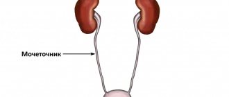
- Localization. The retroperitoneal location of the organs is normal. The right kidney (D) is located just below the left (L). The location of the right kidney is considered normal at the level of the 12th thoracic vertebrae and the 2nd lumbar vertebrae, and the left kidney - at the level of the 11th thoracic and 1st lumbar vertebrae.
- Shape and contours. The bean-shaped shape is considered normal. The structure of the tissue is normal - homogeneous with smooth contours.
- The structure of the renal parenchyma, that is, the tissue that fills the organ. In a healthy person, its thickness ranges from 14 to 26 mm. With age, the parenchyma becomes thinner, and for older people the norm for this indicator is 10-11 mm. An increase in this parameter indicates inflammation or swelling of the organ, a decrease indicates dystrophic changes.
- State of blood flow. When analyzing renal blood flow, a color image is used on the monitor of an ultrasound machine. Dark tones indicate that the patient's blood flow is normal (50-150 cm/sec). Bright spots indicate increased renal blood flow.
Return to contents
Samples (templates) of ultrasound protocols for prostate pathologies
Sonographic signs of nodular goiter of the thyroid gland
Sonographic signs of nodular goiter of the thyroid gland with a demonstration of the scanogram
https://www.youtube.com/watch?v=https:accounts.google.comServiceLogin
Sonographic signs of a large nodular formation of the right lobe of the thyroid gland with a demonstration of the scanogram
Echographic signs of multiple hypoechoic nodular formations of both lobes of the thyroid gland
Sonographic signs of a giant nodular goiter of the thyroid gland with a scanogram demonstration
Sonographic signs of mild diffuse changes in the thyroid gland, such as subacute thyroiditis
Sonographic signs of hypoechoic foci of the left lobe of the thyroid gland (to differentiate inflammatory foci from nodular formations)
Sonographic signs of nodules of both lobes of the thyroid gland
Sonographic signs of nodular formations of both lobes of the thyroid gland with a demonstration of the scanogram
Sonographic signs of diffuse changes in the thyroid gland like diffuse toxic goiter
Sonographic signs of nodular formations of the right lobe of the thyroid gland, having the echostructure of adenomas
Sonographic signs of nodular formations of the right lobe of the thyroid gland, having the echostructure of adenomas with a demonstration of the scanogram
Sonographic signs of calcification of the right lobe of the thyroid gland
Sonographic signs of adenoma of the left lobe of the thyroid gland
https://www.youtube.com/watch?v=ytcreatorsru
Sonographic signs of hypoechoic formations of both lobes of the thyroid gland
Continuation of the section on samples of ultrasound protocols for thyroid pathologies
Part 1. Samples of ultrasound protocols for thyroid pathologyPart 2. Samples of ultrasound protocols for thyroid pathologyPart 3. Samples of ultrasound protocols for thyroid pathologyPart 4. Samples of ultrasound protocols for thyroid pathology
Sonographic signs of prostatic hyperplasia
Sonographic signs of an increase in the volume of the prostate gland, apparently of a stagnant nature
Sonographic signs of congestive prostate gland, a focus of fibrosis in the prostate parenchyma
Sonographic signs of chronic prostatitis, congestive prostate gland, calcified scar in the prostate capsule
Sonographic signs of chronic prostatitis
Sonographic signs of prostatic hyperplasia with intravesical growth
Sonographic signs of chronic prostatitis with calcification in the parenchyma of the central zone against the background of hyperplasia of the middle lobe of the prostate
Echographic signs of an anechoic formation of the caudal pancreas with a scanogram demonstration
How is a prostate ultrasound done? This procedure is performed in two ways: classic examination along the anterior wall of the abdomen (abdominal method) and through the introduction of a special sensor into the rectum (rectal method). Abdominal ultrasound is less informative compared to the second diagnostic method.
https://www.youtube.com/watch?v=ytcopyrightru
In this case, other organs of the lower abdominal cavity interfere with a good examination and assessment of the condition. Such an examination is carried out to identify extensive tumors and neoplasms or in cases where diagnosis is not possible.
Ultrasound with a transabdominal sensor is performed when the bladder is filled with urine, otherwise interpretation of the results will be incorrect. To do this, a man must either not go to the toilet for at least two hours before the test, or drink half a liter of water half an hour before the test.
To perform an ultrasound on the anterior wall of the abdomen, the patient needs to lie on the couch and expose the abdomen and groin. The sonologist runs a sensor over the patient’s abdomen and scans the condition of the organs.
A transcript of what was seen is recorded in a special research protocol. During the examination, the specialist examines the condition in longitudinal and transverse conditions. A rectal ultrasound of the prostate requires not only filling the bladder, but also cleansing the intestines with an enema or laxatives.
This cleansing is performed several hours before the diagnosis, during which the patient is prohibited from eating. During an ultrasound scan of the prostate gland, the patient is placed on the left side through the rectum and a special sensor is inserted into the opening of the anus. In size, this device does not exceed two centimeters in diameter, which corresponds to the volume of a human finger.
The sensor is inserted a few centimeters, which does not cause much discomfort. During the procedure, a man is required to bend his legs at the knees and pull them towards his stomach, i.e., take the “fetal position.” A special condom for ultrasound diagnostics is placed on the sensor, lubricated with a special gel.
The examination is performed abdominally, in a supine position. The specialist first examines cross sections of the organ, moving the sensor from bottom to top (from the pubic bone to the navel).
Next, the transverse position of the bladder is examined, for which the patient should be laid on his side. After this, the man is recommended to go to the toilet to relieve fluid and the diagnosis continues.
The interpretation includes visible changes in the organ after release from urine, the condition of the walls in both positions.
Interpretation of prostate ultrasound data is carried out by the treating specialist. In the ultrasound diagnostic protocol, the sonologist describes the condition and volumes of the prostate and bladder, and indicates whether there are pathologies in their structure. The decoding also includes data on the homogeneous or heterogeneous structure of the membranes, organ contours, the presence of neoplasms and foreign objects (stones, sand).
In a healthy patient, the ultrasound protocol data contains such conclusions as symmetry, regular shape (it can be oval, triangular or semilunar), localization should be along the posterior wall. Correct parameters are considered to be from 16 to 26 mm in thickness, 27–42 mm in width and 24–40 mm in length.
https://www.youtube.com/watch?v=cosamomglavnom
Based on the results of measuring these sizes, an automatic (if the ultrasound machine allows) and manual calculation is carried out, as a result of which the patient receives the size in cubic centimeters. In the absence of pathologies, the size of the prostate should correspond to parameters from 25 to 30 cubic centimeters. The following calculations during ultrasound, which the examination protocol includes, are weight.
An organ is considered healthy if its parameters fall within the norm of 26.5 to 30 grams.
The ultrasound protocol also contains information about existing pathologies. The most common of these is prostate adenoma. On the monitor of an ultrasound machine, this neoplasm looks like a capsule with smooth and clear edges, clearly defined and well visualized.
At the same time, the shape of the adenoma can be different. This pathology occurs quite often, and with age the risk of its occurrence increases several times. In order not to miss the development of adenoma, all men over 40 years of age are recommended to have an ultrasound scan annually.
Adenoma is a benign formation that occurs as a result of the proliferation of the glandular component of prostate tissue, or the joint proliferation of the glandular and connective components. Ultrasound detects two types of adenoma: nodular or diffuse. The first type of neoplasm is more common and occurs in almost 80% of cases.
At the initial stages of development, the size of nodular adenoma is very small, but thanks to ultrasound it is possible to detect neoplasms from 7 mm in diameter. In conditions where the tumor has reached a large size, a description of the adenoma is added to the ultrasound diagnostic protocol.
It may be asymmetrical and have a heterogeneous structure, which is also recorded in the examination results. Thanks to ultrasound, it is possible to detect compression of the urethra by a developed adenoma, but only the rectal method is suitable for this.
If the ultrasound procedure includes Doppler measurements, data on the veins and arteries is also entered into the examination protocol. The next pathology is prostatitis. The appearance of this formation will depend on the stage of its development and on the type of inflammatory processes that led to it.
If the disease is in an acute form, enlarged areas of the prostate will be clearly visible on the ultrasound monitor, and the echogenicity of the organ will be significantly reduced. In the chronic stage, sclerotic areas with increased echogenicity are examined.
If prostate cancer is suspected, a comprehensive examination is carried out: transrectal diagnosis of the tumor and its biopsy. Based on its results, the final diagnosis is made.
The prostate is an exocrine unpaired gland. It is an important part of the male genital organs. The gland is the size of a large chestnut and is located in the pelvis. The urethra runs inside the prostate. The gland does not allow urine to pass through during an erection and is responsible for the synthesis of testosterone.
What is prostate ultrasound? This is an examination of the patient using ultrasound. The method helps to detect changes in the prostate and nearby organs, including the bladder. Ultrasound waves pierce tissues and are reflected from healthy and affected areas in different ways. In this case, not only the presence of the disease is diagnosed, but also its degree.
Types of prostate ultrasound
There are two main types of ultrasound. The transabdominal method is the simplest and most painless for men. The examination is performed over the abdomen. However, this leaves a large distance to the prostate gland, so the accuracy of the method is unsatisfactory.
Its opposite is transrectal ultrasound (TRUS). The sensor is inserted directly into the rectum. This allows you to accurately identify changes in the prostate and obtain complete information about it. However, this study is unacceptable for many men, so the transabdominal method is more often used.
There are two more methods of prostate ultrasound. In the first case, the sensor is simply aimed at the gap between the scrotum and the anus. The second method is transurethral. The study is carried out directly through the urine output channel. These methods are less informative and are used extremely rarely.
Ultrasound results in adult women and men
Diagnosis of kidney condition does not differ between people of different genders. The norms of indicators are the same for both men and women. The normal size of a woman's kidneys is different during pregnancy. The norm is considered to be lengthening of the organ up to 2 cm; slight expansion is allowed along with the pelvis and ureters. The norm for adults when deciphering the results is as follows: thickness - 40-50 mm, length 100-120 mm, width 50-60 mm, thickness of the functional part - 15-25 mm. The sizes of the right and left kidneys differ, but not more than 2 cm. The normal ultrasound scan of the kidneys in an adult is determined by the height indicator. Using the table below, you can determine the normal size of the kidneys relative to a person's height.
| Height | Length, mm | Width, mm | Parenchyma thickness, mm | |||
| Left | Right | Left | Right | Left | Right | |
| 150 | 85 | 82 | 33 | 29 | 13 | 13 |
| 160 | 92 | 90 | 35 | 33 | 14 | 13 |
| 180 | 105 | 100 | 38 | 37 | 17 | 15 |
| 200 | 110 | 105 | 43 | 41 | 18 | 17 |
Return to contents
What indicators does the diagnostician evaluate and record?
The set of characteristics being assessed depends on the organ being studied.
- Liver (size, homogeneity, contours, echogenicity, structure, vertical dimensions of lobes, presence of local neoplasms, portal and hepatic veins, abdominal aorta, nature of the vascular pattern).
- Gallbladder (size, shape, clarity of contours, walls, existing neoplasms, intraluminal formations).
- Bile ducts (common bile duct size, intrahepatic bile ducts).
- Spleen (size, shape, contours, splenic index, structure).
- Pancreas (sizes of head, tail and body, contours, echostructure, echogenicity, presence of pathological neoplasms).
- Stomach (size, contours, structure, echogenicity, presence of neoplasms).
- Kidneys (size, contours, echostructure, location, parenchyma thickness, anatomical structure, presence of abnormal neoplasms, mobility).
- Adrenal glands (size, shape, location in the cavity, contours, structure).
- Heart (size, localization, echostructure, myocardial size, myocardial mass index, wall thickness in diastole, ejection fraction, stroke volume, ventricular wall thickness, size of the ventricles of the heart, size of the septum between the ventricles).
An example of an abdominal ultrasound protocol
Ultrasound results in children
Kidney examinations in children using ultrasound are carried out in the same way as in adults. When prescribing an ultrasound for a baby, the likelihood of abnormalities in the development of organs is taken into account due to heredity, severe pregnancy and childbirth in the mother, resuscitation of the child at birth, and changes in urine tests. Kidney ultrasound in older children is prescribed after urine tests, if there are complaints of pain in the lumbar region or abdomen, due to injuries, or problems with urination. To decipher the results of a child’s ultrasound, a completely different table of indicator norms is used. Features of visualization of the kidney on ultrasound in newborns, since the organs are small and the development of their structure is not complete.
| Age | Right | Left | ||||
| thickness, mm | length, mm | width, mm | thickness, mm | length, mm | width, mm | |
| 1-2 months | 18,0—29,5 | 39,0—68,9 | 15,9—31,5 | 13,6—30,2 | 40,0—71,0 | 15,9—31,0 |
| 3-6 months | 19,1—30,3 | 45,6—70,0 | 18,2—31,8 | 19,0—30,6 | 47,0—72,0 | 17,2—31,0 |
| 1-3 years | 20,4—31,6 | 54,7—82,3 | 20,9—35,3 | 21,2—34,0 | 55,6—84,8 | 19,2—36,4 |
| up to 7 years old | 23,7—38,5 | 66,3—95,5 | 26,2—41,0 | 21,4—42,6 | 67,0—99,4 | 23,5—40,7 |
Return to contents
FAST protocol
The abbreviation FAST stands for Focused Assessment with Sonography for Trauma. Emergency ultrasound is performed for wounds, primarily penetrating ones, with damage to the chest or abdominal cavity. Its purpose is to detect blood inside cavities to determine which organ is affected.
This is a special technique that gives results within a few minutes. To conduct such a study, special preparation of the patient is not required as with routine ultrasound.
Interpretation of ultrasound results and identification of kidney pathologies
An ultrasound machine allows you to measure various parameters of the organ being examined. But the numbers themselves don’t say anything. Only an experienced specialist, namely a urologist, can give a high-quality interpretation of the meanings. Deciphering the results of a kidney study means comparing the obtained indicators with the norms. The size of the kidney is normal if the indicators fall within the range characteristic of a person of a certain age. The patient receives a conclusion after the study. When interpreting ultrasound results, special terminology is used.
Return to contents
Concepts and terminology
An entry such as increased pneumatosis intestinalis indicates that there is a large amount of gases inside the organ, and the result may be distorted. This is due to improper preparation for the ultrasound, and does not mean the presence of the disease.
The quality of renal ultrasound results largely depends on proper preparation of the patient for the procedure.
When examining the structure of the parenchyma, the concept of echogenicity is used. The echogenicity of healthy organ tissue is homogeneous. Hypoechogenicity describes a structure that is darker than the surrounding tissue. A hyperechoic formation appears on the monitor as a white spot. Homogeneous hyperechogenicity is distinguished and heterogeneous, when there is an alternation of normal tissue and tissue with increased echogenicity.
The term nephroptosis indicates strong mobility of the organ, displacement of the kidney from its normal position to the abdomen or pelvis. With this disease, the normal outflow of urine from the renal structures is disrupted, intrarenal pressure increases, and blood circulation in the organ worsens. Nephroptosis of the right kidney is the most common. The left kidney descends much less frequently. A more rare occurrence is the prolapse of a couple of organs at once.
The term microcalculosis means the discovery of sand or small stones in the kidneys that may pass on their own. The abbreviation MKD stands for uric acid diathesis and indicates the presence of urate sand. Ultrasound clearly shows the presence of stones (urolithiasis), as well as other associated pathological manifestations (pyelonephritis, hydronephrosis).
If the ultrasound transcript contains the term space-occupying formations, this may indicate the detection of neoplasms, cysts and abscesses. A formation with clear contours or darkened areas on the kidney will also indicate the presence of cysts. An abscess on ultrasound is perceived as a round-shaped formation with low echogenicity.
Return to contents
Pathological damage
Ultrasound is also used to confirm kidney injuries. There are 5 conventional categories of injuries to this organ, depending on the severity of pathological changes:
- minimal damage to the organ, no ruptures are observed (subcapsular hematoma of the kidney);
- cortical rupture
- cortical tear >1 cm without contrast extravasation;
- rupture with damage to the ureteropelvic segment;
- damage to the vascular pedicle or separation from the vessels and ureter.
Statistics show that ultrasound detects about 97% of kidney diseases. By comparing the obtained ultrasound indicators with the norms determined by many years of practice, a number of pathologies can be diagnosed: nephroptosis, hydronephrosis, dystrophy, amyloidosis, vascular inflammation, urolithiasis, benign or malignant formations, abscesses, diverticula, cysts, kidney hematomas. It is important to remember that only a specialist of a certain qualification can correctly interpret the results, make an accurate diagnosis and prescribe treatment.
etopochki.ru
Samples (templates) of ultrasound protocols for liver pathologies
Polycystic kidney disease
Sonographic signs of calcification in the myometrium of the anterior wall of the uterine body
Sonographic signs of intramural uterine fibroids, uterine endometriosis (fluid cavities in the ovaries, possibly endometrioid cysts)
Sonographic signs of intramural-subserous fibroids of the anterior wall of the uterus with signs of secondary changes (anechoic inclusions in the node) with a demonstration of the scanogram
Sonographic signs of uterine fibroids. Avoid pelvic organ prolapse
Sonographic signs of intramural uterine fibroids
Uterus dimensions 75 x 72 x 47 mm, anteflexio, pear-shaped, smooth contours, myometrium of increased echogenicity, homogeneous, in the projection of the cavity an oval-shaped liquid formation measuring 10 x 8 mm is located, having homogeneous contents without internal acoustic reflections, the structure resembling a fertilized egg
Sonographic signs of intramural-subserous fibroid node near the fundus of the uterus on the left
Sonographic signs of small cystic transformation of both ovaries
Sonographic signs of multifollicular transformation of the ovaries
Sonographic signs of a follicular cyst of the right ovary (to differentiate from an endometrioid cyst) with a scanogram demonstration
Sonographic signs of an intramural-subserous node of fibroids of the posterior wall of the uterus on the right and an intramural node of the anterior wall (fibroids or nodular form of endometriosis) with a demonstration of the scanogram
Sonographic signs of intramural nodular formation of the anterior wall of the uterine body (to differentiate intramural fibroids from the nodular form of uterine endometriosis)
https://www.youtube.com/watch?v=ytaboutru
Sonographic signs of metroendometritis, the presence of fluid in the retrouterine space (possibly in the lumen of the right uterine tube). Transvaginal ultrasound is recommended to clarify the diagnosis
Sonographic signs of the presence of a hyperechoic structure in the projection of the uterine body cavity (to differentiate a fibrous endometrial polyp from calcification in the myometrium of the submucosal region)
Sonographic signs of a neoplasm of the left lobe of the liver, chronic calculous cholecystitis with a scanogram demonstration
Sonographic signs of right-sided scrotal hernia, right-sided chronic epididymitis, left kidney parenchyma cyst, moderate bilateral hydrocele with scanogram demonstration
Sonographic signs of prostatic hyperplasia, simple cyst of the left lobe of the liver
Sonographic signs of chronic hepatitis, chronic prostatitis
Sonographic signs of bladder diverticulum, mild hypertrophy of the bladder wall, mild prostatic hypertrophy, chronic prostatitis with scanogram demonstration
Sonographic signs of a wrinkled right kidney, a calculus (possibly a calcified scar) in the projection of the mouth of the right ureter, local thickening of the mucous membrane of the left lateral wall of the bladder
Sonographic signs of chronic pancreatitis, fatty hepatosis, chronic predominantly left-sided pyelonephritis. No echostructural changes in the gallbladder were detected
Sonographic signs of a wrinkled right kidney, vicarious hypertrophy of the left kidney, prostate adenoma, trabecular structure of the bladder wall
Sonographic signs of microlith of the right kidney, chronic prostatitis, mild prostatic hyperplasia. No echostructural changes in the bladder were detected
Sonographic signs of right kidney stone, prostatic hyperplasia, chronic prostatitis with scanogram demonstration
Sonographic signs of a heterogeneous formation in the lumen of the gallbladder neck (differentiate a tumor of the bladder neck with a hard clot of bile), a disconnected bladder, dilatation of the common bile duct, right-sided nephroptosis with a scanogram demonstration
Sonographic signs of chronic calculous cholecystitis with wrinkling of the gallbladder. Condition after amputation of the uterine body. Echostructural changes in the thyroid gland, kidneys, liver, pancreas and spleen within the age norm with a scanogram demonstration
Sonographic signs of bilateral nephrolithiasis, right-sided nephroptosis, salt crystals in the bladder
Sonographic signs of chronic calculous cholecystitis, chronic hepatitis
Sonographic signs of chronic calculous cholecystitis, fatty hepatosis
Sonographic signs of chronic calculous cholecystitis, congestive gallbladder. Age-related echostructural changes in the kidneys, liver, pancreas
Sonographic signs of gallbladder stone, fatty hepatosis
Sonographic signs of fibroadenomas of both mammary glands (a formation in the left mammary gland at 16 o’clock is differentiated with a cyst filled with echogenic content; a formation in the right mammary gland is differentiated with a fatty lobule)
Echographic signs of a hypoechoic formation in the upper outer quadrant of the left breast, with a structure resembling a fat lobule (differentiate with fibroadenoma)
Sonographic signs of diffuse fibrocystic mastopathy, more pronounced in the left mammary gland (differentiate with additional formations of the mammary glands) with a scanogram demonstration
https://www.youtube.com/watch?v=ytdevru
Sonographic signs of multiple cystic cavities of both mammary glands
Sonographic signs of a cyst of the left breast with a scanogram demonstration
Sonographic signs of focal changes in the mammary glands: in the left mammary gland there is a hypoechoic structure, the structure resembling adenosis; single cystically dilated milk ducts in the tissue of both mammary glands with a scanogram demonstration
Sonographic signs of a cystically dilated milk duct in the right breast
Sonographic signs of acute right-sided orchiepididymitis with scanogram demonstration
Sonographic signs of a simple cyst of the upper pole of the right testicle, mild hydrocele on the left with a demonstration of the scanogram
Sonographic signs of pronounced left-sided chronic epididymitis with exacerbation, left-sided hydrocele with scanogram demonstration
Sonographic signs of hyperplasia of the lymph nodes of the neck (mainly on the right), fluid formation in the upper part of the neck on the right with a demonstration of the scanogram
When is it prescribed?
Any examination, even one as painless and safe as an ultrasound, requires indications for use.
For ultrasound examination of the urinary system in adult patients, the following indications are:
- Monitoring the condition of the kidneys in chronic diseases of the urinary system, for example, pyelonephritis, glamerulonephritis, cystitis, cysts.
- Prevention of diseases or monitoring the condition of organs when there is a threat of disease, for example, in order to prevent the development of pyelonephritis in time for cystitis.
- Regular migraines of unknown origin or with hypertension.
- Endocrine diseases.
- Swelling of the face, legs.
- Congenital anomalies of the genital organs.
- Pain in the lumbar region or previous injuries and bruises in this area.
- Impaired excretory function (frequent urge, incontinence) or painful urination.
- Changes in urine tests.
- Renal colic.
At this time, late toxicosis often occurs, in which deterioration of the condition of the paired organ is a decisive indicator for urgent delivery.
What is the preparation of a patient for an ultrasound scan of the kidneys?
Ultrasound is one of the most informative ways to diagnose various diseases and pathologies of internal organs. Ultrasound examination does not harm the body, so it can be performed many times. Preparing for a kidney ultrasound is quite easy.
How is a kidney ultrasound performed?
Indications for ultrasound include frequent increases in blood pressure, headaches, swelling of the lower extremities and face, pain in the lumbar region and difficulty urinating.
Based on the patient’s complaints, the doctor determines which type of kidney ultrasound will be most effective:
- Ultrasound echography is an ultrasound of the abdominal cavity and kidneys. The principle of its operation is based on the ability of the tissues of internal organs to reflect sound waves. Using echography, you can determine the presence of neoplasms, tumors, cysts and stones.
- Doppler ultrasound or ultrasound of renal vessels. Using Doppler, the doctor can determine changes in the blood flow of blood vessels, the direction and speed of blood movement, the presence of plaques and stenoses of the arteries.
Preparation for an ultrasound by the doctor involves applying a special conductive gel to the skin of the abdomen.
The patient lies on his back. The specialist places the sensor on the body and, starting with the bladder, examines the organs of the urinary system. During the procedure, the patient is asked to turn on one side or the other, hold his breath or inflate his stomach. The folk way to cleanse the kidneys! Our grandmothers were treated using this recipe...
Cleaning your kidneys is easy! You need to add it during meals...
Ultrasound examination of the kidneys allows early detection of the following diseases:
- kidney stones (the presence of single or multiple stones in the renal pelvis);
- pyelonephritis in acute and chronic stages (inflammation of the urinary tract);
- polycystic disease, the presence of a single cyst or tumor;
- congenital defects of the renal vessels, complicating the normal blood supply to organs and the outflow of urine.
Preparing an ultrasound scan of the kidneys
After making an appointment, many patients wonder how to prepare for a kidney ultrasound? The peculiarity of the study is that the aquatic environment transmits sound waves well, and the accumulation of gases and air prevents them. Therefore, there are nuances that will tell you how to properly prepare for an ultrasound.
Three days before your appointment, it is not recommended to consume foods that cause gas formation: black bread, white cabbage, potatoes, raw vegetables and fruits, sweets, alcohol and milk. During these three days, it is advisable to take enterosorbents that reduce the formation of gases and flatulence. Can I eat the night before the test or should I go to bed on an empty stomach? It is possible if the dinner is light and no later than 7 pm.
Keep in mind that you should not eat food on the day of the test. Kidney ultrasound is performed on an empty stomach, but the bladder must be full. 1-1.5 hours before the kidney ultrasound, drink a couple of glasses of clean water. If you need to go to the toilet, try not to empty your bladder completely.
Ultrasound examination of the kidneys on an empty stomach can be performed on both adults and children; it is absolutely safe, as it takes place without exposure to radiation. In addition, there is no negative impact on the place of contact of the body with the sensor and the internal organs of the abdominal cavity.
Pregnancy is also not a contraindication to the study. What to do before an ultrasound in this case? All the same. You should not eat foods that stimulate gas formation, eat food before the procedure, and drink a glass of still water an hour before.
Where to get tested
You can undergo an ultrasound examination of the kidneys in a clinic or specialized medical institution with a nephrological and urological focus. An ideal option if you are being examined by the same specialist for some time. In this case, the doctor is aware of your history and has the opportunity to monitor the organs over time.
Currently, paid ultrasound rooms are widespread. Before making an appointment, make sure that the organization has a license to provide services, and its specialists have the necessary certificates. For patients who cannot move independently, there is a doctor’s home visit service. It is worth considering that portable devices have a minimal set of functions: a small screen, the inability to assess the state of blood flow and the absence of a recording device.
Not every institution is able to provide patients with everything necessary for examination. Just in case, you need to take with you a diaper or a small sheet that can be laid on the couch, as well as a small towel or napkins to wipe off the gel remaining after the ultrasound.
The price of a kidney ultrasound depends on many factors. Thus, the cost of the procedure in provincial city clinics is 200-300 rubles, and in private offices the cost ranges from 500-600 rubles. An examination in a treatment center in the capital can cost you 10-15 times more.
Ultrasound of the kidneys, normal indicators and pathologies
Ultrasound diagnostics reveals the structure, shape, location and size of the organ. If the indicators in the examination report are identical to the norm, then the organ tissue is in order. However, this does not exclude the possibility that the function of one or both kidneys has already been impaired, and the painful symptoms are caused precisely by their pathology.
In adult patients, the normal sizes of the kidneys when interpreted by ultrasound are as follows:
- Length 10-12 cm;
- Width 5-6 cm;
- Thickness 4-5 cm;
- The thickness of the parenchyma can be from 1.1 in an elderly patient to 2.3 cm in a young person.
Normal indicators are characterized by the following signs:
- The right kidney is slightly lower than the left;
- Both buds are bean-shaped;
- The capsule is up to 0.15 cm thick and is hyperechoic;
- The kidneys are identical in size or they do not differ by more than 15 mm;
- The external contour of the organs is clear and even;
- Normal posterior and anterior dimensions of organs during diagnosis are no more than 15 mm;
- The echo density of the kidney capsule is higher than that of its pyramids;
- Displacement of organs during breathing - no more than 20-30 mm;
- PLS, pyelocaliceal system, with a completely filled bladder, is anechoic and cannot be visualized;
- The resistance index (according to Doppler) of the main renal artery at the hilum is approximately 0.7, and in the interlobar veins - from 0.34 to 0.74.
Partial hypertrophy of the renal cortex and “Bertin's columns” detected by ultrasound are not deviations from the norm.
If the indicators are normal, the diagnostic report should not contain the words “echoshadows”, “echogenic neoplasms” and “microcalculosis”. They indicate that stones have formed in the organ. In conclusion, the term “mass formation” should not be contained, which indicates that the kidney is inflamed, there is an abscess or cyst in it.
Samples (templates) of ultrasound protocols for liver pathologies
Sonographic signs of a simple cyst of the right lobe of the liver
Sonographic signs of liver cirrhosis. Sonographic signs of splenomegaly
Sonographic signs of a neoplasm in the left lobe of the liver with a scanogram demonstration
Sonographic signs of simple cysts of the left lobe of the liver
Sonographic signs of compaction of the liver parenchyma
Sonographic signs of fatty hepatosis
Sonographic signs of moderately severe fatty hepatosis
Sonographic signs of fatty hepatosis, focal areas
Sonographic signs of hemangioma of the right lobe of the liver
Diffuse changes in the parenchyma
The results of the ultrasound are deciphered exclusively by the doctor. It takes into account not only the identity of the organ’s condition to normal, but also the anamnesis and the entire clinical picture, taking into account the health status and age of the patient.
When conducting an ultrasound examination of adults, diffuse manifestations on the monitor may look like:
- Pronounced;
- Clearly visible;
- Not clearly visible;
- Moderate;
- Weak.
Seeing DI in the kidneys is a reason to include the following points in the examination protocol:
- Whether space-occupying formations are present or not in the organ. If they exist, their echostructure, echogenicity and location are described;
- Whether stones were found, their dimensions, number, where they were found, whether an acoustic shadow appeared or not;
- Anomalies in the structure of the organ. The doctor should describe whether a cyst, sponginess of the organ, hypoplasia, aplasia, other kidney disorders, etc. are detected.
A photograph of the examination is attached to the written protocol. If diffuse abnormalities are detected in the organs, the laboratory technician who performed the ultrasound shows them with arrows. So the attending physician will draw his own conclusions, because the results of an ultrasound are not an accurate diagnosis, but only a conclusion based on the results of the examination.
The essence of the procedure
For contrast diagnostics using radiography, computed tomography or magnetic resonance imaging, preparations containing iodine or radioisotopes are used - substances that are “caught” by the beam scanner. The sonographic method (ultrasound) differs in principle in that for ultrasonic waves the chemical composition or radioactivity does not matter, but only the density of tissues, their ability to reflect or absorb sound waves.
The least dense gases are ordinary air and any gas; they do not reflect waves, but absorb them, and black areas resembling holes form on the screen. This is why gases are the best contrast for ultrasound. It is dangerous to introduce air itself into cavities, and even more so into vessels. Therefore, air suspensions were invented from gas bubbles with a diameter of 2 microns (0.002 mm), which easily penetrate the capillaries, through their walls and are removed from the body without causing air embolism (blockage of blood vessels).
During an ultrasound of the kidneys and other organs with such contrast, a black air rim is formed around pathological objects, making their image as clear as possible, allowing you to accurately determine the location, shape, size and structure.
What are diffuse changes in the kidneys, how are they detected on ultrasound and why are they dangerous?
The phrase “diffuse changes” in nephrology refers to many abnormalities. They manifest themselves as destruction in the renal capsule, pyelocaliceal system or sinus.
Organ diffusion is called:
- Reducing or increasing its volume and size;
- Drying or expansion of renal tissue;
- Pathologies of adipose tissue and blood vessels.
Diffusion of the maxillary system, sinus and capsule with thickening of the entire kidney is most often provoked by vascular destruction, inflammation of fatty tissue, and developing urolithiasis.
All these symptoms and signs can lead to acute renal failure, a condition in which the patient can only be helped by surgery.
Changes of a diffuse nature give reason to suspect the kidneys:
- An abscess at an early stage, that is, a pathological change in tissue;
- Structural venous pathologies, changes in the size, number and patency of blood vessels;
- Thrombosis;
- Presence of stone-like bodies;
- Fluid present in the pelvis;
- Disturbed processes of reabsorption and metabolism involving sodium compounds.
According to the characteristics and level of damage to the fiber in nephrology, the following diffuse changes in the kidneys are distinguished:
- Thickening or expansion of the capsule;
- Variability in size, change in size, location and normal displacement of the kidneys;
- An increase in volume due to inflammation of the entire system or part of it;
- Thinning of the organ due to chronic pathology;
- Focal and uniform changes in the structure of the sinus.
How to properly prepare for an ultrasound of the abdominal cavity and kidneys
Have you been trying to cure your KIDNEYS for many years?
Head of the Institute of Nephrology: “You will be amazed at how easy it is to heal your kidneys just by taking it every day...
Read more "
Many people are interested in how to prepare for an ultrasound of the abdominal cavity and kidneys when their doctor prescribes such a procedure. There are several rules that must be followed during preparation for this procedure. Its capabilities are quite wide. Ultrasound is one of the most informative diagnostic techniques. But to achieve this, you should properly prepare for the study. Otherwise, you may get false results.
Purpose of ultrasound examination
Ultrasound of the abdominal cavity and kidneys is the most informative procedure that is used in the diagnosis of various diseases that are associated with the functioning of vital organs located in this area. For example, this applies to the spleen, pancreas, gall bladder, liver, and aorta. Recommendations for such a study are given by the doctor, but it is best to undergo an additional procedure once a year in order to identify them in the early stages if pathological changes develop.
In addition to being informative, this method is also painless. In addition, it is absolutely not dangerous to the body. Thanks to this method, it is possible to accurately detect not only the presence of changes, but also the shape, size, location, structure of organs, blood vessels, channels, and various neoplasms. If there are formations of a focal type, they are quite easily visualized by ultrasound. For example, this happens with liver and pancreatic cancer. The procedure is also very effective in the presence of abscesses, cysts, adenomas, hematomas, and calculi. But it will be possible to fully confirm such a diagnosis (especially if it concerns cancer) only after additional histological examination of tissues in this area. Even if a small amount of fluid accumulates (starting from 100 ml), an ultrasound of the abdominal cavity will indicate this.
Ultrasound is not always prescribed. Indications for this procedure are sensations of unpleasant bitterness in the oral cavity, the presence of hypersalivation, a feeling of heaviness on the right side under the ribs. The same applies to epigastric pain, and it should have a pulling or bursting character. Mostly, these sensations appear after eating. Frequent gas formation is also a suspicious symptom.
There are a number of diseases for which it is necessary to perform an ultrasound of the abdominal cavity. This applies to hepatitis, pancreatitis, cirrhosis of the liver, the formation of cysts in the abdominal organs, cholecystitis, stones in the gall bladder, urethra and kidneys.
In some cases, ultrasound is prohibited. For example, such a procedure is contraindicated after fluoroscopy of the digestive tract. After endoscopy of the gastrointestinal tract, you will also have to abandon ultrasound. This also applies to colonoscopy and fibrogastroduodenoscopy. If pneumoperitoneum or laparoscopy were performed before, the procedure is also temporarily prohibited. In the first 2 cases, you will have to postpone it for a couple of days. In the latter case, the delay should be from 4 to 5 days.
If the patient is prescribed an ultrasound, then preparation for this procedure cannot be avoided. Preparatory activities are very important for the accuracy, information content and effectiveness of the procedure itself.
In three days
Preparation for an abdominal ultrasound should begin 3 days before the procedure itself. At this stage it is necessary to change the diet. You are allowed to eat every 4 hours, but the portions should be small. As a result, it turns out that you will have to eat 5 times a day. Additionally, you need to monitor how much liquid a person consumes per day. You need to drink at least 1.5 liters of clean water. As for the diet, you will have to completely remove from it all foods that increase gas formation.
You cannot eat rich flour products or black bread. It is better to exclude fish and meat dishes, especially if they are fatty. You will have to temporarily give up vegetables and fruits, legumes. As for drinks, alcohol, carbonated liquids, juices, and milk are not suitable. What can you eat before the procedure? Cereal porridges are allowed. Suitable cereals include barley, flax, buckwheat, and oats. All porridges should be cooked not in milk, but in plain water. You are allowed to eat a small amount of poultry meat, lean fish, and beef. You can eat 1 egg per day (you need to cook it soft-boiled). All foods must be baked, steamed or boiled.
Sometimes doctors prescribe adsorbents. This is required for people who have a tendency to increased flatulence. In this case, Enterosgel, Espumisan or activated carbon are suitable. In some cases, doctors recommend taking drugs from the enzymatic group. For example, Festal, Creon, Mezim, Pancreatin are suitable. They help digest food and prevent intense gas formation.
The day before the examination
Preparation for an ultrasound of the abdominal cavity the evening before the procedure itself also has some features. Dinner is allowed, but it should not be difficult to digest. You need to have dinner before 20.00. In this case, you need to give up meat and fish dishes. If a person suffers from constipation, then he is prescribed a laxative. The drug should be taken at approximately 16.00. For these purposes, you can use Senade or Senadexin. Before an ultrasound of the abdominal cavity a day before this procedure, doctors sometimes prescribe Simethicone, Meteospasmil, Disflatil, Espumisan. You need to drink them 3 times a day, a couple of capsules (or 2 spoons). But they are prescribed only by a doctor.
You should not take them on your own. If the body reacts poorly to drugs that have laxative properties, then it is better to use Bisacodyl. These are special rectal suppositories. One suppository must be inserted into the rectum. This medicine helps with constipation. If laxatives do not help, the doctor will recommend an enema 12 hours before the procedure.
On the day of abdominal ultrasound
Preparation for an abdominal ultrasound on the day of the procedure itself also has certain features. If the ultrasound of the kidneys and organs takes place in the morning, then you will have to refuse breakfast. If the procedure is scheduled for the second half of the day (after approximately 15.00), then you can have breakfast, but the breakfast itself should be very light. It should be eaten before 11.00. Before an abdominal ultrasound, a couple of hours before, you need to drink 5 to 10 tablets of activated angle. You can replace it with Simethicone. You will need approximately 2 spoons or 2 tablets. If the patient often complains of flatulence, then in the morning you will have to do an enema that will help cleanse the intestines. An enema should be given before the ultrasound of the kidneys. Many people forget the next rule. Before the procedure, you should not chew gum, suck on sweets, lollipops, etc. Smoking is prohibited (this applies to adults). It is prohibited to take any drugs that have an antispasmodic effect.
As for small children, preparing them for an ultrasound of the abdominal organs is much more difficult. If the baby is still an infant, then he should not be fed before the ultrasound 3-4 hours before. You should not give anything to drink an hour before the procedure. Older children (from 1 to 3 years old) are prohibited from feeding 4-5 hours before the procedure. It is forbidden to give drink an hour before. For children over 3 years old, preparation for an abdominal ultrasound will be more rigorous. They should not eat for the last 7-8 hours before the procedure and drink any liquid an hour before the ultrasound.
How is the procedure performed?
The ultrasound procedure is performed in a special way. Usually it includes a mandatory examination of the liver, spleen, blood vessels, pancreas, gall bladder and retroperitoneal cavity. All other organs that are located in the abdominal cavity are considered optional for examination, so they are checked only under certain instructions from the doctor. The standard procedure requires determining the location of internal organs and their sizes. It is imperative to study the structure of the organ itself and its tissues. The doctor should determine the presence of fluids in the peritoneal area. Or rather, it is required to confirm its complete absence. After this, the presence of cysts, tumors, stones, various neoplasms, stones, etc. is checked.
The procedure goes quite quickly. Usually it takes no more than 20-30 minutes. Only an ultrasound specialist can conduct such a study. He may be assisted by a nurse. She fills out special protocols. Ultrasonography is painless; even discomfort and other unpleasant sensations will not arise. A special substance in the form of a gel is applied to the sensor itself. During the study, the person lies on his back. If necessary, the doctor asks you to lie on your side for a more detailed examination of the organs. Sometimes the specialist asks you not to breathe for a couple of seconds. The sensor itself is connected to the monitor of the ultrasound equipment. The doctor moves this sensor over the person's skin in the abdominal area. During the study, the doctor voices various numbers and medical terms. The nurse will enter all data into the protocol. Immediately after completing the study, you can return to your normal lifestyle. You are allowed to eat or drink water. No more restrictions are imposed.
During the procedure itself, it is necessary to examine not only organs and blood vessels, but also to exclude the presence of various diseases and their complications. Using ultrasound, you can detect the presence of tumors, cirrhosis, cysts in the liver, evaluate the bile ducts and bladder, detect stones, polyps, edema, study the structure of the pancreas, stomach, spleen, intestines, kidneys, genitals, and blood vessels.
Conclusion
Before you think about how to prepare for an abdominal ultrasound, you need to visit a doctor. It is he who will tell you why this procedure is needed, for what purpose it is prescribed, what needs to be done to prepare for it. Ultrasound helps determine the presence of focal changes, growths, study the size, structure, location of organs and other parameters. However, it is very important to properly prepare for an ultrasound, otherwise all the results obtained will be unreliable.
Causes and symptoms of renal diffusion
There are several causes of pathological abnormalities in the kidneys.
Changes in the renal capsule and kidney capsule. This often occurs in children. However, this is not considered a pathology. Until the child turns 3 years old, his kidneys have a lobular structure, and therefore the organs look specific on the monitor during an ultrasound examination.
Problems in adults manifest themselves in a sharp deterioration in the condition of organ tissues. First of all, their inner parts, cups and pelvis. Small changes in the pyelocaliceal system occur constantly. They are associated with the penetration of nutrients and oxygen into the kidney during eating and playing sports, physical exercise, taking medications, and hormonal changes.
In the course of pathological destruction, the changes become profound, and the normal size of the kidneys and the condition of the organs change. The risk group includes:
- Alcohol lovers;
- Aged people;
- Diabetics;
- Smokers;
- Fat people.
The likelihood of kidney diffusion is further increased by: metabolic disorders, disruptions in the functioning of the gastrointestinal tract and hereditary predisposition, which often manifests itself only in adults. In addition, a person may have other somatic diseases in which tissue expansion occurs, a change in the size of an organ, or other pathological changes.
The main causes of destruction of the pyelocaliceal system and parenchyma are an unhealthy lifestyle, smoking, alcohol addiction and poor nutrition. Often its appearance is also influenced by:
- Relapses of a completely uncured disease;
- Polyps, tumors and cysts of the kidney;
- Hereditary abnormalities in the structure of the kidneys;
- Acute and chronic kidney destruction, including trauma.
The main symptoms of this condition of the pelvic organs are thickening of their tissue and the asymmetry of the paired organ in volume, which appears visually on ultrasound. Early signs of pathology are swelling due to insufficient vascular permeability and high blood pressure. That is why, if a person has high blood pressure, he needs to monitor not only the cardiovascular system, but also the processes that occur in the kidneys.
The result of expansion of the tissues of the collecting system is an increase in filtering activity and a decrease in the reabsorption of fluid. A person experiences regular problems with urination. It is characterized by a burning sensation during bowel movements, sharp pain and the presence of blood in the urine. In addition, congestion in adults leads to the formation of stones, which can cause blockage in the ureter of a sick person.
A sign of diffuse pathologies in the kidneys in adults is pain in the lower back, on both sides of the spine. In this case, the paired organ increases in volume, and its capsule (parenchyma) stretches.
Indications for cystoscopy
– Cystoscopy may be recommended in cases where urinary tract disorders are suspected. The urinary tract may have structural problems that can cause the flow of urine to become blocked or cause urine to flow back. If left untreated, structural problems can lead to potentially serious complications.
- cancer (malignant tumors) of the prostate gland (prostate) or bladder; - polyps. These are growths of normal tissue or new growths (usually benign) that extend from the mucous membrane or diverticula - packets that form when the mucous membrane pushes through the muscle membrane;
- stones in the bladder. These are calcium crystals that can lead to infection, inflammation and bleeding of the urinary tract or other blockages of the urinary tract; - benign hypertrophy (increase in size), or hyperplasia, or prostate adenoma. It usually occurs in men over 50 years of age.
An enlarged prostate interferes with the normal passage of urine from the bladder. If left untreated, an enlarged prostate can completely prevent the bladder from emptying; - frequent urinary tract infections (UTIs); - blood in the urine; - urinary incontinence, forced release of urine from the bladder; - painful urination;
– congenital anomalies of the urinary tract. Abnormalities of the urinary tract that occur at birth can lead to urine backflow or kidney problems; - traumatic injury to the urinary tract; - there may be other reasons for a doctor to recommend a cystoscopy to a patient.
Cystoscopy may be performed on an outpatient basis or as part of the patient's hospital stay. Typically, cystoscopy proceeds as follows: - The patient is asked to remove clothing, jewelry or other items that may interfere with the procedure. A special gown is usually provided for the procedure.
- An intravenous catheter may be inserted into the patient's arm. - The patient may receive sedation or anesthesia, depending on the specific situation and location of the examination. If the patient is given a sedative or anesthetic, his or her condition will be constantly monitored during the procedure - heart rate, blood pressure, breathing and blood oxygen levels.
– In some cases, a special blue dye may be given to the patient 10-15 minutes before the procedure. During this time, the dye goes to the kidneys, where it mixes with urine. Blue-colored urine helps the doctor check the bladder for blockages. - The patient is placed on the examination table on his back, knees apart.
The legs will be placed in stirrups. - Anesthetic gel is inserted into the urethra using a special catheter. This may be slightly uncomfortable until the area is numb. - Once the urethra is numb and the anesthesia is fully effective, the doctor inserts a cystoscope into the urethra.
During insertion of the cystoscope, the patient may experience some discomfort. - While the cystoscope is passed through the urethra, the doctor checks the lining of the urinary tract for abnormalities or obstructions. The cystoscope will be advanced until it reaches the bladder.
– Once the cystoscope is inserted into the bladder, the doctor may infuse sterile water or saline to dilate the bladder for better visualization. While the bladder is filling, the patient may have an urge to urinate or a feeling of mild discomfort. - The doctor will examine the entire bladder to ensure there are no defects.
Related articles:
– After the procedure, the patient can be admitted to the recovery room for observation if anesthesia or sedation was used during cystoscopy (a modern anesthesia technique that allows you to comfortably endure most unpleasant medical procedures previously performed without anesthesia.
This is a dream-like state of calm, tranquility and equanimity, which is induced with the help of drugs usually used for general anesthesia. Sedation helps patients relax physically and emotionally during medical tests and procedures that may be unpleasant or painful.
The recovery process will vary depending on the type of sedation used. Once the patient's blood pressure, pulse and breathing are stable, the patient is admitted to a hospital ward or discharged home. Cystoscopy is usually done in an outpatient setting. - The patient can resume his normal diet and activities if the doctor allows him.
– The patient should drink more fluids, which dilutes the urine and reduces urinary discomfort. Some burning when urinating in the first few days after the procedure is normal, but it should decrease over time. Warm sitz baths may be recommended to help relieve urinary discomfort.
– After a certain period of time after the procedure, you may notice blood in the urine. The amount of blood gradually decreases over one to two days. - In case of pain or discomfort, it is recommended to take a pain reliever as recommended by your doctor (note that Aspirin and some other pain relievers may increase the likelihood of bleeding). The doctor may give the patient additional recommendations after the procedure, depending on his specific situation.
Pathological processes in the sinuses of the kidneys
The sinuses of the kidneys suffer indirectly due to inflammation, atherosclerosis, stones, and cystosis.
Sinus diffusion characterizes the appearance of several pathologies. This happens due to its complex structure and special location. Too tight an organ entrance may indicate the following:
- A cyst has formed in the sinus. It presses on the vessels, which leads to acute organ dysfunction;
- Stone-like bodies have formed in the pelvis;
- In the human organ, due to the formation of atherosclerotic plaques, the vessels have become denser;
- Against the background of a chronic and sluggish pathology, the walls of the pelvis became denser;
- Heterogeneous diffusion of the sinuses provokes swelling of the kidneys and pain in them, which causes an increase in blood pressure and pain in the heart.
Often, diffusion in the renal sinus occurs due to inflammation of the vascular pedicle and the entrance gate of the organ (fibrolipomatosis). This is accompanied by its sclerotic change. Fibrolipomatosis can appear due to the following problems:
- Prostate adenomas;
- Inflamed renal pelvis;
- Stones formed in the ureter;
- Increased blood pressure in the kidneys.
Urine, when it passes back from the pelvis to the kidney, along with aggressive decay products penetrates into the intermediate tissue of the sinus. This provokes inflammation of blood vessels and lymph nodes with their fibrous and sclerotic lesions.
When the lymphatic drainage is blocked, stagnation occurs. This leads to sharp pain in the lower back, which is similar to sciatica. If such symptoms appear in adults and children, you should definitely consult a doctor.
Signs of diffusion during ultrasound examination
Diagnostic methods such as CT scanning, MRI and ultrasound help urologists and nephrologists assess kidney health.
Ultrasound is the most common, because it is accessible, does not require complex preparation, and provides a lot of information at a low cost of the procedure.
Signs of diffuse changes in the kidneys and their destructive damage during ultrasound examination:
- It is impossible to determine the veins of the organ;
- The kidney tissue is thickened, expanded, and the size and other indicators can be changed both up and down;
- The echogenicity of the system is weakened;
- The sinus is thinned and an echo signal comes from it;
- The parenchymal tissue has an indistinct contour;
- Blood supply to the system is difficult;
- The tissues of the organ are oversaturated with blood vessels;
- The presence of fluid was detected in the pelvis;
- Identification of echostructures is difficult;
- Reverse blood flow in the arteries of the organ.
Any of these signs may be an indication for a person to be hospitalized. Often diffuse changes in the parenchyma are only a symptom, and the pathology in the system is much more serious. An accurate diagnosis can only be made after a comprehensive study of the health of the human urinary system.
uziwiki.ru
Patient reviews of kidney ultrasound
Among the many reviews of people who underwent kidney ultrasound in Moscow, there are only a few in which the study was carried out using a new, contrast-enhanced technology. No special additional sensations compared to traditional kidney ultrasound, or undesirable consequences have been described.
The method is still quite young and is constantly being developed and improved. The use is so far limited only to large modern clinics and leading metropolitan institutes, and examination can only be done by referral from a doctor after a basic ultrasound of the kidneys.