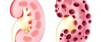
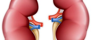
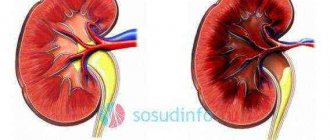
The entire pregnancy proceeded well, the ultrasound did not reveal any pathologies. They did a three-dimensional ultrasound, all organs were complete and without pathologies. At the age of 5, the child underwent an ultrasound of the abdominal cavity and the doctor said that the right kidney was within the normal size for age, but could not find the left one. How is this possible if everything was in place during pregnancy? Again I read the extract from the ultrasound done at 24 weeks (“left kidney is normal, right kidney is normal”)
Woman.ru experts
Find out the opinion of an expert on your topic
Muzik Yana Valerievna
Psychologist, Psychoanalyst. Specialist from the site b17.ru
Semikolennykh Nadezhda Vladimirovna
Psychologist. Specialist from the site b17.ru
Yulia Vadimovna Voronina
Psychologist, bibliotherapist. Specialist from the site b17.ru
Wrzecinska Eva
Psychologist. Specialist from the site b17.ru
Victoria Kiseleva
Psychologist, Gestalt therapist. Specialist from the site b17.ru
Zadoina Lyudmila Alexandrovna
Psychologist. Specialist from the site b17.ru
Andreeva Anna Mikhailovna
Psychologist, Clinical psychologist, oncologist. Specialist from the site b17.ru
Korotina Svetlana Yurievna
Psychotherapist. Specialist from the site b17.ru
Pavlichenko Marina Valentinovna
Psychologist, Psychologist-consultant. Specialist from the site b17.ru
Lukashevskaya Natalya
Psychologist, PERSONAL AND FAMILY. Specialist from the site b17.ru
Amazing. Maybe you should go to another doctor? Maybe their device is old, bad, or something else. This is the first time I've heard this at all.
AS an option - absent in a typical place - soldered upward or fallen into the pelvis or migrated to the midline. Well, or it didn’t exist, in 24 Weeks 0tbaldy wrote so as not to irritate. Or in 5.5 years it has shrunk or lysed. After vaccination with a poorly inactivated strain under the shoulder blade, for example. In any case, if it is not detected without contrast, look with contrast.
Could it actually be a shift? My heart is right-sided, or rather shifted to the right side. But the rest of the organs are in their place. Maybe you have something in this area too?
At week 24 we went for a paid three-dimensional ultrasound, every organ was examined there... we are waiting for a visit to the nephrologist... I hope for a shift. The pediatrician said that sometimes children don’t notice a kidney in their stomach because of gas..
I know such a case, on ultrasound there was a kidney in the womb. the child was born with one ((
Wow. go to another doctor.
Related topics
Our ultrasound specialist is ready to search for kidneys in a couple of months) A friend in America for an assistant! I studied as an uzist with a medical education for two years! Remember this and don't worry, everything will be fine.
Norms for ultrasound of fetal kidneys
Normally, a longitudinal ultrasound scan reveals the fetal kidneys as a bean-shaped or oval formation located in the lumbar region. On a transverse scan they have a round shape and are identified as paired formations on both sides of the spine.
One of the structural units of the kidney is the calyces, which, merging with each other, form one common cavity - the renal pelvis. Gradually narrowing, the pelvis continues into the ureter. The ureter drains into the bladder.
Using an ultrasound machine, the doctor can visualize the pyelocaliceal system starting from the 14th week of intrauterine development. Normally, the diameter of the renal pelvis should not exceed:
- in the second trimester – 4-5 mm;
- in the third trimester – 7 mm.
Examination of the internal structure of the organ becomes possible after 20–24 weeks of pregnancy. During this period, it is already possible to diagnose some developmental anomalies:
- agenesis (absence of one or both kidneys);
- atypical location of the organ (dystopia);
- increase in size;
- expansion of the calyces and (or) renal pelvis;
- cystic changes in the kidneys.
The fetal ureters are not normally visualized.
For both prevention and treatment purposes, nephrologists recommend “positional therapy” to pregnant women. The expectant mother is placed on the side opposite to the diseased kidney in a bent knee-elbow position. The foot end of the bed is raised. This position helps to deflect the pregnant uterus and reduces pressure on the ureters.
Heavy aftertaste
Glomerulonephritis is another inflammatory kidney disease caused by pathogenic bacteria - streptococci. What is noteworthy is that most often this disease occurs after a sore throat or flu. The main danger of the inflammatory process is that when normal urine output stops, blood poisoning begins or attacks of renal colic often recur.
“Glomerulonephritis in pregnant women is manifested by pain in the kidneys and lower back, headaches, and decreased performance,” says nephrologist Tatyana Nefedova. – The main symptom during pregnancy is swelling on the face under the eyes, on the lower extremities, the anterior abdominal wall, and increased blood pressure. Childbirth in mothers with kidney diseases occurs naturally; the need for a caesarean section arises only when the kidney descends into the pelvic area, when the kidneys are fused (“horseshoe kidney”), after plastic surgery to restore the missing bladder wall, when placental abruption due to high blood pressure, fetal hypoxia and in some other cases.”
No man is an island?
“Doctors take into account a lot of circumstances before allowing a woman who has had one kidney removed to give birth,” thinks obstetrician-gynecologist Inna Levina. “This issue is resolved positively if the remaining kidney is absolutely healthy and compensates for the work of the removed one, and if at least two years have passed since the operation.”
Only five years ago, Russian researchers showed that it is possible to carry and give birth to a baby with one kidney. The prognosis for the woman and fetus in most cases is favorable, of course, subject to regular monitoring by a doctor.
“A single kidney may be the result of a congenital developmental defect or remain after the removal of the second kidney due to any disease: pyelonephritis, kidney stones, tumor, injury, etc. – says nephrologist Tatyana Nefedova. – The reserve capacity of the kidney is quite large. Normally, only a quarter of the kidney tissue functions at a time. After the removal of a kidney, the blood supply to the remaining one almost doubles, almost all of the kidney tissue gradually begins to function, and its functional capacity approaches the normal level that existed with two kidneys. The process of compensating for the functions of a lost kidney is lengthy. It ends only one and a half to two years after the operation.”
After the operation, the remaining kidney works with double load, its intense activity gradually leads to some exhaustion. Therefore, mothers who have once undergone a nephrectomy (removal of a kidney) cannot be considered absolutely healthy, even if the second organ seems completely healthy. Since the capabilities of one kidney are limited, it reacts sensitively to various influences, such as pregnancy, infection, etc.
The most favorable time for pregnancy to occur is from two to four years after nephrectomy, when the functional restructuring of the organ is completed. The physiological functions of the remaining kidney are usually normal during pregnancy, and urination is not impaired. Protein in the urine of pregnant women after nephrectomy is as insignificant as in healthy women.
“I would like to warn you that women who have undergone nephrectomy often develop a urinary tract infection (for example, cystitis) during pregnancy,” says nephrologist Tatyana Nefedova. – This complication occurs in more than half of pregnant women. However, the functioning of the kidney suffers little: it does not deteriorate significantly either during pregnancy or after childbirth.”
“I want to reassure expectant mothers who have previously undergone this difficult operation,” reassures obstetrician-gynecologist Inna Levina. – The absence of one kidney does not affect the duration of pregnancy and is not a cause of premature birth or miscarriage. The postpartum period in most cases proceeds quite well: obstetric complications and deterioration of the urinary organs are rare and, as a rule, are not associated with the previous intervention.”
The power of stereotypes
“Unfortunately, there are often situations when, having seen a serious “renal” diagnosis in the card, women are often offered to terminate the pregnancy. Such advice can often be heard from an obstetrician-gynecologist observing a “problem” mother, regrets obstetrician-gynecologist Anna Skorobogatova
. – This is a rather stereotypical and incorrect approach. As if only healthy women can reproduce, which, by the way, are becoming fewer and fewer. Only the pregnant woman herself and, perhaps, her closest relatives have the right to decide whether to be a mother. But to what extent this is possible should be determined by a council of specialists, consisting, in addition to a gynecologist, at least of an experienced therapist and, when it comes to the kidneys, a nephrologist.”
Termination of pregnancy is indicated for:
- combination of pyelonephritis with severe forms of gestosis;
- lack of effect from the treatment;
- acute renal failure;
- hypoxia (lack of oxygen) in the fetus.
“The best option, if you are sent for an interruption, is to visit one or more specialists and carefully discuss the situation again,” the specialist urges. – Don’t despair ahead of time, remember, you shouldn’t worry. Be sure to go to a nephrologist. Now the level of medicine allows us to solve very complex problems. Pregnancy monitoring tactics are developed and the necessary treatment methods are selected. They often make it possible to improve the situation and minimize the consequences of kidney disease for both the mother and the child.”
Doctors recommend
“An expectant mother with any kidney pathology should be surrounded by the attention of not only a gynecologist, but also a nephrologist,” insists nephrologist Tatyana Nefedova. “Pregnancy, of course, is not a disease, but in this situation you will have to take care especially carefully. Here are a few points: walking a lot is not forbidden, but you need to do it slowly. Under no circumstances should you participate in marathon races (swimming and other sports competitions - just kidding!). But seriously, the lifestyle of such a woman should be calmer in everything. Don’t neglect your daytime rest, it’s time to fulfill your dream of getting plenty of sleep! Try to protect yourself as much as possible from nervous overload, no matter how difficult it may be at times. To worry less, you can use sedatives allowed during pregnancy (valerian, for example).”
If you cannot avoid swelling and protein in the urine, you will have to switch to a low-salt diet: it does not require a complete abstinence from salt, but if the taste of your favorite dish does not suffer too much, then it is better to do without it. And, of course, mothers with kidney disease are advised to think less often about typical “pregnant” delicacies, such as pickles, smoked meats and marinades.
“However, in an effort to give birth to a child at all costs, you should not neglect the danger to his and your own health and life,” obstetrician-gynecologist Inna Levina strongly recommends. – After all, if significant contraindications for bearing a baby are discovered already during pregnancy, it will have to be interrupted. If pregnancy is not contraindicated, immediately after its discovery, contact an antenatal clinic. Since it is important for an obstetrician-gynecologist, nephrologist and urologist to know what kidney function was at the beginning of the first trimester in order to correctly judge its changes as pregnancy progresses. The expectant mother must be constantly monitored and must go to the hospital repeatedly for examination and treatment. Remember, hospitalization is mandatory, and under no circumstances self-medicate!”
In medical practice, unfortunately, there are such serious changes in kidney function that pregnancy becomes an unbearable burden for them. The inability to bear and give birth to a healthy child is one of the most terrible sentences for most women. However, the consequences of pregnancy for some (not all, mind you!) kidney diseases are so formidable that with mature reflection, any woman will understand: medical contraindications are not the whim of doctors and not “reinsurance”! Knowledge, even bitter, is always better than ignorance, which can lead to the most tragic consequences.
I will only add that I would strongly recommend diluting any common sense with a fair amount of optimism: after all, the possibilities of medicine are now very great.
Possible abnormalities in kidney development in the fetus
The main method for detecting pathology in the fetus is ultrasound. It can be used to diagnose malformations in the earliest stages of pregnancy.
What are the most common abnormalities in the development of the fetal urinary system that a doctor can see on an ultrasound?
Pyeelectasis
The most common pathology. It can occur in one kidney or in both at once. There are:
- isolated dilatation of the pelvis – pyelectasia;
- dilation of both the pelvis and ureters - pyeloureteroectasia;
- simultaneous expansion of the pelvis and calyces - pyelocalicoectasia (or hydronephrotic transformation).
Note! A slight increase in the size of the cups and pelvis (up to 1 mm) is eliminated independently. Dilation of more than 2 mm requires dynamic ultrasound monitoring in order to promptly diagnose hydronephrosis.
Hydronephrosis
What are the symptoms that a woman has chronic pyelonephritis?
The apparent reasons are varied. If you have chronic pyelonephritis, you may not know about it for some time (before diagnosis). Pain is felt in the lumbar region. Aching or dull and weak. When it's cold or damp outside, they get worse. Women experience frequent urination and even urinary incontinence. Blood pressure increases in patients. Women feel pain when urinating.
How intense will the disease manifest itself? It depends on whether it is 1 kidney or both and how long ago? If a woman has chronic pyelonephritis, then during the period of remission she will not feel much pain and will decide that she is healthy. Painful sensations will become noticeable during the acute stage of the disease.
What causes the exacerbation? Visible reasons: people have weak immunity. It happens after eating spicy foods, if you often drink alcohol in any form, or if you get hypothermic somewhere. Symptoms of the disease:
Your temperature is above +38 °C; You feel a nagging pain in your lower back. There are also pains in the peritoneal area, but less often. If you stand somewhere for a long time or play sports, they will remind you of themselves. You get tired faster than usual and often feel weak; Headache; Muscle pain is felt; You feel sick; The face and limbs swell; Urination becomes more frequent, persistent frequent urge; You feel pain when urinating; Urine is cloudy; There was blood in the urine.
Indications for examination
X-ray or ultrasound of the kidneys is carried out based on the patient's complaints and the attending physician's suspicions of the presence of pathological changes in the organs.
X-rays or ultrasound of the kidneys are carried out on the basis of the patient's complaints and the attending physician's suspicions of the presence of pathological changes in the organs. So, the main reasons for ultrasound are:
- Pain in the lumbar region;
- Injuries to the kidneys or peritoneum;
- Consolidation or enlargement of the kidneys that are clearly palpable on palpation;
- Presence of blood in the urine;
- Changes in diuresis (more/less urge to urinate or lack thereof);
- Constantly elevated blood pressure that cannot be adjusted with medication;
- Suspicion of cancer.
Reasons for the absence of one kidney
Before going to the urologist, we took blood and urine tests - the usual ones, at the clinic. 1. There is no urgency in the absence of complaints and normal urine tests. 2. As in any other organ: no more and no less. 2. What can a child complain about so as not to miss a symptom indicating a kidney problem? 3. If the kidney still exists, but does not perform its functions, can removal surgery be avoided?
Which of these methods is more informative in the case of a non-visualized kidney? Which is safer for a child? The kidney could not disappear. You need a consultation with an experienced pediatric nephrologist-urologist. A cat that walks on its own doesn’t surprise anyone, but when the kidney starts doing the same thing, it’s no longer okay.
After ultrasound
It is worth understanding that the ultrasound protocol is not yet a diagnosis. This is only a preliminary examination of the kidneys for possible pathologies. If the attending physician has certain suspicions, he will definitely refer the patient for additional instrumental studies:
- CT and MRI;
- Contrast radiography;
- Angiography, etc.
Only after collecting a detailed history and conducting all the necessary studies, a specialist makes a diagnosis and prescribes effective treatment. It is important to follow all doctor's recommendations. After a while, the specialist may prescribe a repeat ultrasound to monitor the pathology over time.
Important: Ultrasound is the most informative, accessible and painless method for examining the kidneys. That is why, when prescribing this procedure, it is worth undergoing hardware diagnostics in order to be able to get rid of the pathology of the urinary system in a timely manner.


