Radionuclide (radioisotope) diagnostics, the methods of which include nephroscintigraphy, became widespread at the end of the twentieth century. This study complements traditional kidney disease screening efforts. In terms of accuracy and information content, it can compete even with excretory urography and often surpasses it.
- Video: what is radionuclide diagnostics
- Sensitivity of scintigraphic examination
- Static nephroscintigraphy of the kidneys
- Dynamic study
Table: time elapsed after drug administration and marker location
- Indications and contraindications for renal scintigraphy
- Preparation for the procedure
- How is dynamic nephroscintigraphy of the kidneys performed?
- Decoding images
What the study shows
Types
Scintigraphy happens:
- Dynamic.
- Static.
- Express.
Widely used, dynamic nephroscintigraphy visualizes kidney function in detail. The radionuclide study method consists of penetration of a radiological contrast agent into the cells of an organ through the blood vessels. An important point is to evaluate the results when the drug enters the structures of the bladder. Nephroscintigraphy is a highly informative method for examining the joint activity of the kidneys and bladder.
If there is a suspicion of the disease, renoscintigraphy is performed even in childhood. To improve the reliability of the results, a separate sample is taken and a specific substance is used.
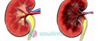
Static scintigraphy is indicated in case of deterioration in organ function or the presence of pathological kidney damage. Such nephroscintigraphy clearly determines the shape, location, and size of the organs being examined. In addition, structural changes are detected and blood circulation is assessed.
On this topic
- Onconephrology
How long do they live after surgery for kidney cancer?
- Olga Vladimirovna Khazova
- February 28, 2020
This method is more informative than ultrasound or x-ray. In this case, static scintigraphy is done only in combination with other diagnostic studies.
The third type of procedure (circuloscintigraphy) is used for a detailed study of renal circulation, which changes significantly in diseases.
Renal scintigraphy
Renal scintigraphy (nephroscintigraphy) is a radiation research technique used to assess the functionality of renal structures. The basis of the technique is the use of a radiodrug, without which it is impossible to carry out diagnostics. There is a completely incorrect opinion about the dangers of such drugs, which is often associated with the banal illiteracy of ordinary people.
These drugs have minimal levels of radioactivity, they are safe and do not cause allergic reactions.
The radiodrug Hippuran is most often used in the procedure. Scintigraphic kidney diagnostics is a highly informative and effective diagnostic and treatment method, with which pathology can be detected a year earlier than with other studies. But, unfortunately, due to the high cost of equipment and the shortage of radiopharmaceuticals, such a procedure is not available to everyone.
Indications
Dynamic renoscintigraphy is a more informative method that is used much more often than other types. To carry out this diagnostic procedure, the following indications are required:
- Various deterioration in the functional activity of the kidneys.
- The second or third stage of expansion of the pelvis, calyx. The development of hydronephrosis is affected by impaired urination.
- Abnormal organ structure.
- The presence of cysts and other neoplasms. Dynamic scintigraphy is used to determine the degree of malignancy.
- Planning transplant .
- Surgical interventions that preserve the organ.
- Determination of metastasis in the genitourinary system.
Static nephroscintigraphy is used for abnormal arrangement of organs, impaired development of the urinary system, pyelonephritis, strictures or arterial hypertension.
Kinds
Nephroscintigraphy can be performed in two ways: dynamic or static. Static diagnostics is an additional method after radiography that determines the general parameters of the kidneys, their location and shape, size, etc. Static diagnostics cannot determine the presence of functional disorders in the urinary system, and therefore does not show the full picture of the existing pathology.
Dynamic nephroscintigraphy is performed after administration of the radiodrug. At various intervals, the entry of the drug into the renal structures, ureters and bladder tissues is recorded. The images obtained as a result of dynamic nephroscintigraphy display the processes of urine formation and urination step by step, which allows you to study in detail the functionality of each kidney separately or both organs together.
Contraindications
Despite its safety, renoscintigraphy is not indicated for everyone. Since the procedure is lengthy, for patients in serious condition, the manipulation will cause a lot of discomfort. It is not recommended to carry out such diagnostics on pregnant women, since the study has a detrimental effect on the development of the fetus. In such a situation, scintigraphy is performed in emergency cases.
During the breastfeeding period, the child is transferred to artificial feeding for several days. This is done to completely remove the contrast agent from the mother’s body.
To determine cellular metabolism, radionuclide testing is used. In the presence of an oncological tumor, scintigraphy is performed only a month after chemotherapy, several months after irradiation.
Consequences
Almost all manipulations related to nuclear medicine do not pose a danger to human health. Therefore, kidney scintigraphy can be prescribed not only to adult patients, but also to children. It is much more dangerous to leave pathological processes unstudied and untreated.
Adverse reactions after diagnosis are rare, in contrast to the negative symptoms that occur after tomography and radiography. Pharmaceutical substances used in the research process are removed from the body in no more than a day. However, they do not have a negative effect on other organs and systems. To speed up the release period of the concentrating agent, doctors recommend drinking more fluids and being physically active after scintigraphy.
Long-term practice of using scintigraphy confirms its safety. The procedure can be carried out without fear even daily, if there are indications for this. The study makes it possible to detect renal abnormalities several years earlier than radiography.
The only drawback of the diagnostic procedure can be considered cost and inaccessibility. Diagnostics are not performed in all medical institutions, and its price can be up to 7,000 rubles.
Preparation process
Diagnostic manipulation does not require any special preparation. However, before scintigraphy you need to eat and drink several glasses of still water. In this case, you should not drink tea or coffee, as these drinks can affect the results of the study.
As for nutrition, marinades, smoked meats and spicy foods should be excluded from the diet. A day before the diagnosis with a pharmacological test, hypertensive patients should stop taking diuretics, and a week before the examination it is recommended to stop taking ACE inhibitors. Such events are dangerous due to crises, so scintigraphy is performed in a hospital setting.
The main difference of the method
The essence of the technique is to assess the state of functioning of internal organs using a radiopharmaceutical drug. Conducting a study without a chemical substance (RPF) is impossible.
A radiopharmaceutical has a number of fundamental characteristics that determine its properties. The only disadvantage of the substance is a small dose of radioactivity.
Once in the human body, the radiopharmaceutical does not provoke allergies or side complications. To carry out the manipulation, a tiny amount of the drug is required, which, after administration, affects only the organ being examined.
Attention! A tiny amount of a chemical with a low dose of radioactivity cannot cause side effects. The idea that this product can cause radiation exposure is false.
Thanks to the ability of scintigraphy to diagnose disorders in tissues and organs at the stage of pathology formation, the method is recognized in Europe and the USA. In Russia, the technique is less widespread due to a lack of equipment.
Carrying out the procedure
The manipulation is done in one or more gamma cameras. The patient is given an intravenous injection of a radioisotope substance that emits gamma rays. The camera detects the radiation and then displays a visual picture on the monitor. It is important to remove metal items from the patient before starting scintigraphy. Sometimes the patient is given a special contrast liquid to drink.
During the examination, specialists leave the room. The scan evaluates the state of renal blood flow in each organ. During the entire procedure, the patient should not move or speak. To determine the mobility of the kidneys, on the advice of a doctor, it is necessary to change position or sit down. Diuretics are used to study obstruction, and antihypertensive drugs are used to study the renal arteries. Most often, the procedure is performed on an empty stomach with an empty or full bladder.
How is nephroscintigraphy performed?
Diagnostic time can vary significantly, but manipulation always comes down to the following algorithm:
- The patient is positioned on a table in front of the gamma camera and a carefully calculated dose of the drug is administered.
- The diagnostic specialists leave the room and the scintillator begins to detect radiation. This does not apply to static examinations, when contrast is introduced some time before the start of scanning.
- During the session, a person is prohibited from talking or moving, but if nausea, dizziness or tachycardia occurs, he must inform the staff about this by pressing a special button.
- After completing the examination, it is recommended to drink more water, accelerating the conclusion of the radiopharmaceutical.
Nephroscintigraphy is an informative and safe examination with a minimum of contraindications. It is invaluable in identifying malignant pathologies and initial subtle changes in tissue or kidney function. The main disadvantages are high cost and lack of prevalence.
results
After the procedure is carried out in several stages, the results obtained are deciphered. The specialist must evaluate the shape, topography and size of the organs, the degree of their activity, and the intensity of blood flow. The structure of the parenchyma is also determined. After this, the examination is assessed according to pathological areas. Doctors analyze the condition of each kidney and determine the amount of radioisotope substance. Decoding this data allows you to accurately assess kidney function.
To determine excretory and secretory activity, an analysis of two examined areas is carried out. This is done to determine the degree of development of kidney diseases. At the end of the decoding, specialists study the area of each organ to determine the actual functionality.
Varieties of techniques
There are several types of renal scintigraphy. The most common are three types:
- Renoscintigraphy is a dynamic and most used method with wide practical significance.
- Nephroscintigraphy – both dynamic and static examination of the kidneys is possible.
- There is a third type of high-speed scintigraphy, which is not widely used - circuloscintigraphy for studying the blood circulation of the area under study and the kidneys.
Purpose of the examination
The main purpose is to determine renal function, but it can be caused by a wide variety of urological or nephrological indications.
The most common are:
- kidney injuries;
- renal failure;
- congenital diseases;
- kidney transplant;
- hypertension and other diseases.
The range of indications for prescribing a study is much wider. This is explained by the high efficiency of the study.
You can see all the failures and disorders: the speed of blood flow in the kidneys, notice the difference in the functioning of the left and right kidneys, see scars from past inflammatory processes and much more.
Its main advantage is accuracy, harmlessness and absolute painlessness.
When is it appointed?
There are the following indications for kidney scintigraphy:
- Congenital anomalies.
- Injuries.
- Acute and chronic pathologies.
- Preparing for transplantation.
- Assessment of renal blood flow.
- Detection of dilation of the urinary tract and pyelocaliceal system.
- Renal infarctions.
- Detection of urinary tract blockages.
- Reflux detection.
- Confirmation of the diagnosis that the kidneys are the source of high blood pressure.
- Identification of complications after kidney transplantation.
Decoding the information received
A radiologist interprets the data immediately after the procedure. Obtaining analysis results is step-by-step:
- the condition and size of the kidneys are assessed;
- the functioning of the organ and the intensity of blood supply are examined;
- the degree of damage to existing foci, renal obstruction is determined with a detailed study of the right and left organs;
- the functions of both kidneys are compared;
- Each segment of the examined organ is studied together with secretory and excretory activity.
By studying these parameters, the doctor can identify where the inflammatory process is occurring, whether vesicoureteral reflux is involved in the pathology, how impaired urination is, and whether there are prerequisites for the development of urolithiasis or the growth of a dangerous tumor.
If the study shows urolithiasis, severe damage to the parenchyma or kidney tissue, then the patient may be offered surgery.