Pathological anatomy / Pathological anatomy Kidney diseases
Clinical manifestations. In most patients, poststreptococcal glomerulonephritis begins suddenly, acutely in the form of malaise, fever and nausea 7-14 days after tonsillitis, with the rapid development of nephritic syndrome. Oliguria appears (a sharp decrease in urine volume) and the urine darkens (the color of “meat slop”) as a result of hematuria. An objective examination reveals swelling on the face, especially periorbital, and mild hypertension. In the blood, an increase in the titer of antistreptolysin O and a significant decrease in the complement C3 fraction are determined. CEC tests are usually positive. An increase in the content of nitrogenous wastes (uric acid and creatinine) in the blood indicates impaired renal function. Urine tests determine hematuria, the number of leukocytes increases, casts appear, and proteinuria of varying severity is also present. When examining the throat and skin for microflora, streptococcus is usually not detected, because by this time the infection has usually resolved.
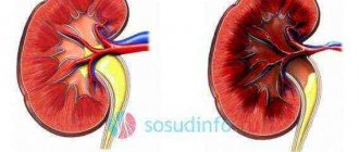
Pathological anatomy. Macroscopically, the kidneys are slightly enlarged due to edema. The capsule is easily removed, revealing a smooth, even surface. In severe cases, there may be numerous petechiae (red specks) on the surface.
Microscopic examination reveals diffuse glomerulonephritis. In the initial stage, the glomeruli are enlarged and the number of cells in them is increased (Fig. 4). An increase in the number of cells occurs as a result of proliferation of mesangial, endothelial and epithelial cells of the outer layer of the glomerular capsule, as well as varying degrees of infiltration by polymorphonuclear leukocytes. Severe edema and swelling of the endothelium leads to a narrowing of the lumen of the capillaries.
Light microscopy can reveal immune complexes in the form of characteristic “clumps,” especially with trichrome stains. On electron microscopy they appear as large, electron-dense, dome-shaped deposits on the epithelial side of the basement membrane (subepithelial “humps”) (Fig. 4). Deposits of immune complexes in the mesangium, subendothelial and intramembrane regions are also often detected. Immunofluorescence reveals granular deposits of IgG and C3 along the glomerular basement membrane and in the mesangium.
Forecast. With the use of anti-inflammatory and immunosuppressive therapy, 95% of patients clinically recover within 6 weeks. Normal kidney function is restored within a year. Sometimes an increase in the number of cells in the mesangium can persist for many months and even years. Disturbances in urine sediment may persist for several years. A small number of patients experience progression with rapid development of renal failure within 1-2 years. These patients exhibit the presence of multiple crescents (subacute glomerulonephritis - see below).
Long-term prognosis remains a controversial issue. According to some studies, children have a good prognosis. A worse prognosis is determined in adults, in whom chronic renal failure develops in 30-50% of cases. However, it should be noted that these may be cases of acute onset of chronic glomerulonephritis.
Rapidly progressive (subacute) malignant glomerulonephritis is a rare disease that reflects severe damage to the glomeruli. Subacute glomerulonephritis is characterized by:
the presence of epithelial crescents in more than 70% of glomeruli. Half Moon,
filling Bowman's capsule, consist of epithelial cells and macrophages that proliferate in response to fibrin effusion from damaged glomeruli. Crescents represent irreversible damage to the glomerulus, leading to
Urolithiasis (urolithiasis)
Urolithiasis (urolithiasis) is a metabolic disease caused by various causes, often of a hereditary nature, characterized by the formation of stones in the urinary system (kidneys, ureters, bladder or urethra). Stones can form at any level of the urinary tract, from the renal parenchyma, in the ureters, in the bladder and ending with the urethra.
The disease can be asymptomatic, manifested by pain of varying intensity in the lumbar region or renal colic.
The history of the names of urinary stones is very fascinating. For example, struvite (or tripyelophosphate), named after the Russian diplomat and naturalist G. H. von Struve (1772-1851). Previously, these stones were called guanites because they were often found in bats.
Stones made from calcium oxalate dihydrate (oxalates) are often called weddelites because. the same stones are found in rock samples taken from the bottom of the Weddell Sea in Antarctica.
Characteristic symptoms
The primary symptom of the development of the syndrome is a significant decrease in urine excreted from the body. The daily volume of waste liquid release is reduced to close to 400 ml. The appearance of protein elements is visually observed in the structure of urine.
The patient begins to suffer from growing weakness. Apathy, loss of appetite, dizziness, and drowsiness appear. A pronounced aroma of ammonia can be heard from the oral cavity. A person susceptible to developing the syndrome is bothered by constant thirst. Periodic attacks of nausea and vomiting occur.
Over time, certain areas of the body become swollen. An increased concentration of potassium in the blood due to impaired kidney function causes a tingling sensation in the limbs. Convulsive phenomena occur periodically. Tendon reflexes worsen. The concentration of toxins in the body leads to depression of the nervous system, the appearance of hallucinations, and delusions.
Kidney tuberculosis
The acute miliary form of renal tuberculosis is a local manifestation of the hematogenous generalization of the tuberculosis process. Multiple gray tubercles are formed in the cortex of both kidneys. The surface of the bud becomes dark red and seems to be strewn with semolina.
During histological examination, among the convoluted tubules, glomeruli, in the walls of blood vessels, and sometimes in their lumen, it is possible to see typical lymphoid or epithelioid cellular tubercles with characteristic giant cells, and sometimes with small areas of necrosis in the center of the tubercle.
Chronic tuberculosis of the kidney manifests itself in the form of caseous-cavernous, tubercular and fibrous-indurative forms.
Early stage of chronic tuberculosis
In the early stage of chronic tuberculosis, the tubercles are located predominantly in the cortex. Due to the relative resistance of the cortex to tuberculosis infection, they often undergo scarring. The tubercles in the medulla, located mainly in the region of the renal papilla, sinus or calyx, merge and undergo caseous decomposition. The renal tissue softens, and the caseous process spreads towards the cortex. Separate, delimited cavities with uneven, corroded edges are formed, filled with a yellowish-looking cheesy mass.
The process can also spread throughout the mucous membrane and affect the necks of the calyces, causing ulceration, scarring, narrowing and obliteration. Lesions on the papillae can be single or independently affect several calyxes.
Spread of tuberculosis infection
Gradually, the tuberculous process from the edge of the cavity spreads to other areas of the renal parenchyma, where destruction also occurs. The spread of tuberculosis infection occurs along the length and also retrogradely through the blood and lymphatic vessels and through the urinary canaliculi. Newly formed foci of cheesy necrosis undergo melting, and new cavities form. As long as the primary small focus of decay in the medulla does not communicate with the calyx or pelvis, it can exist for long periods without clinically manifesting itself. In most cases, the contents of the cavity break through a thin fistulous tract into the urinary tract. From such a focus of Mycobacterium tuberculosis, pus enters the renal pelvis, ureter, and bladder.
Thus, with reduced reactivity of the body, a whole series of isolated or fused cavities are formed in the kidney, sometimes occupying most of the renal parenchyma (see Fig.).
| Rice. Tuberculosis of the kidney and ureter |
Progressive tuberculous destruction can lead to complete destruction of the kidney, to tuberculous pyonephrosis. An enlarged kidney is a thin-walled sac filled with thick pus, cheesy masses, or consisting of separate cavities separated by bridges of sclerotic renal tissue, protruding onto the surface of the kidney in the form of lumpy, fluctuating areas.
This picture is typical for predominantly exudative kidney tuberculosis. With a predominantly productive reaction of the body in the area of the papilla and the vaults of the calyces, one, rarely more, tuberculous cheesy nodule 0.5-1 cm in diameter appears - the so-called focal tuberculosis of the kidney. This isolated node is delimited from the surrounding parenchyma by a demarcation line with specific and nonspecific granulation tissue, which gradually matures and turns into a fibrous membrane, encapsulating the cheesy focus described above. In some cases, such a focus is subjected to petrification.
In rare cases, when the exudative reaction gives way to a productive one, cheesy foci in the kidney, pelvis and ureter are gradually delimited from the remaining tissue by a fibrous capsule and undergo petrification. In other cases, due to stenosis of the neck of one or more calyces, part of the kidney is completely switched off, being delimited from the rest of the parenchyma by a dense connective tissue capsule, the so-called switched off kidney tuberculosis.
Ulceration and subsequent scarring of the ureter lead to obliteration of its lumen, the communication between the kidney and the urinary tract is completely stopped. Caseous decomposition in the disabled kidney continues, the contents of the caverns may undergo calcification, the so-called putty-like, or neutered, kidney.
Changes in the kidneys in chronic tuberculosis
Histological examination of chronic tuberculosis can reveal changes of various types.
In cases where the process is predominantly destructive, foci of cheesy necrosis are noted. Along the periphery of the necrotic tissue, a clearly expressed picture of a specific inflammatory process is noted. Accumulations of elements of young granulation tissue are visible, consisting of epithelioid and lymphoid elements with an admixture of typical Langhans cells and areas of cheesy necrosis.
In other fields of view, well-formed epithelial-cell tubercles with giant cells are visible among the tubules.
One can also observe a number of areas where the inflammatory infiltrate is not typical for tuberculous granulation tissue. Such foci resemble the picture of the so-called chronic interstitial nephritis, when between individual preserved tubules the infiltrate contains a significant amount of lymphoid and histiocytic elements, and some foci consist entirely of accumulations of decaying neutrophilic leukocytes, forming a picture of microabscesses.
Thus, in the same preparation it is often possible to observe different stages of the tuberculosis process. While in some fields of view there are extensive foci of cheesy necrosis and accumulation of exudate, in others one can see a histological picture characteristic of predominantly productive inflammation with the presence of groups of fibrous tubercles.
Sometimes the cortical and medulla layers of the kidney are evenly or uniformly dotted with tuberculous tubercles, adjacent to each other without signs of ulceration and cheesy decay. Neither ulcers nor cavities are formed. Caseosis can be detected in the center of some tubercles, but complete melting of many tubercles is not observed. This kind of tubercular form of tuberculosis is sometimes found in large areas of the kidney; in rare cases, the entire kidney is riddled with tubercles.
Tuberculous mycobacteria cause not only specific changes in the kidney tissue in the form of tubercles, caseosis, ulcers, and cavities. Near and even far from tuberculosis foci, areas of nonspecific changes can be found in the form of vascular sclerosis, inflammation, hyalinosis, cicatricial growth and desolation of the glomeruli, granular and fatty degeneration of the tubular epithelium, lymphoid and small cell infiltration of interstitial tissue and proliferation of connective tissue, which leads to atrophy and cicatricial replacement of the parenchyma, both directly and through compression of the arterial trunks and malnutrition.
The ratio of specific and nonspecific changes in the kidney is different: there are cases when the phenomena of chronic nephrocirrhosis are so dominant in the anatomical picture that tuberculous elements cannot be detected either macro- or microscopically (indurative form of renal tuberculosis).
The proliferation of connective tissue and areas of wrinkling make one suspect that in these cases there was also an effect of nonspecific inflammation.
Indeed, in isolated cases of chronic kidney tuberculosis, chronic pyelonephritis can join the tuberculosis process.
The renal pelvis and calyx are involved in the tuberculosis process in a secondary descending manner. Small tubercles are formed on their mucous membrane, surrounded by a zone of hyperemia. Along with the tubercles, proliferation of granulation tissue and ulcerations at the site of caseous degenerated tubercles are observed. The submucosal and muscular layers of the pelvis are involved in the pathological process, which is accompanied by infiltration of its wall. The pelvis becomes thickened, swollen, and roughly folded.
In some cases, under the influence of tuberculosis toxin, even before the appearance of specific tuberculosis changes, atony and expansion of the calyces of the pelvis and ureter occur.
Specific changes in the ureter are similar to those in the pelvis. The ureter expands, its wall thickens and infiltrates. In addition to tubercles, ulcerations, and proliferation of granulation tissue, narrowing of the lumen of the ureter may occur due to developed infiltration, granulation, or scar tissue. Tuberculosis lesions occur not only in the wall of the ureter, but also in the surrounding tissue.
The latter becomes sclerotic, turning the ureter into a dense shortened cord fused to the surrounding tissues.
Tuberculosis of the bladder is usually associated with tuberculosis of the kidney, very rarely with tuberculosis of the prostate and seminal vesicles, but is never primary in the genitourinary system. The first foci of tuberculosis usually develop in the area of the ureteric orifice and rarely in the area of the bottom or wall of the bladder.
Miliary tubercles on the mucous membrane of the bladder, surrounded by a rim of hyperemia, become necrotic and, merging, form ulcers, usually elongated, irregular in shape with a yellowish bottom and a rash of tubercles along the periphery. The edges of the ulcer are hyperemic and edematous.
Bladder ulcers, initially single, small and superficial, later become multiple, larger and deeper, penetrating into the muscle layer. Merging, in advanced cases they affect significant areas of the cystic wall and, undergoing partial scarring, cause wrinkling and rigidity. The capacity of the bubble sometimes decreases to 20-30 cm3. Cicatricial changes in the area of the ureteric orifice in some cases lead to a crater-like gaping of the orifice, in others to strictures or obliteration.
In rare cases, tuberculous bladder ulcers rupture into the pelvic cavity, peritoneum, rectum or vagina.
In the vicinity of the mouths of the ureters or in the fundus there are growths of tuberculous granulations, sometimes reaching significant sizes and similar in appearance to papilloma.
Pathological anatomy pyelonephritis
It is characterized by the involvement of the renal pelvis, calyces and interstitial tissue in the infectious and inflammatory process.
Etiology and pathogenesis
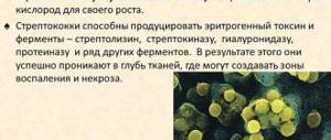
Refers to infectious diseases. The main causative agents of this disease: Escherichia coli, staphylococcus, streptococcus - enter the kidneys in three ways:
1. Through a hematogenous descending route, the infection enters the kidneys during sore throat, influenza, and sepsis.
2. Lymphogenous introduction of infection is observed in pathologies of the large intestine, as well as the genital organs.
- Urogenic ascending infection of the pelvis and calyces occurs from the underlying parts of the excretory system in the presence of stones, tumors of the urethra, and, accordingly, stagnation of urine.
However, infection of the pelvis and calyces of the kidney is not enough for the development of pyelonephritis. A corresponding restructuring of the body's reactivity is necessary.
Clinical and morphological forms of pyelonephritis
- Spicy
- Chronic, recurrent in the form of acute attacks.
Acute pyelonephritis
Can be one- or two-sided. The medulla is affected more significantly than the cortex.
Macroscopy:
On examination, the kidneys are enlarged and full of blood. Wide pelvis and calyces are filled with cloudy urine or pus. There are foci of hemorrhage on the mucous membrane. Abscesses are observed.
Microscopy:
In the mucous membrane of the pelvis and calyces the following are detected:
- Plethora
- Leukocyte infiltration
- Foci of necrosis
- Microabscesses.
With ascending acute pyelonephritis
microorganisms penetrate the epithelium of the pelvis and rise into the interstitial tissue of the collecting duct zone, where microabscesses, leukocyte infiltration, and tissue swelling appear.
For hematogenous (primary) acute pyelonephritis
multiple small abscesses in the peritubular stroma can merge into large ones. Scars develop in place of the destroyed kidney tubules.
Chronic pyelonephritis
It is a chronic disease leading to kidney failure, often accompanied by hypertension.
Urine with this disease may be sterile, but often contains small amounts of protein.
In chronic pyelonephritis, great importance is attached to immune mechanisms (high titer of antibodies during exacerbation).
Features of a pyelonephritis wrinkled kidney
- Uneven scarring
- Tight adhesion of kidney tissue to the capsule
- Sclerosis of the pelvis and pelvic tissue
- Asymmetry of changes in both kidneys.
Complication of pyelonephritis
Spicy:
- Formation of kidney carbuncles as a result of the fusion of large abscesses
- Pyonephrosis - the formation of communications of purulent cavities with the pelvis
- Perinephritis - transition of a purulent process to the kidney capsule
- Paranephritis – transition of the process to the perinephric tissue
- Papillonecrosis – necrosis of the pyramidal papillae
Chronic:
- Development of nephrogenic hypertension
- Development of arteriolosclerosis in the second intact kidney
- Pyelonephritic renal shrinkage and the development of chronic renal failure.
Outcomes of pyelonephritis
Spicy:
- Recovery
- Death from noted complications
Chronic:
- Uremia due to kidney shrinkage
- In arterial hypertension, death occurs from cerebral hemorrhage or myocardial infarction.
Diseases of the endocrine organs
The endocrine system is scattered throughout the body. It is presented:
- highly specialized secretory organs (VS),
- hormone-producing cells of non-endocrine organs (digestion, respiration, excretion).
Among the VHS there are:
A. Central regulatory entities:
- hypothalamus
- pituitary
- pineal gland
B. Peripheral endocrine glands:
- thyroid
- parathyroid glands
- adrenal glands
B. Organs performing endocrine and non-endocrine functions:
- gonads (testes, ovaries)
- pancreas (PG)
- placenta
The main purpose of the endocrine system is related to the regulation of homeostasis.
The function of endocrine organs is controlled by the hypothalamus. In its mediobasal part there are neurosecretory nuclei that produce hormones: liberins and statins.
These hormones enter the anterior pituitary gland (APG) through the vascular system. Liberins stimulate the secretion of certain PDH hormones, while statins inhibit secretion.
In turn, PDH cells secrete hormones that regulate the secretory activity of peripheral endocrine organs.
The function of individual VVS, as well as the interaction of the peripheral endocrine glands, hypothalamus and pituitary gland, is carried out using a positive and negative feedback mechanism.
The development of VVS diseases is associated with:
- imbalance of their regulation
- direct damage to the glands under the influence of endogenous and exogenous factors
VVS diseases can manifest themselves
- hypofunction
- hyperfunction
- dysfunction
Morphological changes
The endocrine glands contain:
- dystrophic
- atrophic
- hypo- and hyperplastic processes
- sclerosis
- structural adjustment
- tumors
Source: https://worldwantedperfume.com/patanatomija-pielonefrit/
Treatment of the disease Pyelonephritis
Pyelonephritis is usually treated with antibiotics. They are prescribed either empirically (i.e., already tested), or based on the results of sensitivity to antibiotics of the microorganism isolated during urine culture. Of the recommended drugs, preference is given to antibiotics with a broad spectrum of action. Usually, for uncomplicated pyelonephritis, antibiotics are prescribed orally. The duration of the course of treatment is at least two weeks, although often the symptoms of the disease begin to disappear in the first 3-4 days of treatment.
Antibacterial drugs for the empirical treatment of bacterial infections of the urinary system, such as Biseptol, fluoroquinolones, ciprofloxacin, levofloxacin, are widely known. Other antibacterial drugs for the treatment of pilonephritis are applicable if the identified bacteria are insensitive to proven drugs or if the patient is allergic to them. After completion of antibiotic treatment, a control urine test is required to determine the effectiveness of therapy. It includes urine analysis for the presence of bacteria, protein and normalization of white blood cell counts.
There are the following indications for hospitalization of the patient: high body temperature, chills or repeated vomiting, a real threat of dehydration. In such patients, fluid replacement and antibiotics are prescribed intravenously. You need to know that a pronounced increase in body temperature and chills require close attention. They can be harbingers of generalization of infection (bacteria entering the bloodstream and spreading throughout the body).
If the examination reveals a blockage of one of the ureters with a “pebble” that has “blocked” the flow of urine from the kidney, then “minor surgery” comes into play. The troublemaker is removed using a special device inserted through the urethra into the bladder and then into the ureter.
After the end of antibacterial therapy and the disappearance of symptoms of pyelonephritis, the doctor may prescribe additional tests, which include:
– intravenous urography (introduction of contrast into the venous system with its release by the kidneys and, accordingly, X-ray contrast of their collecting system);
– ultrasound examination of the urinary system;
– cystoscopy (examination of the bladder through a cystoscope - a special optical device inserted into the lumen of the bladder through the urethra).
How to protect yourself from pyelonephritis, how to remove the risk of “catch” this disease? There are very simple and, most importantly, effective methods that we recommend adopting.
First, wash away bacteria frequently. Don't let them settle and multiply. To do this, you need to include more liquids in your daily diet. There should be at least half a liter of water among them. Water “nutrition” increases urine formation and its renewal. Bacteria do not have time to “catch” on you. At the same time, this step is the prevention of urolithiasis, one of the provocateurs of pyelonephritis.
Secondly, constantly drink cranberry or other acidic juices rich in vitamin C. It acidifies the urine, which is not good for bacteria. They grow slowly.
Thirdly, during hygiene in the perineal area, women must follow the wiping rule: front to back. This reduces the risk of bacteria entering the urinary tract from the skin and rectum.
Fourthly, before intimacy, toilet of the external genitalia is necessary. This reduces the risk of bacteria entering the urinary tract during sexual intercourse.
Fifthly, if the fourth is not done, then after sexual intercourse you should urinate and in this way remove bacteria that have entered the urinary tract.
Sixthly, for women with frequent dysuric disorders due to urinary tract infections, it is better to take an antibacterial drug (once or twice) after sexual intercourse.
In cases of stagnation of urine due to structural disorders of the urinary system, surgical treatment that restores normal outflow is also a preventive measure aimed at reducing the risk of relapses of pyelonephritis.
What does pyelonephritis threaten our body with? What can we ultimately expect from him? Uncomplicated pyelonephritis rarely leads to severe kidney damage. Unless, of course, patients have concomitant diseases.
But in patients at risk (children, diabetics, patients with structural disorders of the kidneys and urinary tract, disorders of the nervous regulation of the functioning of the bladder), chronic pyelonephritis is possible. Over time, it can develop into chronic renal failure (the inability of the kidneys to remove waste from the body). These are the “pies” you get.
Purulent pyelonephritis
Apostematous pyelonephritis is a disease in which purulent inflammation of the kidneys is observed. Many small pustules appear on the organs. This pathology is a consequence of the development of acute pyelonephritis, its last stage. It can leak from one side or simultaneously from both. How is the disease treated?
Our readers successfully use Renon Duo to treat kidneys. Seeing how popular this product is, we decided to bring it to your attention. Read more here...
Shock kidney: pathogenesis
The mechanism of occurrence of the pathological syndrome is quite complex. For the purpose of describing a shock kidney, a whole range of causes should be noted. The ischemic factor is of decisive importance. Serious inhibition of the heart muscle leads to a decrease in stroke volume. The consequence is a decline in renal blood flow. The disorder causes poor filtration of fluids that pass through the organ.
Often, a shock kidney makes itself felt against the background of spasm of local blood vessels under the influence of changes at the neurohormonal level. A similar picture occurs when the blood ducts are exposed to excessive amounts of serotonin and histamine. The occurrence of a problem leads to the destruction of cellular structures.
Among the main troubles that cause the development of shock kidney syndrome is the effect of nephrotoxins on local tissues. The accumulation of toxic substances occurs due to insufficient quality of organ functioning. Toxins that are poorly excreted from the body destroy beneficial enzymes and damage cell membranes. The result is often necrosis of the renal structures. There is blockage of the organ tubules. The kidneys partially lose their ability to remove urine from the body.