Excretory urography is a diagnostic method that allows you to obtain information about the state of the human urinary system from the kidneys to the urethra. Using an X-ray contrast agent, you can clearly see the entire urinary tract in the images and detect possible disorders, anomalies, neoplasms, etc.
Excretory urography of the kidneys
When is the study indicated?
The procedure is used when a detailed study of the organ is necessary. The result is a clear picture, without the slightest inaccuracies.
The reason for the event may be:
- abnormal location of the kidneys, the inability of the doctor to carefully examine the structure of the organs;
- the presence of protein and blood in the urine;
- pain in the abdomen, back, and urinary system;
- repeated inflammations and relapses of chronic ailments;
- urolithiasis (carried out at the stage of detailed diagnosis of the disease);
- disturbance of urinary outflow (its complete absence);
- complications after surgery;
- serious injuries to the kidneys and peritoneum.
Take into account! In some cases, excretory urography is prescribed for suspected pelvic cancer. An x-ray is taken at the preoperative stage.
Features of infusion urography
Intravenous infusion urography is a variation of the previous diagnostic method and allows you to obtain images of the urinary tract even with decreased renal function. Indications for its implementation are the following pathological conditions:
- abnormalities in the structural structure and development of the kidneys and other organs;
- violation of their functions;
- inflammatory processes, both acute and chronic;
- kidney prolapse;
- urolithiasis disease;
- tumor neoplasms.
How is the examination carried out?
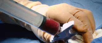
Before the procedure, a special contrast agent, Urografin, is injected into the patient’s vein. On an x-ray, this composition is visualized more clearly than the vessels and surrounding tissues. The amount of contrast depends on the patient's weight. In the case of Urografin, the dose used is calculated based on the ratio of 1 g per 1 kg of body weight. During the procedure, the specialist monitors the patient’s condition and the penetration of the contrast agent into the kidneys. The device records the moments of interaction of contrast with the patient’s organs - X-ray images are obtained that are used to make a diagnosis. The procedure lasts about half an hour. In rare cases, x-rays can take up to an hour and a half (in case of serious pathologies).
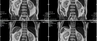
The first picture is taken 3 minutes after using contrast, the second – after 7 minutes, the third – after a quarter of an hour. If necessary, the number of shots is increased.
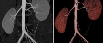
The urogram shows a clear shadow, which makes it possible to recognize all obstacles to the urinary outflow. New growths and wall defects can be examined. The doctor assesses the condition of the patient’s kidneys, their outline, structure.
During the examination, the patient is in a lying position. The exception is cases of suspected organ prolapse - the patient will need to stand for a while to determine the degree of displacement of the kidney.
What is the basis for obtaining information during the study?
These two words: “excretory” and “urography” are the main essence of the method. Excretory means “the release of a substance,” and urography means that the release occurs through the kidneys, and the whole process is recorded in separate images (graphy), or sequentially and dynamically (scopy) using an X-ray machine. This is only possible when the substance, filtered by the kidneys and then entering the urinary canals, is contrasting, that is, able to clearly stand out in the picture against the background of all other internal organs.
Its movement through the blood vessels after intravenous administration occurs very quickly, and within 5–10 minutes the contrast enters the renal arteries and then the kidneys. Filtering in the renal glomeruli, the contrast agent becomes an integral component of urine, begins to move through the ureters, fills the bladder and is then excreted through the urethra during urination.
During this entire process, individual moments of which are recorded by an X-ray apparatus at the marks of 5–7 minutes, 12–15 and 20–25 minutes, as well as at 46 and 60 minutes according to indications, contrast makes it possible to determine all kinds of pathological conditions of the urinary organs: defects of the anatomical or functional in nature. Therefore, excretory urography is indicated for a wide range of patient complaints and is quite common in adults and children to confirm or exclude various diseases of the kidneys, ureters, bladder, and urethra.
With excretory urography, all urinary organs are assessed
In addition to obtaining this data, the signs identified from x-rays will help the specialist learn about the condition of the organs adjacent to the kidneys and bladder. This is especially important if the doctor suspects a negative effect on the urinary system.
In order for the information to be as reliable and useful for diagnosis as possible, it is very important to choose a contrast agent that does not have a negative effect on the body of the subject.
Preparatory stage: recommendations on the topic
Preparation for the examination begins with a medical consultation - the doctor gives directions for taking the necessary tests. Immediately before the procedure, the patient will have to undergo a number of activities:
- A few days before using a contrast agent, you must avoid eating foods that can disrupt the functioning of the gastrointestinal tract and kidneys (legumes, sweets, fresh fruits and vegetables).
- 8 hours before the x-ray you should refuse food and cleanse the intestines with an enema.
- At the same time, you need to drink a lot of fluid. If the patient has eaten a product that caused gas formation, it is worth drinking charcoal in accordance with the attached instructions.
- In the morning before the examination, you cannot have breakfast; if necessary, you can do an enema. For children under one year old, morning feeding can be replaced with a warm drink.
- Before the x-ray, the doctor takes the patient's blood for analysis and questions him about recent medication use.
- In case of emergency urography, the patient is introduced to the list of possible risks and receives written consent for medical procedures.
- If the patient is excitable, preparation for the examination also includes taking sedatives.
It is important to know! Specialists, like the patient, must prepare for excretory urography of the kidneys - take a test to determine the patient’s sensitivity to the contrast agent. 1 ml of the composition is administered, and doctors monitor the patient’s condition for a couple of minutes. In the absence of allergic reactions, further use of the drug for its intended purpose is permitted. If an allergy appears, they resort to using alternative research methods.
Preparation for excretory urography
In order for the diagnosis to be successful and show the most accurate result, you need to prepare for it. 4 days before the examination, the patient is recommended to donate blood for biochemistry to assess the condition and performance of the kidneys.
After contrast is administered, the kidneys accumulate it and filter it into urine. If the organ is not functioning well, the drug will be excreted slowly. There is a threat of acute renal failure.
On the eve of the procedure (after 24:00), you should not eat or drink water or other liquids. The doctor also prescribes the laxative Duphalac or a cleansing enema. If the patient has type 2 diabetes, he should stop taking metformin a couple of days before the urography.
Because this substance in combination with contrast negatively affects the functioning of the kidneys.
More about the results
Formulating the results of the examination, the doctor analyzes the location and shape of the organs and notes the speed at which the contrast agent is removed. The specialist assesses the condition of the parenchymal structures and the pyelocaliceal system. Obstacles to the outflow of urine are determined, and the degree of fullness of the pelvis is determined.
Urography can also detect kidney stones. They are noticeable in the final images, because at the beginning of the process, kidney stones are darkened by contrast. If the injected substance does not move through the organ, the kidney may be absent, have an underdeveloped structure, or be blocked by a stone.
What diagnostic data can be obtained from the study?
According to the accepted methodology for performing urography with a contrast agent, a series of x-rays are taken at certain time intervals, then their description is made. Thus, the first overview photograph can be taken either before the administration of contrast, or immediately after it. Next, the image is obtained at 5–7, 12–15, 20–25 minutes, taking into account the physiological norm of complete removal of iodine-containing substances 30 minutes after injection. According to indications (older age, diagnosed renal pathologies), photographs can be taken later, since excretion occurs more slowly.
These time periods were not set randomly. They indicate a certain stage of the spread of contrast through the kidneys and urinary canals. The survey image evaluates the location of the kidneys, their number, the location of the ureters and bladder, and the course of the urethra. Already at this stage, it is possible to ascertain doubling or hypoplasia of the kidneys, nephroptosis, the presence of stones, abnormal structure or location of the bladder and other parts of the system. In addition, the presence of free gas outside the intestine, in the abdominal cavity, is determined.
Filling of the kidneys and ureters with contrast is determined within 10–15 minutes of the study
The next stage (5–7 minutes) means the penetration of the contrast into the kidneys. The image clearly shows the renal calyces. Continuing to be secreted (excreted) by the kidneys, the contrast agent fills all the calyces and pelvis (12–15 minutes) and begins to move through the ureters. An image taken at 20–25 minutes shows the filling of the bladder. As a result, a series of images allows the doctor to draw a conclusion not only about the patency and anatomy of the urinary tract, but also about the state of the excretory function of the kidneys, which is assessed by the time of excretion of iodine-containing compounds.
List of contraindications
Despite the increased effectiveness of excretory urography of the kidneys, this diagnostic method has a number of significant contraindications:
- Kidney (liver) failure.
- Pregnancy, breastfeeding period.
- Inflammation of kidney tissue.
- Thyroid diseases.
- Patient intolerance to iodine.
- Pheochromocytoma.
- Problems with blood clotting.
- Diabetes (type 1, 2).
- Autoimmune diseases.
- Previous circulatory disorder (heart attack, stroke).
- Multiple organ failure.
- The patient’s serious condition does not allow him to remain in one position for a certain period of time.
The presence of any of the above circumstances precludes the examination. The doctor may rely on alternative, albeit less reliable, diagnostic methods. These include computed tomography, magnetic resonance imaging, and ultrasound.
List of adverse reactions and complications
As is the case with other methods of medical examination, the described manipulation involves a number of side effects. More often, unpleasant manifestations bother the patient exclusively during the X-ray process. Among them:
- metallic taste in the mouth;
- hot flashes;
- dizziness;
- nausea;
- burning sensation in a vein.
All of the above conditions are considered normal. The patient’s well-being improves without taking additional measures.
In the case of the described procedure, there may be options for delayed complications. They usually appear one day after urography. Such phenomena are recorded very rarely and can be local or general.
The “provocateur” of the first type of complications is the puncture of a vein, carried out for the purpose of introducing a contrast agent. The puncture site turns into a hematoma, and phlebitis may develop (the development of an inflammatory process in the venous wall in the injection area).
General complications are discussed in case of incorrect analysis of the patient’s contraindications for X-ray examination. The list of complicated conditions of the patient is given in the table below:
| Complication | Description | Risk group |
| Allergic reaction to contrast | Cough, rash, seizures | Patients suffering from bronchial asthma and other allergic diseases |
| Hemodynamic disorders | A sharp increase in blood pressure | Patients suffering from nephrotic hypertension |
| Kidney failure | Occurs due to increased stress on the kidneys. Manifested by pain in the lumbar region, increased levels of urea, creatinine and hypoproteinemia in the blood. Threatens to develop into uremic coma. | Patients with kidney and urinary system diseases |
All of the common complications mentioned require immediate medical attention.
Side effects
Despite the fact that with proper preparation and under the close supervision of experienced physicians, the procedure is quite safe, side effects may occur after it is performed.
Side effects include the following:
- after the procedure, the patient may feel a taste of iron in the mouth;
- in some cases, a rash may be observed on the patient’s skin;
- after the procedure, the patient may feel very thirsty and dry mouth;
- slight swelling of the lips is a rather rare pathology after urography;
- a contrast agent can lead to tachycardia (rapid heartbeat), which soon stops and the person notices the rhythm of the heart muscle that is familiar to him;
- during urography, as well as after its completion, the patient’s blood pressure may drop significantly;
- the most severe and dangerous consequence after the procedure is the appearance of liver failure (even if the patient has never previously complained of problems with the body’s main barrier – the liver).
Since the side effects are quite significant, it is worth noting once again that intravenous urography must be carried out under the close supervision of experienced doctors and all prescribed recommendations must be followed. If you feel unwell or have complications after urography, you must immediately notify your doctor.
Features of the study in children
Excretory urography of the kidneys is safe for the child’s body. The mechanism of its implementation in the case of children is similar to the principle of x-ray examination in adults. The dose of the contrast agent used is determined very carefully: it is calculated based on the child’s body weight, the condition of the patient’s kidneys and liver.
Small children rarely manage to stay in one position for a long time, so the length of medical manipulation is reduced. In order to minimize the likelihood of allergic reactions, the child is prescribed antihistamines before the study.
Patient preparation
The technique requires certain preparation, which should begin 3 days before the scheduled urography. Not only the information content of the procedure, but also the safety of the patient depends on compliance with the recommendations, so compliance with the instructions is mandatory.
Preparation for intravenous urography:
- Anamnesis collection.
- Cleaning the intestines from feces and gases (washing, enema). The procedure must be done twice - in the evening, on the eve of the examination, and 3 hours before the appointed time.
- In 3 days you need to switch to a diet that prevents increased gas formation. It is necessary to exclude baked goods, confectionery products, carbonated drinks, fresh vegetables and fruits, fermented milk products, and legumes.
- The day before the test, limit the amount of liquid you drink - this will increase the concentration of urinary sediment.
- 12 hours before the procedure, take activated charcoal, which will reduce the likelihood of gas accumulation in the intestines.
- On the day of urography, a light snack is allowed, excluding foods that are too high in calories and dishes that increase gas formation.
- If the patient is anxious or afraid of manipulation, he is prescribed sedatives in individual dosages.
Preparation is necessary to obtain highly accurate data and minimize the risk of complications during contrast fluid administration. Measures before urography are aimed at preparing the patient and are complex not only because of the multi-stage nature, but also because of the individual characteristics of each person.
Points to pay attention to:
- Bedridden patients swallow a large amount of air, so they are recommended to be in an upright position more often before the procedure.
- Diet during the preparation phase is important for young people.
- Elderly people and patients with intestinal atony require cleansing enemas for a quality diagnosis.
The use of iodine-based products impairs the liver's ability to neutralize gases - this must be taken into account in the period after the examination. After the diagnostic procedure, it is recommended to drink plenty of fluids, which will speed up the removal of contrast from the patient’s body.
What do patients think about the procedure?
Adult patients who, according to strict indications, underwent kidney urography using contrast, recognized the real need for medical manipulation. They adhere to this opinion, despite the amount of radiation exposure and the possibility of allergic reactions after the procedure. The majority of parents of the children surveyed also responded positively.
Periodic improvement of methods for diagnosing diseases of the kidneys and urinary system does not lead to the movement of excretory urography to the background. This type of x-ray examination is called the “gold standard” for identifying most specialized ailments. According to forecasts, the procedure will not give up its positions for a long time. Such an advantageous position of urography was ensured by the safety, accessibility and ease of its implementation.
( 10 ratings, average: 4.80 out of 5)