Which method of diagnosing an aberrant artery to choose: MRI, CT, angiography
Selection method
- Often, an aberrant artery is diagnosed accidentally
- CT may be considered as the method of choice.
Is MSCT of neck vessels informative for aberrant carotid artery?
- Soft tissue structure in the hypothymlanic region
- Absence of a vertical segment of the ICA
- Expansion of the tympanic tubule
- A defect is visualized in the bone plate between the middle ear cavity and the horizontal segment of the carotid canal.
What conventional and MR angiography will show
- The ectopic vessel narrows at its entry into the base of the skull
- This vessel (aberrant ICA) is located laterally and posteriorly compared to the typical location.
Palpation
Palpation and examination of the subclavian artery (palpation) is carried out according to the pattern of palpation of the apical impulse, that is, with three or two fingers. First, the arteries at the edge of the sternocleidomastoid muscles above the collarbones are examined. Then a transition is made to the area of the depth of the subclavian fossae under the collarbones at the edges of its deltoid muscles. The examination is carried out very carefully, using the method of applying fingers and pressing on the soft tissue in the area of the externally examined area.
In a healthy person who is at rest, the subclavian arteries will not be palpated, or their pulsation will be subtle. This is explained by their sufficient depth. You can feel a strong pulsation in people with poor development of the muscle tissue of the shoulder and neck, after physical exertion, emotional shock, as well as in asthenic patients.
With pathology of the subclavian artery, its pulsation is clearly manifested. This phenomenon can be observed with aortic insufficiency and hyperkinetic type of hemodynamics. With a vascular aneurysm, pulsation is usually felt in the supraclavicular area, slightly limited (2-3 cm). The weakening of the pulsation of these arteries can be accurately assessed by palpating them simultaneously using both hands. This may be due to a violation of their patency (thrombosis, compression, atheromatosis) or if there is an anomaly - an aberrant right subclavian artery.
What would the attending physician want to know?
- When planning surgical intervention in this area, the operating surgeon must be aware of the presence of an ectopic vessel in the surgical field.
Physiological (a) and ectopic (b) variants of the structure of the ICA (cited from: Lo et al; with modifications).
a Normally, the petrous part of the ICA consists of vertical and horizontal segments. The inferior tympanic artery, arising from the ascending pharyngeal artery, passes through the tympanic canal and in the region of the promontory (promontorium ) forms an anastomosis with the carotid tympanic artery, which, in turn, passes through the carotid tympanic canal.
b With aplasia of the cervical segment of the ICA, compensation of blood circulation is carried out by increasing the intensity of blood flow through the dilated inferior tympanic and carotid tympanic arteries.
The vertical segment of the carotid canal is absent.
All about the kidney vessels
The vessels of the kidneys, unlike the arteries and veins of other organs and tissues, experience a double functional load.
They transport and drain blood not only to nourish the organ, but also to provide one of its most important functions - filtering blood from metabolic products and toxins.
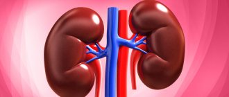
Human kidneys
In addition, the kidneys are able to evaluate the pressure of the fluid entering them through the vessels and thereby control the level of blood pressure.
For these reasons, disturbances in the blood supply to the kidneys affect the entire body in one way or another.
In order to understand how the kidneys function and what importance their adequate blood supply has for the body, it is necessary to at least generally understand the anatomical structure of this organ and the vascular structures surrounding it.
The kidneys are a paired bean-shaped organ located in the retroperitoneal space - in other words, their projection to the surface will be on the back of the lower back at the edge of the ribs.
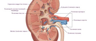
The renal arteries depart directly from the aorta, which go to the “gate” area of the kidney. Already immediately at the “gate” the artery is divided into several branches.
Further division leads to the formation of an extensive vascular network within the organ.
This network (more precisely, its arterial section) can be divided into two groups of vessels.
Some are designed to do the same thing as all arterial capillaries in the body - to transfer oxygen and nutrients to the kidney tissues. The other part of the arterioles goes to the functional units of the organ - nephrons.
In the nephron glomerulus, blood is filtered to form the so-called primary urine.
In this way, the main function of the kidneys, which is generally known to most people, is carried out - purifying the blood with the formation of excretory products (urine).
From the tissues and glomeruli of nephrons, vessels carrying blood that does not contain oxygen - venules - depart. They gradually merge with each other, forming the renal vein at the portal of the organ.
It then drains into the inferior vena cava.
Renal vascular disorders and diseases
All pathologies that affect the blood supply to the kidneys can be divided into two large groups - congenital and acquired.
Vascular pathology of the kidneys
The first include diseases that were caused by improper formation of organs during intrauterine development.
Poor ecology and bad habits of the expectant mother can have a detrimental effect on the health of her baby - he may develop an accessory renal artery, multiple arteries, arteriovenous fistulas, and stenoses.
All this can often be detected during routine ultrasound monitoring of a pregnant woman, and this condition can be corrected immediately after birth.
Consequences of congenital anomalies
Anomalies in the development of the renal vessels can lead to a condition where one of the accessory branches of the artery can partially compress the ureter. This makes it difficult for urine to flow out of the renal pelvis, and it gradually stretches due to stagnation and fluid accumulation.
In this case, the main substance of the kidneys begins to be compressed by the growing pelvis on one side and the connective tissue capsule on the other.
Changes in the blood vessels of the kidneys
Pressure leads to the death of nephrons and an increase in the symptoms of renal failure - then doctors call this condition hydronephrosis.
In addition to the described scenario, the presence of congenital anomalies of the renal vessels increases the risk of developing urolithiasis and inflammatory lesions of the urinary tract.
Renal artery stenosis
In an adult, renal vessels most often suffer from stenosis - narrowing of the lumen due to atherosclerosis, the consequences of fibrotic inflammation, and thrombus formation.
Kidney pathologies
Renal artery stenosis is especially dangerous - this condition leads to a decrease in blood flow to the organ.
Experiencing a lack of nutrients and oxygen, the kidney gradually loses nephrons, which are replaced by connective tissue.
The appearance of the organ changes - it becomes smaller in size, lumpy, and wrinkled. The phenomena of its deficiency are also increasing - however, something else is dangerous for the body at this stage.
The thing is that the kidneys contain receptors that control blood pressure and the number of red blood cells in it.
With stenosis, less blood enters the renal vasculature, which they regard as a lack of pressure and red blood cells. In addition to the filtration function, these organs secrete some hormones - renin and erythropoietin.
The first promotes a strong increase in blood pressure, while the second stimulates the formation of new red blood cells in the red bone marrow. Their release occurs when blood pressure drops and anemia occurs, respectively.
With stenosis, the death of nephrons is accompanied by colossal secretions of these hormones.
As a result, a person’s blood pressure will increase, and it is not possible to reduce it with a number of medications.
At the same time, the number of red blood cells and hemoglobin in the blood begins to increase - this makes the blood thicker (as experts say, the rheological properties of blood deteriorate), and it becomes more and more difficult for it to flow through narrow capillaries.
Tissue nutrition is disrupted, the load on the heart and blood vessels increases greatly, and the risk of thrombosis and associated heart attacks and strokes increases.
Diagnosis of renal vascular disorders
Medical specialists have learned to determine renal vascular damage using a variety of indirect methods.
For example, a combination of such manifestations as high blood pressure, erythrocytosis and reduced diuresis (daily volume of urine) indicates renal artery stenosis.
However, indirect signs are indirect because they do not provide a 100% guarantee of the correct result.
For this reason, various medical imaging techniques have been developed to identify such diseases.
Kidney ultrasound
The simplest and most widespread method for diagnosing kidney diseases and examining its vessels is ultrasound examination (ultrasound).
This technique involves irradiating organs with very high-frequency sound vibrations - the waves are reflected from areas of different densities and are recorded by the device, creating an image.
To diagnose the kidneys, a simple ultrasound is sufficient, while to study the vessels, special equipment is needed - a Doppler scanner.
This device not only creates a “picture” of the state of the internal organs, but also determines the direction of fluid flow (in this case, blood) and its speed.
With its help, you can see in great detail the stenosis of the artery or any of its additional branches; determine at what speed the blood enters the organ, whether there are turbulences and areas with reverse flow.
The only disadvantage of this method is that it will not be able to visualize stationary or very slowly moving fluid (as happens with severe stenoses).
MRI
Another, more troublesome and expensive method of examining the renal vessels is the use of contrast agents. Actually, this is a whole group of methods.
For example, by introducing an iodine-based substance into the blood (it does not transmit x-rays), you can see stenosis and other vascular disorders using simple radiography, digital fluoroscopy, and computed tomography.
And when using similar compounds based on gallium and gadolinium, it is possible to perform contrast magnetic resonance imaging (MRI) and see the structure of the kidney vessels in full detail and in three-dimensional format.
At the moment, these are the most advanced and accurate methods for examining the renal vessels.
It is important to remember that violations of these formations are very dangerous for the entire body, so you should listen to your body. Especially if you experience symptoms such as:
- headache and increased blood pressure;
- lower back pain;
- changes in the color, quantity and other properties of urine;
- swelling on the face in the morning.
All this may be a sign of damage to the kidneys or their blood vessels.
In such a situation, do not neglect visiting a specialist - in the early stages of many pathologies, treatment can quickly and permanently improve a person’s condition.
Source: https://promoipochki.ru/anatomiya-pochek/vse-o-sosudax-pochek.html
What diseases have symptoms similar to aberrant internal carotid artery
Dehiscence (non-fusion) of the jugular bulb
— Protrusion of the jugular bulb into the middle ear cavity is determined
— Increase in the size of the jugular fossa
— Defect at the base of the hypotympanic area
Tumor of the tympanic glomus
— Focal effect of soft tissue space-occupying formation in the middle ear cavity
Tumor of the jugular glomus
- Widening and erosion of the jugular fossa and, in rare cases, also of the petrous bone
Cholesterol granuloma
— Effect of soft tissue space-occupying formation with destruction of surrounding bone structures
— TI-weighted imaging typically shows increased signal intensity
Treatment method
A disease such as stenosis can be treated with medication, in its mild form, interventionally and surgically. But the main methods of therapy, according to experts, are bypass surgery and stenting. These treatments have been used for a very long time and have an excellent success rate during the procedure.
Bypass surgery
If stenosis is detected in the 2nd section of the artery, bypass surgery is indicated. If the ipsilateral common carotid artery is damaged, crossover shunting is preferred. This method of surgical intervention does not injure the patient’s tissues and organs, does not require the use of general anesthesia, takes little time and does not cause serious postoperative complications. Before performing it, it is necessary to perform an ultrasound.
If the great subclavian artery is damaged on the left or on both sides, then its reconstruction in the affected area will first be necessary. If the operation is unsuccessful, repeated intervention is difficult. Contralateral lesions of the subclavian vessels require preliminary elimination of the style syndrome, only then can bypass surgery be started. Reconstruction of the damaged section of the artery is possible only with non-regressive vertebrobasilar insufficiency. All surgical interventions, be it bypass surgery, stenting and others, are not carried out without a complete preliminary examination of the patient and an accurate diagnosis.
Stenting
This method is indicated for patients who have a hypersthenic physique and a special topography of their subclavian arteries. The first section of the artery in such people is difficult to feel. The stenting method is very convenient and significantly prevails over surgical abdominal intervention. During this gentle process, no changes occur in the arteries, and body tissues are not injured.
Using stenting, doctors increase the lumen of the affected vessel. For this purpose, a catheter and a balloon-shaped stent are used. All procedures are performed under local anesthesia. The movement of the stent through the artery occurs under the supervision of an experienced specialist who regulates its location. Having reached the narrowing area, the device opens. If the stent is not open enough, angioplasty is performed. The total operation time is no more than 2 hours.
Tips and mistakes
Due to the similarity of imaging results, in some cases a glomus tumor or any other tumor of the middle ear cavity may be misdiagnosed, which is associated with significant risks in terms of treatment tactics.
Aberrant ICA. Unenhanced CT (bone window), axial projection (a), MRA in M1P mode with contrast (b) and DSA (c). The horizontal segment (arrowheads) of the ectopic right ICA protrudes into the middle ear cavity without evidence of cortical covering (a). The vertical segment (bifurcated arrow) is narrower than on the opposite side (b, c) because the ectopic vessel enters the petrous bone through the tympanic canaliculus.
Departments and functions
The surface location of this vessel is very convenient for puncture. Subclavian artery catheterization is also often performed in this area of the neck. Experts give preference to this area because it is accessible, due to its anatomical features, the artery has a more than suitable lumen diameter and a stable position.
During catheterization, the placed catheter will not come into contact with the walls of the vessel, and the drug that will be administered through it will quickly reach its target, actively influencing hemodynamics.
The main sections of the subclavian artery are three sections:
- Interstitial space. The vertebral and paired arteries depart from it;
- Costocervical trunk;
- Branching of the transverse cervical artery.
The subclavian vessel, located in the 1st section, passes into the skull. Its function is to supply blood to the brain and neck muscles. The internal mammary artery supplies blood to the thyroid gland, diaphragm and bronchi. It is divided into the overhanging intercostal vessel and other adjacent arteries.
Complications
Although such operations cannot be called complex, they still have a fairly long rehabilitation period. After stenting, it is recommended to take painkillers, since the puncture sites and incisions of the soft tissues and arteries may hurt. Postoperative complications are extremely rare, since before the procedure the patient undergoes a complete examination of the entire body (ultrasound, etc.). But still, the body’s reaction under certain circumstances can be unpredictable (for example, if there is a defect - an aberrant subclavian artery).
After stenting, the patient may experience:
- Allergy to drugs;
- Temperature increase;
- Headache;
- Wound infection;
- Air embolism;
- Stent migration;
- Bleeding at puncture sites;
- Arterial thrombosis;
- Neurological complications.
Interventional therapy of stenosis and other diseases of the subclavian arteries using stenting and agioplasty is a modern minimally invasive measure. Such effective procedures are carried out in a very short time and do not require long-term hospitalization. It is enough to first undergo an ultrasound and pass the necessary tests.
Vessels
Artery Meningeal Middle: Groove
Feb 06, 2020 Kokh V. A.
5709
Vessels
Thoracic Internal Artery: Topography
Feb 06, 2020 Kokh V. A.
7718
Vessels
Immediate results of surgical treatment of patients with aortic arch anomalies
We assessed the immediate results of surgical treatment of patients in all study groups based on survival rates and the incidence of various types of postoperative complications.
In groups 1 and 3 of the study, the immediate results of treatment were also assessed based on control Doppler ultrasound of the arteries of the lower extremities and determination of the blood pressure gradient between the upper and lower extremities. We considered it necessary to begin the analysis of the immediate results of surgical treatment of patients in the three study groups by studying a number of intraoperative factors that, in our opinion, influence the incidence of postoperative complications. We included the above-mentioned factors: the type of operation performed, primary or repeated reconstruction of the thoracic aorta and its branches, time of complete clamping of the aorta, volume of intraoperative blood loss, the need for simultaneous clamping of both subclavian arteries, the need to use artificial circulation and perfusion time.
In addition to the listed intraoperative factors, the immediate results of surgical treatment were studied taking into account a number of initial indicators - the age of the patients, the type of congenital anomaly of the aortic arch and concomitant pathology of the thoracic aorta, the initial pressure gradient in the area of narrowing of the aorta, and the presence of concomitant diseases.
This chapter provides a comparative description and statistical processing of the initial indicators of the patients' condition, features of the surgical process and immediate results of treatment in patients of all study groups.
The study groups (No. 1 and No. 3) also did not have statistically significant differences in the frequency of repeated surgical interventions on the thoracic aorta (p 0.05).
In the main group of the study, a higher frequency of extra-anatomical aorto-aortic bypass grafting from the ascending aorta to the descending aorta was revealed (6 in the main group and 1 in the control group 3), however, no statistically significant difference was obtained for this indicator either (p 0.05) .
It should be noted that the time of aortic clamping meant only the time of complete clamping of the aorta; the time of parietal clamping of the aorta, for example, when performing aorto-aortic bypass surgery or parietal clamping of the aorta during implantation of the subclavian arteries was not taken into account.
In the main group (group No. 1), 31 patients were operated on under conditions of complete aortic clamping, and the exact time of aortic clamping was established in 21 patients from the main study group; in 7 cases, operations were performed with parietal aortic clamping. In group No. 3, complete cross-clamping of the aorta was performed in 39 patients, excluding one patient who underwent aorto-aortic bypass surgery from the ascending aorta to the descending aorta; the exact time of aortic cross-clamping was established in 100% of cases.
From the table above (Table 4.2) it is clear that in group No. 3 (control group), operations were performed somewhat more often under conditions of artificial circulation (10 cases, versus 6 cases of CPB in the main group), however, in this indicator, between groups No. 1 and No. 3 no statistically significant difference was obtained.
Such intraoperative indicators as the average time of aortic clamping, the volume of intraoperative blood loss and the time of artificial circulation in patients of groups 1 and 3 did not differ statistically significantly (p 0.05).
Among the initial factors that determine the technical features of the operation, as well as prognostic factors in terms of the development of spinal ischemia, one should highlight such an indicator as the systolic pressure gradient in the area of narrowing of the aorta. When comparing the average pressure gradient in the area of the aortic arch or isthmus, as well as the number of patients with a blood pressure gradient less than 40 mm Hg. Art. in groups 1 and 3 we also did not find a statistically significant difference (P 0.05).
It should be noted that in some patients who did not have a pressure gradient between the upper and lower extremities when measured using the Korotkoff method (with the origin of both subclavian arteries distal to the zone of aortic narrowing), a direct gradient in the narrowing zone was detected during angiographic examination and tonometry.
Summarizing the above, we can conclude that the groups of patients with pathology of the thoracic aorta we studied (No. 1 and No. 3) did not differ statistically significantly in most indicators of the operational process that influence the immediate results of surgical treatment and in the most important indicators of the initial condition.
As features of the surgical treatment of patients of the main group in comparison with patients of groups 2 and 3 (Fig. 47), the following can be noted: in the main group, extra-anatomical aorto-aortic bypass surgery from the ascending aorta to the descending aorta was more often performed - 15.8% in the main study group and in 2.5% of patients in group No. 3.
Only in the main group of patients was there a need for simultaneous clamping of both subclavian arteries, which was required in 39.5% of patients (n=15) of group 1; in groups No. 2 and No. 3, simultaneous clamping of both subclavian arteries was not performed. In 10% of patients in group 3 (n=4), simultaneous clamping of the left subclavian and common carotid arteries was required to adequately perform the operation.
the frequent need to perform surgery through right thoracotomy is 23.7% (n=9); in groups 2 and 3, operations through right thoracotomy were performed in 20% (n=2) and 2.5% (n=1), respectively. in the main study group, in 18.4% of cases there was a need for various manipulations on the subclavian arteries, such as prosthetics (n=1), reimplantation into a new orifice (n=4) or resection (n=2), in group 3 of the study prosthesis of the left subclavian artery was performed in 5% of cases, and its reimplantation into a new ostium in 2.5%.
Patient gender: female
Patient age: 25 years
VARIANT ANATOMY OF THE AORTIC ARCH:
ARTERIA LUSORIA
Chernykh A.V.
Doctor of Medical Sciences, Professor, Head of the Department of Operative Surgery with Topographic Anatomy, Voronezh State Medical University named after. N.N. Burdenko of the Russian Ministry of Health
Zakurdaev E.I.
Candidate of Medical Sciences, Assistant at the Department of Operative Surgery with Topographic Anatomy of the Voronezh State Medical University named after V. N.N. Burdenko of the Russian Ministry of Health
Yakusheva N.V.
Candidate of Medical Sciences, Associate Professor of the Department of Operative Surgery with Topographic Anatomy of the VSMU (Voronezh State Medical University) named after. N.N. Burdenko of the Russian Ministry of Health
Sudakov D.V.
Candidate of Medical Sciences, Assistant at the Department of Operative Surgery with Topographic Anatomy of the Voronezh State Medical University named after V. N.N. Burdenko of the Ministry of Health of Russia, surgeon of the BUZ VOKB No. 1 (Voronezh Regional Clinical Hospital No. 1).
annotation
During topographic-anatomical dissection of the fixed corpse of a man who died at the age of 62 years, an aberrant right subclavian artery was discovered. It was located behind the branches of the aortic arch and the organs of the neck. The close location of the right aberrant subclavian artery with numerous organs, blood vessels and nerve trunks can cause various complications during surgical interventions.
Key words:
aortic arch, aberrant right subclavian artery, vascular anomalies.
Relevance of
the problem
Interest in studying the variant anatomy of blood vessels is determined by the need to minimize the risk of intraoperative complications. The most common anomaly of the aortic arch is the aberrant right subclavian artery - arteria
lusoria
. This anomaly occurs in 0.4-2.0% of cases[1][3]. Typically, the aberrant right subclavian artery arises from the aortic arch distal to the orifice of the left subclavian artery and is located behind the great vessels and the esophagus[9]. Rarely are cases of an aberrant vessel located between the esophagus and trachea. The aberrant right subclavian artery can also be located anterior to the trachea[10].
Materials and methods
The fixed corpse of a man who died at the age of 62 was examined. A topographic-anatomical preparation of the anterior neck and mediastinum was performed. Access to the organs and vessels of the neck was performed through two horizontal and one vertical incisions[7][8]. The upper horizontal incision connected the apices of the mastoid processes of the temporal bone and was carried out along the lower edge of the lower jaw. The lower horizontal incision connected the bases of the acromial processes of the scapula and was carried out along the upper edge of the clavicles and the manubrium of the sternum [2] [4]. The vertical incision connected the horizontal ones and corresponded to the midline. A median sternotomy was performed to examine the anterior mediastinum[7].
Research results and discussion
In the case studied, the aortic arch gave off four branches, whereas in typical situations there are three. The first branch of the aortic arch, counting from right to left, was the right common carotid artery. The second and third branches of the aortic arch were the left common carotid and subclavian arteries. The fourth branch of the aortic arch was the aberrant right subclavian artery – arteria
lusoria
(Fig. 1).
Rice. 1. Aberrant topography of the right subclavian artery.
Designations:
1 - aortic arch, 2 - right common carotid artery, 3 - trachea, 4 - esophagus, 5 - left common carotid artery, 6 - left subclavian artery, 7 - aberrant right subclavian artery (
arteria lusoria
).
The abnormal vessel originated from the posterior surface of the aortic arch 0.6 cm distal to the orifice of the left subclavian artery[6]. From left to right, the aberrant right subclavian artery crossed the remaining branches of the aortic arch and the esophagus along the posterior surface. The initial segment of the aberrant blood vessel was located in the left scalenovertebral space, the main trunk was in the retrovisceral cellular space of the neck[5][14]. Next, the aberrant artery penetrated into the right scalenovertebral space, where it gave off the vertebral and internal thoracic arteries.
The data obtained in the study are somewhat at odds with the literature. Thus, on the studied sectional material, the diameter of the superficial epigastric artery at the level of the inguinal ligament was 1.7±0.2 mm, and the vein – 2.1±0.4 mm. It follows from this that the diameter of the studied blood vessels meets the requirements of plastic surgeons for performing microvascular anastomoses.
Conclusion
1. A rather rare variant of the aortic arch, the aberrant right subclavian artery, has been identified and described. In this case, the anomalous vessel originated from the aortic arch distal to the origin of the left subclavian artery and was directed from left to right through the retrovisceral cellular space of the neck.
Literature:
1. Treatment of purulent wounds in patients with diabetes mellitus using electrically activated aqueous solutions / D.V. Sudakov, E.A. Fursova, M.V. Frolov, O.V. Sudakov // System analysis and management in biomedical systems. 2011. T. 10. No. 2. P. 427-429.
2. New data in the treatment of purulent wounds in microsurgical patients // D.V. Sudakov, A.V. Chernykh, E.I. Zakurdaev, A.N. Tikhonov // Central Scientific Bulletin. 2020. T. 2. No. 1 (18). pp. 9-11.
3. Assessing the information content of clinical examination indicators in patients with type 2 diabetes mellitus and purulent wounds / D.V. Sudakov, E.V. Starodubtseva, O.V. Sudakov, V.N. Snopkov // System analysis and management in biomedical systems. 2013. T. 12. No. 4. P. 1163-1165.
4. Experience of autotransplantation of the main nerve trunks of the forearm with the sural nerve against the background of bite wounds of the upper extremities / D.V. Sudakov, A.V. Chernykh, N.V. Yakusheva, A.N. Tikhonov // Central Scientific Bulletin. 2020. T. 2. No. 1 (18). pp. 12-14.
5. Constructing a forecast for the effectiveness of autotransplantation with the sural nerve in microsurgery of the upper extremities in patients with type 2 diabetes mellitus / D.V. Sudakov, A.V. Chernykh, N.V. Yakusheva, N.O. Vasiliev // In the collection: Current issues of modern medicine. Collection of scientific papers based on the results of the III international scientific and practical conference. 2020. pp. 122-124.
6. The problem of antibiotic resistance in microsurgery / D.V. Sudakov, A.V. Chernykh, E.I. Zakurdaev, A.N. Tikhonov // Central Scientific Bulletin. 2020. T. 2. No. 1 (18). pp. 15-17.
7. A rare case of a giant sliding inguinal-scrotal hernia of the bladder / A.V. Chernykh, E.N. Lyubykh, Yu.V. Maleev, E.I. Zakurdaev, A.N. Shevtsov, V.V. Spitsin // Journal of Anatomy and Histopathology. 2013. T. 2. No. 3 (7). pp. 67-69.
8. Sudakov D.V. Analysis of the composition of the microbial flora in patients with type 2 diabetes mellitus after autotransplantation of damaged main nerves of the forearm with the sural nerve / D.V. Sudakov, A.V. Chernykh, N.V. Yakusheva // In the collection: Modern medicine: current issues and development prospects. Collection of scientific papers based on the results of the international scientific and practical conference. Innovation Center for the Development of Education and Science. 2020. pp. 134-138.
9. Sudakov D.V. Modeling the process of treating purulent wounds in patients with diabetes mellitus / D.V. Sudakov // System analysis and management in biomedical systems. 2013. T. 12. No. 1. P. 327-330.
10. Sudakov D.V. Constructing a forecast for the effectiveness of using the Ilizarov wrist extrafocal compression-distraction apparatus in microsurgery / D.V. Sudakov, A.V. Chernykh, N.V. Yakusheva // In the collection: Modern medicine: current issues and development prospects. Collection of scientific papers based on the results of the international scientific and practical conference. Innovation Center for the Development of Education and Science. 2020. pp. 138-143.
11. Chernykh A.V., Vitchinkin V.G., Yakusheva N.V., Maleev Yu.V., Zakurdaev E.I., Bolotskikh V.A., Spitsyn V.V. High origin of the radial and ulnar arteries // Journal of Anatomy and Histopathology. – 2014. – T. 3, No. 3. – P. 63-65.
12. Chernykh A.V., Zakurdaev E.I., Yakusheva N.V., Vitchinkin V.G., Maleev Yu.V., Zakurdaeva M.P., Andrianova K.A., Lazareva O.A. Applied aspects of variant anatomy of the inferior epigastric artery // Journal of Anatomy and Histopathology. – 2020. – T. 5, No. 4. – P. 74-78.
13. Chernykh A.V. Autotransplantation of the sural nerve in microsurgery of the upper extremities in patients with type 2 diabetes mellitus / A.V. Chernykh, D.V. Sudakov, N.V. Yakusheva // Applied information aspects of medicine. 2020. T. 19. No. 3. pp. 107-112.
14. Chernykh A.V. Problems and prospects for studying the topographic anatomy of the parathyroid glands / A.V. Chernykh, Yu.V. Maleev, A.N. Shevtsov // Journal of Anatomy and Histopathology. 2013. T. 2. No. 2 (6). pp. 15-23.
Information about authors:
Chernykh Alexander Vasilievich
– Doctor of Medical Sciences, Professor, Head of the Department of Operative Surgery with Topographic Anatomy of the Federal State Budgetary Educational Institution of Higher Education VSMU named after N.N. Burdenko of the Russian Ministry of Health.
Zakurdaev Evgeniy Ivanovich
– Candidate of Medical Sciences, assistant at the Department of Operative Surgery with Topographic Anatomy of the Federal State Budgetary Educational Institution of Higher Education VSMU named after N.N. Burdenko of the Russian Ministry of Health.
Yakusheva Natalya Vladimirovna
– Candidate of Medical Sciences, Associate Professor of the Department of Operative Surgery with Topographic Anatomy of the Federal State Budgetary Educational Institution of Higher Education VSMU named after. N.N. Burdenko of the Russian Ministry of Health.
Sudakov Dmitry Valerievich –
Candidate of Medical Sciences, Assistant of the Department of Operative Surgery with Topographic Anatomy of the Federal State Budgetary Educational Institution of Higher Education VSMU named after N.N. Burdenko of the Ministry of Health of Russia, doctor-surgeon of the BUZ VOKB No. 1.
VARIANT ANATOMY OF THE AORTIC ARCH: ARTERIA LUSORIA
Chernyh AV, Zakurdaev EI, Yakusheva NV, Sudakov DV
Summary
When topographic anatomical dissection of a fixed cadaver of a man, who died at the age of 62 years, found an aberrant right subclavian artery. It was located behind the branches of the aortic arch and organs of neck. The proximity of the aberrant right subclavian artery with multiple organs, blood vessels and nerve trunks can cause a variety of complications during surgery.
Keywords:
aortic arch, aberrant right subclavian artery, vascular abnormalities.
References:
1. Treatment of purulent wounds in patients with diabetes mellitus using electro-aqueous solutions / DV Sudakov, EA Fursova, MV Frolov, OV Sudakov // System analysis and control in biomedical systems. 2011. V. 10. No. 2. P. 427-429.
2. New data in the treatment of purulent wounds in patients with microsurgical Profile / DV Sudakov, AV Chernyh, EI Zakurdaev, AN Tikhonov // Central Scientific Gazette. 2020. V. 2. No. 1 (18). P. 9-11.
3. Evaluation of informative indicators of clinical examination of patients with type 2 diabetes and purulent wounds / DV Sudakov, EV Starodubceva, OV Sudakov, VN Snopkov // System analysis and control in biomedical systems. 2013. V. 12. No. 4. P. 1163-1165.
4. Experience sural nerve autografting main nerve trunks of the forearm against the backdrop of bite wounds upper limb / DV Sudakov, AV Chernyh, NV Yakusheva, AN Tikhonov // Central Scientific Gazette. 2020. V. 2. No. 1 (18). P. 12-14.
5. Building efficiency forecast sural nerve autografting in microsurgery of the upper extremities in patients with type 2 diabetes / DV Sudakov, AV Chernyh, NV Yakusheva, NO Vasiliev // In: Actual problems of modern medicine. Collection of scientific papers on the results of the III international scientific — practical conference. 2020. P. 122-124.
6. The problem of antibiotic resistance in microsurgery / DV Sudakov, AV Chernyh, EI Zakurdaev, AN Tikhonov // Central Scientific Gazette. 2020. V. 2. No. 1 (18). P. 15-17.
7. A rare case of giant sliding inguinal-scrotal hernia bladder / AV Chernykh, EN Lubih, YV Maleev, EI Zakurdaev, AN Shevtsov, VV Spitsin // Journal of anatomy and histopathology. V. 2. No. 3. 2013. 3 (7). P. 67-69.
8. Sudakov DV Analysis of the composition of the microbial flora in patients with type 2 diabetes after autologous transplantation sural nerve trunk damaged forearm nerves / DV Sudakov, AV Chernyh, NV Yakusheva // In: Modern medicine: current issues and prospects. Collection of scientific papers on the results of the international scientific-practical conference. Innovation Center for Development of Education and Science. 2020. P.134-138.
9. Sudakov DV Simulation of the process of treatment of purulent wounds in patients with diabetes / DV Sudakov // System analysis and control in biomedical systems. 2013. V.12. No. 1. P. 327-330.
10. Sudakov DV Construction of the forecast of effectiveness of use of carpal extrafocal compression – distraction apparatus Ilizarov in microsurgery / DV Sudakov, AV Chernyh, NV Yakusheva // In: Modern medicine: current issues and prospects. Collection of scientific papers on the results of the international scientific-practical conference. Innovation Center for Development of Education and Science. 2020. P. 138-143.
11. Chernyh AV, Vitchinkin VG, Yakusheva NV, Maleev Yu.V., Zakurdaev EI, Bolotskikh VA, Spitsin VV Case report of high origin of radial and ulnar arteries // Journal of anatomy and histopathology. 2014. V. 3. No. 3. P. 63-65.
12. Chernykh AV, Zakurdaev EI, Yakusheva NV, Vitchinkin VG, Maleev Yu.V., Zakurdaeva MP, Andrianova KA, Lazareva OA // Journal of anatomy and histopathology. 2014. V. 5. No. 4. P. 74-78.
13. Chernyh AV Autotransplantation microsurgery sural nerve in the upper limbs in patients with type 2 diabetes / AV Chernyh, DV Sudakov, NV Yakusheva // Applied information aspects of medicine. 2020. V. 19. No. 3. P. 107-112.
14. Chernyh AV Problems and prospects of the study of topographic anatomy of the parathyroid glands / AV Chernyh, YV Maleev, AN Shevtsov // Journal of anatomy and histopathology. 2013. V. 1. no. 2 (6). P. 15-23.
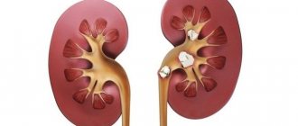
Accessory renal artery: what it is, types, diagnosis and treatment
According to statistics, about 35% of the world's population has diseases of the urinary system. Approximately 30% of all cases are associated with abnormalities in the structure and functioning of the kidneys. For example, accessory renal arteries are often found.
Accessory renal artery
The kidneys perform a vital function: they filter the blood and remove all harmful substances and toxins along with urine. The renal system has a complex structure. Each element of it performs certain functions. An artery delivers blood to an organ. The paired blood vessel supplies the cortex and medulla.
Accessory arteries are a common type of renal vascular pathology. They occur in 80% of all kidney diseases. One vein is observed in 19.2%, two – in 2.1%, three – in 0.7% of patients. This anomaly is more common in women than in men. Usually the defect is diagnosed on the right.
What is this
The accessory artery is the renal artery, which, together with the main artery, supplies the organ with blood. An additional vessel rushes to the lower and upper segment of the kidney. The diameter of this vein is smaller than the main one. Multiple arteries disrupt blood flow and urodynamics. Therefore, in certain situations you need to get rid of them.
Kinds
Accessory arteries are classified by number into:
These varieties do not always lead to pathology. But they are often combined with other renal anomalies. For example, with polycystic, double, horseshoe-shaped, dysplastic or dystrophic kidney.
Reasons for appearance
Deviations from the normal development of an organ are established during embryogenesis. Exactly what factors lead to the appearance of this pathology have not yet been precisely established. Only a hereditary predisposition to the abnormal structure of the bloodstream has been identified. A connection between the disease and the influence of various teratogens on the embryo has also been established.
There is an assumption that bad habits of a pregnant woman and poor ecology can lead to the formation of an additional renal vein in the child.
Symptoms
Usually the pathology does not have pronounced manifestations. Unpleasant symptoms develop only if the accessory artery crosses the urinary tract. This leads to difficulty in the flow of urine from the kidneys. The following symptoms appear:
- Hypertension. A jump in pressure occurs due to the fact that fluid accumulates in the body, blood vessels narrow and blood circulation worsens.
- Hydronephrosis. This condition is caused by a violation of the outflow of urine.
- Thrombosis, bleeding in the intersection area.
- Kidney infarction. Due to prolonged hydronephrosis, the renal parenchyma begins to atrophy. This subsequently causes a heart attack.
- Reducing the daily volume of urine.
- Kidney enlargement.
- The appearance of blood impurities in urine.
- Frequent and painful urination.
- Pain in the lumbar area.
- Formation of conglomerates in the genitourinary organs.
- Nephritis.
- Attacks of renal colic during palpation and physical exertion. Sometimes pain occurs even at rest.
Diagnostics
To detect an additional renal artery, different diagnostic methods are used:
The last method is considered the most accurate. Dopplerography gives a complete picture of the condition of the left and right kidneys, and also monitors blood flow (pressure, direction). But, if the fluid flow is slow, the device will not be able to record its movement. Aortography reveals abnormalities of the solitary artery.