Features of pyelonephritis
Pyelonephritis is the most common kidney disease. The pathology is based on the inflammatory process occurring in the upper parts of the urinary tract. The most common cause is the penetration of pathogenic bacteria into the kidney tissue.
Pyelonephritis can occur in two different forms: in an acute form with pronounced symptoms and in a chronic form with a series of exacerbations and subsidence of the pathological process. Inflammation of the kidneys is quite often combined with anomalies of their anatomical structure and occurs during pregnancy and urolithiasis.
In pyelonephritis, the focus of inflammation is in the calyces and pelvis of the kidneys
Causes of the disease
- constant hypothermia of the body;
- decreased protective function of the body;
- the occurrence of problems with the outflow of urine, this could be: prostate adenoma, ureteral stricture, stones;
- infectious diseases of the urinary system, for example, urethritis, vesiculitis, cystitis or prostatitis;
- a decrease in the level of blood supply to the renal tissue, for example, diseases that contribute to the decrease: diabetes mellitus, renal artery stenosis;
- completed instrumental diagnostics using the following method: cystoscopy, ureteroscopy, ureteroscopy, retrograde urography or bladder catheterization;
- postoperative period regarding pathologies of the genitourinary tract.
Diagnostic ultrasound: principle of the method
The human body is a collection of organs and tissues that have different densities. The kidneys contain a cortex, fluid-filled pelvises, and a large number of blood vessels. The only way to assess the condition of all these anatomical components of an organ is to do an ultrasound.
The method is based on high-frequency mechanical waves produced by an ultrasonic sensor. They spread at different speeds in the structures of the human body, after which they return back to the sensor. The received signals are converted into an inverted visual picture on the device screen.
Ultrasound - sound vibrations with a frequency of more than 20,000 Hertz
Depending on their density (echogenicity), tissues look different during the study. The liquid is reflected on the screen in the form of dark areas, dense structures have a lighter shade. White color indicates stones located inside the urinary tract.
A separate type of ultrasound is Doppler examination of blood flow in the vascular bed of the kidneys. The device's sensor sends a signal that is reflected from moving blood cells and returned back. In this case, on the device screen, the specialist sees a picture of blue and red areas. The first denotes the blood flow moving in the direction away from the sensor. In the second case, the blood in the vessels flows towards the ultrasound source.
Doppler study allows you to evaluate blood flow in the vessels
What is the structure of kidney tissue?
In the normal state, the parenchyma is heterogeneous, and lighter and darker areas are visible on ultrasound. This is due to the structure of the parenchymal tissue itself, which acts as a filter for impurities contained in the blood. The parenchyma consists of pyramids, the inner parts of the tissue adjacent to the renal calyces, and the outer layer, which on ultrasound appears as less dense, light-colored tissue.
If pathological changes are present in the organ system, the parenchymal tissue will be changed, enlarged, or heterogeneous in whole or in part. The structure of the tissue is also related to the fact that cysts and tumors most often arise in this area of the organ, and during ultrasound diagnostics, the first changes, signs and symptoms of the disease can be identified by the condition of the parenchyma.
The outlines of the kidneys themselves should be smooth and clear. A blurred boundary, which is detected on ultrasound, signals the development of inflammation in the tissues and requires additional diagnostics.
Indications for the study
For pyelonephritis, the study can be performed several times. The doctor will prescribe an ultrasound in the following cases:
- the presence of clinical signs of pyelonephritis: fever, pain in the lower back, changes in the nature of urine;
- results of laboratory tests of blood and urine characteristic of pyelonephritis;
- suspected formation of stones in the kidneys or urinary tract;
Suspicion of kidney stones - indication for diagnostic ultrasound
- the need to monitor the effectiveness of treatment measures for kidney inflammation;
- routine preventive examination for chronic forms of the disease;
- checking the restoration of urine outflow after surgery to remove stones.
Ultrasound of the kidneys - video
Who is more likely to be prescribed an ultrasound by a doctor?
If you feel pain in the abdomen and lower back. For no apparent reason, you have a high body temperature for quite a long time. A general blood test shows leukocytosis, increased ESR, leukemia is shifted to the left, anemia is observed; Biochemical analysis shows that creatinine has increased, as has urea, potassium, and blood serum.
These indicators are especially important for making a diagnosis if you do not yet know exactly which organ is affected? The kidneys' ability to remove urine is impaired. At night you have a frequent urge to urinate. At the same time, you feel pain. Over the course of a day, there was less or more urine, and swelling appeared. There is more or less urine, but its specific gravity is less than it was before. Blood with a high protein content, a lot of bacteria, urate salts, with phosphates, and a lot of leukocytes appeared in the urine.
How is an ultrasound examination of the kidneys performed? The patient is asked to take off his clothes. expose your back. They put sensors on the place where the kidneys are located, move them and look on the screen in what condition is the organ?
Now you know how the kidneys are examined and that pyelonephritis is visible on ultrasound. It can be in acute or chronic form. All that remains is to be examined using ultrasound equipment and treated. How long will the course take? It's different for everyone.
Click the Sign up button and we will find you an ultrasound specialist or another doctor in 10 minutes.
Pyelonephritis accounts for up to 70% of all cases of infectious and inflammatory kidney diseases. To clarify the diagnosis, a whole range of studies is carried out: laboratory, instrumental. Ultrasound of the kidneys for pyelonephritis is performed at the first signs of renal pathology.
Advantages and disadvantages of the method
Ultrasound diagnosis of various forms of pyelonephritis has many advantages:
- simple preparation for the study;
- carried out on an outpatient basis without hospitalization;
- no pain during the examination;
- the possibility of performing ultrasound at any age, including newborns;
Ultrasound examinations can be performed on children of any age.
- availability of kidney research for a patient in any condition, including after surgery;
- no punctures or cuts;
- informative in the diagnosis of kidney inflammation, abnormalities of their structure, urolithiasis;
- no need for pain relief;
- informative value in diagnosing complications of pyelonephritis;
- the possibility of conducting the study repeatedly during the treatment of the disease;
- no side effects or harmful effects;
- the possibility of conducting research during pregnancy.
Ultrasound examination during pregnancy is a safe diagnostic method for mother and fetus
Ultrasound does not have any harmful effects on body tissues, so this research method has virtually no contraindications. However, there are a number of diagnostic features:
- ultrasound does not provide information about the work and functionality of the kidneys;
Ultrasound does not provide information about the ability of the kidneys to filter blood
- Ultrasound is not able to determine the type of bacteria that caused the infectious inflammation.
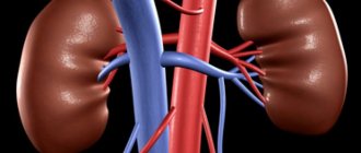
Internal structure of the kidneys
The pyelocaliceal apparatus, which is responsible for the accumulation and removal of harmful substances from the human body. It is here that secondary urine accumulates, which will soon be removed from the body through the ureters and bladder.
The calyces are responsible for storing urine. There can be up to 10 of them inside one organ, 4-6 small and 3-4 large. Large calyxes are adjacent to the pelvis, cavities where urine accumulates. When the pelvis is partially filled, the muscle fibers surrounding the kidney contract and the accumulated urine is discharged into the bladder through the ureter.
When performing ultrasound diagnostics, the doctor examines the internal cavities especially carefully. The most common types of diagnosis include:
- Urolithiasis, that is, improper functioning of the body, due to which salts are deposited in the kidneys, forming stones and sand. This pathology is dangerous because the stone blocks the flow of urine to the ureter, and excess fluid compresses the parenchymal tissue, which atrophies from prolonged compression. In addition, urolithiasis gives the patient a lot of anxiety and discomfort;
- The development of tumors and cysts, which can also compress tissues and blood vessels and disrupt the functioning of individual parts of the kidney or the organ as a whole;
- Narrowing or curvature of the ureter, which leads to delayed urine flow and improper functioning of the entire system. This pathology leads to stagnation of fluid in the pelvis and accumulation of harmful substances in the body.
The ureter itself is examined no less carefully. It may look on the monitor screen like a tube less than 1 cm in diameter and up to 30 cm long. It is hollow inside, which means that on an ultrasound this organ will appear as a tube with light walls and a dark middle. If the ureter is clogged, for example, due to urolithiasis, when a stone comes out, then during ultrasound diagnostics a light spot will be noticeable.
Preparation and conduct of the study
When examining the kidneys using ultrasound, the doctor gets a good picture even without special preparatory measures. However, for a more accurate result, a number of requirements must be met:
- three days before the study, it is necessary to exclude legumes, brown bread, fresh vegetables and sweets from the diet;
Fresh vegetables cause increased gas formation in the intestines
- have dinner the night before the ultrasound no later than seven o’clock in the evening;
- Drinking and eating on the day of the procedure are not limited.
If a simultaneous examination of the abdominal organs and kidneys is planned, the procedure is performed on an empty stomach.
The kidney examination is carried out in several positions: lying, on the side, standing. To improve contact between the device's sensor and the skin, a special gel is used, which can then be removed with a regular napkin.
How is a kidney ultrasound performed?
Ultrasound of the kidneys can often be difficult due to the position of this organ.
Since ultrasound passes freely through soft tissues, but is scattered in the air and does not penetrate bones, it is possible to examine the kidneys in detail only from one side.
In order for the study to be accurate, it must be carefully prepared. First of all, before an ultrasound scan of the kidneys, you should not eat foods that cause gas formation, since ultrasound will not penetrate through the gas-filled intestines and the diagnosis will be uninformative.
Before an ultrasound, you should not eat cabbage, legumes, brown bread, drink beer or carbonated drinks. It is recommended to follow the diet for three days. If the patient suffers from constipation, he is recommended to cleanse the intestines, take a laxative or do an enema before the study.
During the diagnosis, the patient lies on the couch, on his back. The laboratory technician applies the gel to the skin to better glide the sensor and remove air. A transducer that receives reflected ultrasound moves across the skin of the abdomen, creating an accurate black-and-white image of the kidney on a screen in front of the doctor. It is forbidden to perform an ultrasound while standing, as incorrect positioning of the patient may affect the accuracy of the diagnosis and not show the results. The only option in which an ultrasound can be performed standing is if the patient is not feeling well and cannot lie down.
Diagnosis is carried out in a similar way when the patient turns on his back, on his right and left side.
Ultrasound picture of various types of pyelonephritis
The image obtained using ultrasound on the device screen may differ depending on the type, stage of the disease and the presence of complications.
Acute pyelonephritis
In case of acute inflammatory process in the kidneys, the doctor, when conducting an examination, will first of all note an increase in the size of the affected organ. However, it must be remembered that normal indicators are determined using special tables based on the gender and age of the patient. In addition, in some cases, pyelonephritis occurs without changes in the size of the kidneys.
Normal kidney sizes in adults depending on height - table
| Height | Length, mm | Width, mm | Parenchyma thickness, mm | |||
| Left | Right | Left | Right | Left | Right | |
| 150 | 85 | 82 | 33 | 29 | 13 | 13 |
| 160 | 92 | 90 | 35 | 33 | 14 | 13 |
| 180 | 105 | 100 | 38 | 37 | 17 | 15 |
| 200 | 110 | 105 | 43 | 41 | 18 | 17 |
Normal kidney sizes in children depending on age - table
| Age | Right | Left | ||||
| thickness, mm | length, mm | width, mm | thickness, mm | length, mm | width, mm | |
| 1-2 months | 18,0—29,5 | 39,0—68,9 | 15,9—31,5 | 13,6—30,2 | 40,0—71,0 | 15,9—31,0 |
| 3-6 months | 19,1—30,3 | 45,6—70,0 | 18,2—31,8 | 19,0—30,6 | 47,0—72,0 | 17,2—31,0 |
| 1-3 years | 20,4—31,6 | 54,7—82,3 | 20,9—35,3 | 21,2—34,0 | 55,6—84,8 | 19,2—36,4 |
| up to 7 years | 23,7—38,5 | 66,3—95,5 | 26,2—41,0 | 21,4—42,6 | 67,0—99,4 | 23,5—40,7 |
Normally, when moving from a horizontal to a vertical position, the organ can move up to one and a half centimeters. The mobility of the kidney with pyelonephritis is significantly limited.
The ultrasound picture of the elements of the inflamed kidney differs from normal. Usually, in the cortical layer, the pyramids that turn into cups stand out in a darker color. An acute inflammatory process erases these differences. As you recover, they reappear.
In acute pyelonephritis, there is no discernible difference between the cortex and pyramids
When examining the renal vessels using the Doppler method, the absence of blue or red color in the peripheral areas of the cortical layer is noted. Such changes indicate a pronounced decrease in blood flow in the kidney against the background of an acute inflammatory process.
The blood supply to the kidney with pyelonephritis is significantly impaired
Local inflammation in the kidney
Apostematous (pustular) nephritis looks very peculiar on ultrasound examination. The size of the kidneys in this disease may be increased or normal. In the cortex of the organ there are multiple dark areas in which an inflammatory purulent process occurs. Pyramids in apostematous nephritis are not clearly defined. The contour of the kidney is blurred, tuberosity is noted.
With apostematous nephritis, ultrasound reveals many dark areas - abscesses
Kidney carbuncle is another type of acute pyelonephritis, which is characterized by a special ultrasound picture. In the cortex there is an area of increased density of a lighter shade - a carbuncle. As it develops, it acquires a darker color compared to the surrounding tissues. When examining the area of the carbuncle using the Doppler method, a complete absence of blood flow in this area is detected.
A kidney carbuncle appears as a dark area on an ultrasound image.
Chronic pyelonephritis
Chronic inflammation leads to many significant changes. With this type of pyelonephritis, the size of the organ tends to decrease, and gradual shrinkage of the kidney occurs (nephrosclerosis). This situation is characterized by expansion of the pelvis. The thickness of the renal cortex decreases. The extreme degree of expansion of the pelvis, in which the latter has gigantic dimensions, is called hydronephrosis.
Hydronephrosis is an extreme degree of dilation of the renal pelvis
The kidney pyramids also change against the background of long-term inflammation. Their echogenicity gradually increases, and therefore they acquire a lighter shade on the device screen. A peculiar white rim appears around the pyramids - calcium deposition (nephrocalcinosis).
Doppler examination reveals a marked decrease in blood flow in the affected organ. This process especially concerns the renal cortex.
Abnormalities of the kidney structure
Quite often, pyelonephritis occurs against the background of congenital anatomical abnormalities of the kidney structure. Ultrasound diagnostics will provide information about the presence of predisposing factors for the development of the disease.
Quite often there is a situation where there are two kidneys in the body, but their placement differs significantly from the norm. An organ that is usually located in the lumbar region can be localized by a specialist at a much lower level - in the area of the sacral spine.
Pyelonephritis can occur against the background of kidney duplication. In this case, the specialist sees a picture of the presence of two pyelocaliceal systems, supplied with blood by independent vessels. One of the types of structural anomalies is a horseshoe kidney. Such an organ is more susceptible to pyelonephritis, hydronephrosis and urolithiasis.
Horseshoe kidney is a congenital anomaly consisting in the fusion of the lower or upper poles of both kidneys with each other to form an isthmus
Cysts in the kidneys are another common anomaly in the structure of the organ, implying the appearance of cavities containing fluid in it. The ultrasound picture in this case is characterized by the presence of dark areas against the background of a lighter cortical substance. Such formations can fill the entire kidney. This disease is hereditary and is called polycystic disease.
Cysts in the kidneys appear as dark areas on an ultrasound image.
Pyelonephritis with urolithiasis
Kidney stones (calculi) are a frequent companion to the inflammatory process. They have a high density, so they look like light-colored areas on the ultrasound machine screen. A specialist may detect one or more stones in the kidney. In rare cases, the calculus occupies the entire pelvis and is called coral-shaped. Using a modern ultrasound machine, a specialist can identify stones larger than three millimeters in size.
Coral stone occupies the entire space of the renal pelvis
What does a kidney ultrasound show?
Ultrasound diagnostics is considered one of the most accurate research methods that currently exist, and therefore if pathological changes are detected on ultrasound, then with a high degree of probability it can be said that the diagnosis is correct. But only a doctor can identify pathological changes in the system and explain their nature.
What does an ultrasound show:
Kidney sizes. This is one of the first parameters that the doctor and laboratory technician pay attention to. Standard sizes are as follows:
- 100-200 mm length;
- 50-60 mm width;
- 30-50 mm thickness;
- The thickness of the external tissue, parenchyma – up to 25 mm;
- Capsule size – up to 1.5 mm;
- The total weight of one organ is up to 200 grams.
If the size of the kidneys is outside the normal range, the doctor can diagnose a tumor, tissue hypoplasia, or identify other pathology. Most often, the kidneys enlarge in the presence of inflammatory processes, and the decrease in tissue is associated with the patient’s age, and the parenchymal layer usually decreases.
Tumors and cysts most often occur in the parenchyma. Due to its structure, this tissue is looser, softer, and subject to changes, including pathological ones. The opposite effect may also occur: if one kidney was removed, the parenchymal tissue of the other will develop twice as strong.