Diet and medications for kidney stones
For various types of metabolic disorders, it is important to prescribe pathogenetic treatment that affects these processes:
1. In case of disorders of purine metabolism and urate stones, it is recommended to use a diet that helps reduce uric acid levels, drug therapy: xanthine oxidase inhibitors (allopurinol), uricuretics (benzbromarone) and citrate mixtures (blemarene, uralite U).
2. If there is a violation of oxalic acid metabolism and calcium oxalate stones , also use a diet with limited administration of oxalic acid, medications - vitamin B6, magnesium preparations.
3. For hypercalciuria, it is recommended to prescribe thiazides (hydrochlorothiazide) and diphosphonates (ethidronic acid).
4. Treatment of phosphaturia involves a diet low in calcium, using bisphosphonates (ethidronic acid), hydrochloric acid, benzoic acid, ammonium chloride.
5. For phosphate crystalluria and cystine stones - 3 liters of liquid daily, citrate mixtures are the “gold standard” of conservative therapy, providing dose-dependent alkalization of urine without changing the acid-base balance of the blood, antispasmodics and analgesics as indicated.
Studies in laboratory mice have shown a positive effect of L-cystine dimethyl ester (CDME) as an inhibitor of cystine stone growth, making further investigation of CDME viable for the treatment of cystine urolithiasis.
Phosphorus-calcium metabolism. What disorders occur in chronic kidney disease?
The kidneys perform a vital function in maintaining self-regulation processes. The human skeleton contains about 99% of the total amount (Ca) of our body and another 85% of phosphorus (P).
The remaining amount is in the blood plasma in the form of ions and in the form of compounds with protein molecules and acids. Phosphorus, in addition, in nucleic acids is involved in the storage and transmission of hereditary information, and in phospholipids - in performing the functions of nervous tissues and transport of fats.
Interesting fact. Only 0.5% of the total calcium content in our body is subject to metabolism.
In a healthy person, well-functioning self-regulation mechanisms successfully compensate for fluctuations in the content of mineral composition. The exchange of Ca and P normally maintains dynamic equilibrium. The clinical appearance of changes in their metabolism is an unfavorable syndrome of development of chronic renal failure (CRF). Already at the early stages of development, disturbances in metabolic processes are observed.
In severe forms of chronic renal failure, the compensatory level of kidney function is significantly reduced. Changes in the metabolism of Ca and P cause the appearance of certain symptoms and diseases.
Interesting fact. Our body absorbs from 70 to 90% of the phosphorus that comes from food.
Prescribing and following a diet with a low phosphorus content can temporarily stop the compensatory increase in hormone production. With more serious renal dysfunction, using a diet to achieve a balance of Ca and P is no longer enough. It is necessary to prescribe special drugs to bind phosphates ingested with food products. Dialysis helps cleanse the blood. Without appropriate treatment, disorders of the mineral composition of bone tissue can progress. Such situations cause the formation of calcifications, pain in the bones, bone fragility and pathological fractures.
Disturbances in the system of bone-mineral metabolism as a result of CKD are characterized by certain symptoms or their combinations.
- It should be understood that disturbances in mineral metabolic processes are not an isolated phenomenon. They are closely related to the general metabolism in the body. In particular, the homeostasis of calcium and phosphorus is interconnected with protein metabolism and the synthesis of parathyroid hormones. Therapeutic treatment in such situations is complex.
- Thanks to the participation of vitamin D, protein molecules that bind calcium are synthesized, absorbed in the small intestine and converted into a compound suitable for participation in the process of bone formation. With chronic renal failure, kidney cells cease to synthesize the active metabolite of vitamin D in sufficient quantities, which ensures complete regulation of calcium and phosphorus metabolism. Thus, a decrease in vitamin synthesis in kidney cells inhibits phosphorus excretion and leads to the development of hyperphosphatemia.
Interesting fact. Over the past 15 years, the number of patients with chronic kidney disease receiving renal replacement therapy has quadrupled worldwide.
- Increasing vitamin D deficiency leads to impaired mineralization of bone tissue. There is a change in metabolic processes in the skeleton, which leads to a disruption of such important indicators as the balance of mineral composition, changes in the total volume of bones, their linear dimensions and a decrease in strength, that is, renal osteodystrophy. These conditions can cause bone pain or cause a fracture.
- Severe forms of renal failure are characterized by a complex interaction between the functional state of the renal tubular filtering system, the activity of vitamin D, and the production of parathyroid hormones. Thus, a decrease in calcium levels against the background of an increase in phosphates and an increasing deficiency of vitamin D causes secondary hyperparathyroidism - increased production of hormones that help regulate the metabolism of phosphorus and calcium, and subsequent degeneration of the parathyroid glands. Such tissue is already insensitive to the influence of metabolic processes.
It is known that the number of active receptors in the nodes of the affected parathyroid gland decreases by almost 60%.
- Calcification of tissues and blood vessels is a serious complication of chronic renal failure. A critical level of phosphorus and calcium balance leads to the deposition of salts outside the skeletal tissues. In this case, calcium accumulates in the tissues of small vessels, which can cause ischemic changes in the extremities. It is important to control such situations by regulating indicators of phosphorus-calcium homeostasis.
Monitoring regular correction of phosphorus-calcium metabolism parameters and timely treatment of compensatory shifts in hormonal functions is the main component of successful treatment of chronic renal failure, which helps prevent serious consequences of the disease.
Methods for removing kidney stones
All surgical techniques are divided into: remote shock wave lithotripsy, percutaneous and transurethral endoscopic surgery, laparoscopic stone removal, open surgery. There is also a classification according to treatment methods depending on the size of the stone (more or less than 20 mm), its qualities (X-ray positive, infected, uric acid/urate, cystine) and the level of localization in the urinary tract.
X-ray endoscopic methods for stone removal are widely used (transurethral X-ray endoscopic endosurgery (ureterolithoextraction, for example, using a Zeiss loop and Dormia basket, transurethral ureterolithotripsy and lithoextraction, percutaneous X-ray endoscopic surgery (nephrolithotripsy and nephrolithoextraction) and remote shock wave lithotripsy.
Ultrasound crushing of kidney stones
Since the 1980s, the method of extracorporeal shock wave lithotripsy (ESWL) has been developed, which has achieved high results, but the possibility of its use is limited by the size, location, weight and composition of stones, as well as the individual characteristics of the patient. The high effectiveness of treatment was also determined when using the drug tamsulosin after EVL, which allows reducing the use of analgesics after this procedure.
Surgical removal of kidney stones
Among surgical treatment methods, endourological techniques occupy a special place, for example, percutaneous nephrolithotomy, incl. 2-stage ultrasound Doppler-controlled dilatation of the urinary tract, ureterorenoscopy. Open and laparoscopic surgery are also used, which are more suitable in some clinical cases.
Various surgical techniques are used, including pyelolithotomy, nephrolithotomy (including sectional; experiments have been conducted on the use of ultrasound-controlled balloon dilation for percutaneous nephrolithotomy (PCNL), ureterolithotomy.
Removing kidney stones with laser
Laser methods for destroying stones are rapidly developing, especially those that use the optical-acoustic effect (destruction of the stone by cavitation of a bubble of water vapor, which is formed when laser radiation interacts with the surface of the stone). When this effect is used, laser radiation is harmless to surrounding tissues, which sharply reduces the risk of damage to the kidneys or the mucous membrane of the bladder and ureter.
LECTURE NOTES No. 3
Violation of water metabolism.
Violation of acid-base metabolism,
Formation of stones, their types.
Violation of mineral metabolism, K. Na. Ca. R.
Urinary tract and gall bladder stones.
Ca - a violation of mineral metabolism is important when rickets occurs. osteoporosis. It is most often caused by pathology of the parathyroid glands.
K and Na are necessary for the functioning of the heart and kidneys, and the cause of metabolic disorders of these minerals is usually kidney pathology.
P affects brain activity. And it is necessary to remember that the ratio Ca\P = 1\2 is important. Only with this ratio of these minerals will they be normally absorbed by the body.
If mineral metabolism is disrupted, stones can occur in the gall bladder and urinary tract. The composition of gallstones is calcifications, bilirubinates and cholesterol.
The composition of urinary stones is calcifications, phosphates and urates.
Stones can have a radiant or layered structure. They can lead to disruption of the outflow of bile and urine. Thus, their presence can lead to cholecystitis or hydronephrosis.
Acidosis is a disorder that results in excess amounts of acidic compounds. CO2 accumulates in the blood,
(for breathing problems, fever, infections),
which leads to a decrease in the release of CO2 by the lungs.
Alkalosis can also be gas (respiratory) and non-gas (with prolonged consumption of plant foods, toxicosis of pregnant women, poisoning. With alkalosis, a decrease in blood volume occurs, blood flow in the kidneys is disrupted.
Hypohydration occurs with complete water starvation (a person lives for 7-10 days), diabetes insipidus, glomerulonephritis, which leads to heart failure and convulsive conditions.
Overhydration – occurs when there is an excess of water consumption, a decrease in the osmotic pressure of the blood plasma occurs, up to coma. Overhydration leads to edema (ascites, anasarca, hydrothorax, hydropericardium)
Edema, its types – osmotic, oncotic, membranogenic. Edema fluid (transudate) compresses tissues, impairs blood circulation and can lead to the death of the patient
Hydrocele (vacuolization) is the appearance of a large amount of water in the protoplasm or nucleus of a cell, which can lead to destruction of the cell and swelling of the organ. This can occur due to poisoning, water imbalance
ASCITIS
HYDROTHORAX
HYDROPERICARD ANASARCA
1 Necrosis,
definition, causes and mechanisms of development
2. Morphological signs of necrosis: changes in the structure of the cell, nucleus, cytoplasm and tissue.
3. Types of necrosis: 4. Gangrene, its types: bedsores. 5. Heart attack. 6. Outcomes of necrosis. Forms: bedsores, sequestration, heart attack.
7. Atrophy, aplasia, agenesis, cachexia, neurogenic atrophy. 8. Violation of energy metabolism.
9. Disorders of basal metabolism, mechanisms (cellular and extracellular).
Necrosis,
definition, causes and mechanisms of development
necrosis. Necrosis is the death of individual cells. The causes of necrosis can be different: injuries, poisoning with acids and alkalis, toxins, temperature effects, etc.
Morphological signs of necrosis: changes in the structure of the cell, nucleus, cytoplasm and tissue.
Karyopyknosis (cells lose water, nuclei shrink and become denser).
Karyorrhexis (nucleic acids leave the nucleus into the cytoplasm.
Karyolysis - nuclear matter dissolves.
Cytolysis - the entire cell dissolves, a structureless homogeneous mass is formed, called necrotic detritus
, it differs from the surrounding ones in consistency, color, smell (sometimes fetid).A “demarcation line” appears around the focus of necrosis, which is a zone of inflammation
Types of necrosis: direct and indirect; traumatic, toxic, vascular, allergic, neurotrophic, coagulation, collaction. Necrosis may be dry (
coagulation) and
wet
(colliquation). As a result of liquefaction of necrotic masses, a cavity is formed - a cyst.
Gangrene, its types: a special type of necrosis develops due to the ingress of putrefactive flora, and, in contact with the external environment, forms dry gangrene
(lower limbs).
If the tissues are rich in fluid, wet gangrene forms.
Anaerobic or gas gangrene occurs when anaerobes enter the body.
Bedsores (a type of gangrene) are an area of necrosis of the skin, subcutaneous tissue, mucous membranes - formed with constant pressure on this area .
Sequestrum is a section of dead tissue freely located among living tissue: bone in osteomyelitis. The presence of sequestration supports purulent inflammation
A heart attack is tissue necrosis resulting from an acute circulatory disorder . Myocardial infarction may be
red,
(in the lungs),
white (
heart, kidneys).
9. Violation of energy metabolism . The energy contained in chemical bonds is converted into ATP.
TISSUE WATER FOOD CHANGE is the oxidation of substrates to CO2
with the formation of ATP (anaerobic glycolysis).
In a cell, energy is used in two main directions: muscle contraction
and
synthesis of cellular structures.
Thus, the cell is preserved as an integral structure in life-threatening conditions. Violation of the energy supply of cells due to a disorder in the utilization of ATP energy is less common, mainly in the heart.
The basal metabolism is characterized by the minimum amount of energy necessary to maintain life in a state of complete physical rest on an empty stomach at a comfortable temperature. The amount of basal metabolism depends on gender, age, height, weight. Basal metabolism increases with illness, increased blood circulation and respiration. A decrease in basal metabolism is observed with paralysis, hypofunction of the adrenal gland, thyroid gland and fasting.
Prevention of kidney stones
It has been established that after surgical treatment in 7-10% of patients, relapse occurs within a year, in 35% - within 5 years, and in 50% of patients, relapse is diagnosed within 10 years after stone elimination. These data indicate the need for mandatory postoperative diagnosis of metabolic disorders, appropriate therapy and observation.
This is confirmed by the following study: in patients who received correction of metabolic disorders, relapse was observed in only 15% within 10 years with single kidney stones and in 30% with initially multiple stones. According to other authors, relapse was recorded in 34% of patients within 10 years with drug correction and in 61% of patients following only dietary recommendations. The results of these studies indicate the importance of rational, selective metaphylaxis of nephrolithiasis, based on the diagnosis of metabolic disorders and aimed at normalizing biochemical parameters in the urine.
To prevent the recurrence of kidney stones, it is recommended to determine the chemical composition of the stones, as well as to investigate possible metabolic disorders in the body and correct them with the help of diet and appropriate medications.
Information sources:
- “Urinolithiasis: relevance of the issue and prospects for its development”, Belay S.I., Dovbysh M.A., Belay I.M., Bulletin of Vitebsk State Medical University, 2020, T.15, No. 5, P.19-26 .
- “Modern approaches to pro- and metaphylaxis of urolithiasis”, V.V. Chernenko, L.M. Shtilvaser, N.I. Zheltovskaya, Institute of Urology of the Academy of Medical Sciences of Ukraine (director - Academician of the National Academy of Medical Sciences and the Academy of Medical Sciences of Ukraine A.F. Vozianov). Published in the journal: “Medical Estate”, 2007, No. 3, p. 20-23.
Read more:
- Kidney teas (traditional medicine recipes)
- Kidney teas for kidney diseases and urolithiasis
- Cranberry is a miracle berry. Cranberries and cranberry juice for kidney disease
- Traditional recipes for treating kidneys with herbs
- Lingonberry - evergreen healer
- Herbal medicine for urolithiasis and other kidney diseases
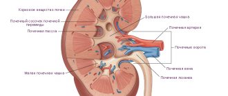
Kidney functions
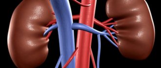
Kidneys
The kidneys perform several functions in the body in accordance with the division into three types of biochemical processes.
All these phenomena are quite complex and are of great importance for normal human life. The biochemical functionality of the kidneys is carried out during processes such as:
- urine formation is the excretory function of the organ;
- the formation of other substances is a homeostatic function;
- regulation of balance – metabolic function.