Lithotripsy (stone destruction) is a method of treating urolithiasis, which involves remote (without direct intervention in the body) or contact destruction of stones using different types of energy. Minimally invasive operations to remove kidney stones are carried out by breaking the stones into small fragments, which are removed directly during the operation and/or are eliminated from the body naturally.
External beam lithotripsy (ESL) or extracorporeal shock wave lithotripsy (ESWL) are synonyms for the same treatment method. The main point of the procedure is to destroy (disintegrate) a stone in the kidney or ureter using a shock wave, which is generated in the therapeutic head of the device using different methods of generating a discharge (electromagnetic, electrohydraulic, piezoelectric lithotripters).
Pulses focused on the stone using an ultrasound probe and/or X-ray guidance gradually destroy the internal structure of the stone, causing it to break up into smaller fragments. The stones are destroyed to the state of sand and small fragments, followed by spontaneous passage with urine through the urinary tract.
Types of lithotripsy according to the method of influencing the stone
External lithotripsy. A method of treating urolithiasis using special complex and high-tech devices - lithotripters. Lithotripters generate a shock wave, which, when focused on the stone using ultrasound or x-rays, leads to its destruction. The painless and low-traumatic operation is most often performed under intravenous anesthesia and is well tolerated by patients.
Contact lithotripsy (KULT - contact ureterolithotripsy). In this endoscopic operation, ureteroscopes are used - special instruments that are brought directly to the stone located in the ureter or kidney through the natural urinary tract. Once the stone is visualized, it is destroyed using laser, ultrasonic or pneumatic energy. Spinal anesthesia is the gold standard for these operations. When using this treatment method, the probability of complete destruction of the stone reaches almost 100%. Sometimes the operation is completed with the installation of a ureteral catheter stent, which is removed after 2–4 weeks.
Percutaneous (percutaneous) nephrolithotomy (PCNL). The operation involves creating a puncture in the lumbar region to insert an endoscope directly into the kidney, followed by destruction of the stones and removal of their fragments. This type of surgical treatment is used for coral stones and large stones. Percutaneous nephrolithotripsy is performed under general or spinal anesthesia.
The above-described minimally invasive operations as an alternative to surgery can be performed in our clinic. The operations are performed by qualified specialists using modern medical equipment. You can find out how much it costs to remove stones from the kidneys and ureters using one of the treatment methods from the price list or by phone.
Indications for surgery
Indications for extracorporeal lithotripsy: stones ranging in size from 5 to 20 mm, located in the calyces of the kidneys, in the pelvis or in the ureter.
For larger stones, other treatment methods are used.
Indications for contact lithotripsy: stones in the ureter, up to 20 mm in size.
Indications for percutaneous nephrolithotripsy: kidney stones larger than 20 mm, coral stones.
The appropriateness of using a particular treatment method is determined by the attending physician during a consultation after examining the patient and familiarizing himself with the results of his tests and examinations.
External shock wave lithotripsy
The number of shock wave pulses depends on the type of lithotripter and the power used. The gradual (rather than sudden) increase in power during the procedure prevents damage to the kidney tissue. In many cases, repeated crushing sessions are necessary, the intervals between which can range from 1 to 7-10 days or more. External lithotripsy of kidney stones is performed under close X-ray or ultrasound guidance and is an operator-dependent method, that is, the effectiveness and safety of the procedure depends on the experience of the doctor performing it.
In patients with “infectious” stones (usually struvite and carbonate apatite), in the presence of bacteria in the urine or a foreign body in the urinary tract (internal stent, nephrostomy), it is advisable to carry out antibacterial prophylaxis (taking into account the results of bacteriological examination of urine).
Contraindications to lithotripsy
General contraindications to lithotripsy:
- acute pyelonephritis;
- acute renal failure or progression of chronic renal failure;
- low blood clotting (hypocoagulation);
- spinal deformities;
- obesity;
- cardiovascular diseases in the acute stage;
- pregnancy;
- aneurysm of the renal artery and aorta;
- acute gastrointestinal diseases;
- atrial fibrillation;
- menstruation;
- kidney tumors.
The choice of a specific treatment method depends on the individual characteristics of the patient’s disease.
- narrowing of the ureter below the stone;
- violation of the excretory function of the kidney;
- acute pyelonephritis or urinary tract infection in the acute stage;
- the size of the stone is more than 20 mm or its high density.
Contraindications to contact lithotripsy:
- urinary system infection in the acute stage;
- ureteral stricture.
Contraindications to percutaneous nephrolithotripsy:
- patient's somatic status
Preparing for surgery
Before performing lithotripsy, a comprehensive laboratory and instrumental examination is necessary:
- clinical blood and urine tests, biochemical blood test, coagulogram, urine culture, blood test for hepatitis B and C, syphilis, HIV, blood group determination;
- CT scan of the retroperitoneal organs with contrast enhancement, ultrasound of the kidneys, ureters, prostate gland (in men), ECG, chest x-ray;
- esophagogastroduodenoscopy and duplex scanning of the veins of the lower extremities (if necessary);
- treatment of concomitant diseases during their exacerbation;
- drainage of the upper urinary tract (if necessary);
- carrying out antibacterial prophylaxis of urinary tract infections.
Stages of the operation
A session of remote lithotripsy lasts about 1 hour. If the stone fragmentation is poor, repeated crushing sessions may be required. A combination with other types of treatment, in particular contact lithotripsy, is possible. If there are large stone fragments, the patient may have a ureteral catheter stent installed. The duration of stay of a ureteral stent varies on average from 2 to 4 weeks.
Contact lithotripsy (laser, ultrasound, pneumatic) lasts from 30 to 60 minutes. The probability of complete destruction of the stone (especially if it is located in the lower parts of the ureter) reaches 100%. With this method of treatment, the destruction of the stone is performed under visual control, allowing the doctor to fragment the stone into a fine mass and remove large fragments.
Stages of intervention
Lithotripsy of stones in the ureter or kidney requires a preliminary comprehensive examination. If necessary, you need to undergo therapy for concomitant diseases and antibacterial treatment to eliminate infectious processes.
Remote and contact lithotripsy lasts up to 1 hour. In the first case, if large stone fragments are present, the patient is advised to have a catheter installed for 2-4 weeks. In the second case, the calculus is destroyed into a finely dispersed mass, which makes it easier to remove from the body.
With percutaneous nephrolithotripsy, the duration of the intervention is up to several hours. It depends on the location and characteristics of the stone. Upon completion of the operation, installation of a nephrostomy drain is necessary.
What is included in the cost of surgery to remove kidney stones?
The cost of operations to destroy stones in the kidney and/or ureter depends directly on the chosen method of surgical treatment, the type of anesthesia, the length of hospital stay and the need for preoperative instrumental and laboratory examination.
Having found out the price of stone removal surgery in our clinic, rest assured that the price will not change after the end of treatment. We have no hidden surcharges; our rates include consultation and examination by a urologist, anesthesiologist, instrumental and laboratory diagnostics, and anesthesia.
Complications in the treatment of kidney stones
Complications with extracorporeal lithotripsy are much less common than with other methods of surgical removal of kidney stones (ureteroscopy, percutaneous nephrolithotripsy). Complications of lithotripsy may be associated with the process of removal of fragments (recurrent renal colic, the formation of a “stone path”), with the inflammatory process (pyelonephritis, sepsis) and with damage to the renal tissue (hematoma formation). Recurrent renal colic can be observed in 2-4% of patients after crushing, the formation of a “stone path” - in 4-7% of patients. According to statistics, severe infectious and inflammatory complications (sepsis) can develop in 1.0-2.7% of patients. Asymptomatic hematomas are diagnosed in 4-19% of cases, and symptomatic hematomas are diagnosed in no more than 1%. To date, there is no convincing evidence of persistent long-term complications of extracorporeal lithotripsy.
Technical improvements, including miniaturization of endoscopes, flexible and disposable instruments, and improved optics, have led to increased use of transurethral endoscopic procedures (ureterorenoscopy) for both ureteral and kidney stones. The greatest progress has been made in retrograde intrarenal surgery. Recent clinical trials for the treatment of large (> 2 cm) kidney stones have demonstrated the high effectiveness of transurethral endoscopic stone removal (91% stone-free rate in 1.45 procedures per patient) with an acceptable risk of complications.
In most cases, ureterorenoscopy is performed under general anesthesia, although spinal anesthesia is also possible.
In order to achieve maximum efficiency and safety, ureterorenoscopy is performed under mandatory x-ray control. The use of modern disposable instruments, such as flexible guidewires, ureteral dilators, ureteral sheaths, baskets and extractors for removing stones or fragments, makes the procedure even more effective.
Rehabilitation
The duration of rehabilitation after stone removal depends on the lithotripsy method and the characteristics of the patient’s somatic condition. When crushed stones pass, they can cause renal colic or lead to exacerbation of calculous pyelonephritis.
After performing extracorporeal lithotripsy, patients are usually discharged on days 2–3. After discharge from the hospital, follow-up with a urologist is required for 2 weeks. On the 10-14th day, it is necessary to perform an ultrasound or x-ray, and take clinical (biochemical) blood and urine tests.
After contact lithotripsy, blood in the urine, moderate burning and pain when urinating or in the lumbar region may be observed for several days. In the absence of pronounced pain, the patient is discharged on the 2-3rd day.
The rehabilitation period of percutaneous lithotripsy takes longer. The average length of hospital stay ranges from 5 to 7 or more days. The nephrostomy drain is usually removed 2-4 days after surgery.
To increase the effectiveness of treatment and prevent the development of relapses of urolithiasis, patients who have undergone lithotripsy must take a responsible approach to the rehabilitation period and strictly follow the doctor’s recommendations.
Prescriptions in the postoperative period:
- dietary nutrition (excluding too salty and spicy foods);
- increased drinking regime (more than 3.0 liters/day);
- antibacterial therapy;
- consumption of vitamins;
- physical activity;
- herbal, laser, vibration therapy, etc.
Undoubtedly, Moscow opens up a lot of opportunities for treatment, incl. performing lithotripsy. But the price of the operation, queues, quality of service, staff responsiveness, efficiency and other criteria may not meet your requirements.
The effectiveness of treatment of urolithiasis in the Federal Scientific and Clinical Center of the Federal Medical and Biological Agency is guaranteed. The clinic employs highly qualified specialists; there are candidates of sciences on staff. The modern department, comfortable wards and operating rooms are equipped with high-tech equipment from global manufacturers. The Urology Department has all types of modern equipment (cystoscopes, ureteroscopes, nephroscopes; lithotriptors: laser, ultrasound, pneumatic and remote) for minimally invasive endoscopic removal of stones of any location in any part of the urinary system. Prices for lithotripsy of kidney/ureter stones using laser, ultrasound and other methods are affordable.
Urolithiasis occupies one of the leading positions among urological diseases. Almost every day, patients who are experiencing one or another stage of this pathological process come to see a urologist.
Often the onset of the disease is an attack of renal colic, which indicates the advancement of a stone along the urethra. There is always a threat of blockage of the lumen of the ureter with a calculus, which requires immediate surgical intervention.
Currently, extensive operations on the urinary system have been replaced by non-invasive or minimally invasive methods of treating urolithiasis and removing stones. Thanks to this, the effectiveness of the therapy has increased many times, and the long-term results and prognosis of the disease have also improved.
Contact lithotripsy is a modern method of minimally invasive surgical intervention, which crushes stones located at different levels of the ureter or in the renal pelvis.
Destroy without touching. How will extracorporeal lithotripsy help with urolithiasis?
According to statistics, 3 to 5% of the world's population suffer from urolithiasis. One of the non-contact methods of treating this disease is extracorporeal lithotripsy. What kind of manipulation is this? How is it carried out? In what cases can it be used? Stepan Petrovich Sidorov, urologist-andrologist, surgeon at Clinic Expert Irkutsk, answers these and other questions.
- Stepan Petrovich, extracorporeal lithotripsy - what kind of manipulation is this? Does it have other names?
— Remote lithotripsy (otherwise, ELT) is a method of treating patients with urolithiasis, which allows you to destroy stones (calculi) in the urinary system without direct contact with them, but by exposing them to directed ultrasonic waves. A more complete name for this manipulation, which can be found in the scientific literature and publications, is external shock wave lithotripsy.
Depending on the type of shock wave pulse generator, there are piezoelectric, electrohydraulic and electromagnetic types of lithotripsy devices.
— What are the advantages of extracorporeal lithotripsy? After all, there are other methods of destroying stones.
— Its main advantage is that it relieves the patient of a kidney stone without making an incision, that is, without violating the integrity of the skin and other tissues. The minimal trauma of the intervention significantly reduces the rehabilitation period after it.
— What are the indications for performing extracorporeal lithotripsy of kidney and ureteral stones?
— The procedure is possible for stone sizes up to 1.5-2 cm, a certain density, and also if it is possible to see the stone using ultrasound or X-ray.
External shock wave lithotripsy procedure
— How is external shock wave lithotripsy performed?
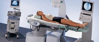
— The procedure is performed on an outpatient basis. The patient lies down on the couch. Using ultrasound or x-ray, we determine the position of the stone and focus it. To avoid kidney injury, ultrasound and x-ray monitoring is carried out throughout the procedure. The procedure usually does not require anesthesia.
The manipulation begins by generating low-power waves with large time intervals between them. Then the intensity and frequency are gradually increased. Fragments of stones and sand are gradually released in the urine. High-density stones do not always lend themselves to crushing immediately, so sometimes the manipulation has to be repeated. The procedure lasts on average 0.5-1.5 hours. This depends on the size and placement of the stones.
— What recommendations should patients follow in the postoperative period?
— In the first days after extracorporeal lithotripsy, urine should be collected in a container to monitor the release of stone fragments. Drink at least 1.5 liters of water per day. Take all medications prescribed by your doctor, which may include antispasmodics, painkillers and antibacterial agents.
It is important to avoid hypothermia, avoid overload and not make sudden movements. Visiting the bathhouse, sauna, solarium is strictly prohibited.
— Are there any contraindications to performing extracorporeal lithotripsy? If yes, which ones?
— Such manipulation is contraindicated in the presence of high-density stones, the size of which exceeds 2 cm. Among the general somatic contraindications, I would like to note acute inflammatory and infectious processes, pathologies of the cardiovascular system, excess weight, and pregnancy.
— What complications can develop and why can be observed after extracorporeal lithotripsy?
— One of the complications after the procedure is renal colic, which develops when fragments of stones pass. The patient may observe blood in the urine due to damage to the urinary tract from sharp stone fragments. Sometimes, after extracorporeal lithotripsy, there is a problem with urination, to eliminate which a catheter is installed.
It is not recommended to perform this manipulation when stones are located at the pelvic level, as this can lead to complications such as bleeding, damage to the kidney parenchyma and nearby organs.
— Is it possible to re-form kidney stones after extracorporeal lithotripsy?
- Yes. Lithotripsy has no effect on the prevention of urolithiasis. With this manipulation we destroy only the existing stone. Re-formation of stones depends on the individual characteristics of the patient and the lifestyle he leads.
— Stepan Petrovich, what needs to be done to stop the stones from appearing again? Is it possible to reliably prevent their formation or is it more correct to talk about reducing this probability if certain rules are followed?
— Unfortunately, there is no universal remedy for preventing the formation of kidney stones. Here, in my opinion, it is appropriate to talk about prevention, which involves following a diet, drinking regimen and an active lifestyle.
You can make an appointment with a urologist at the Expert Clinic Irkutsk here
The editors recommend:
Laser on stones! How can lithotripsy help with urolithiasis?
For reference:
Sidorov Stepan Petrovich
In 2009 he graduated from Irkutsk State Medical University with a degree in General Medicine.
In 2010, he completed his internship in General Surgery.
In 2012, he completed his residency training in Urology.
From 2013 to 2020, he received specialization in endosurgery, ultrasound diagnostics, laparoscopy and endovideosurgery.
Currently holds the position of urologist-andrologist, surgeon at the Expert Clinic Irkutsk. Receives at the address: st. Kozhova, 9A.
What is the procedure and its main advantages?
To carry out the procedure, special devices (lithotripters) are used, which are inserted through the lumen of an endoscope (ureteropyeloscope) into the urethra, bladder and ureter, to the immediate location of the calculus. Then, using an ultrasonic wave, pneumatic pulses or laser action, it is crushed into small fragments (down to dust particles).
These small parts are capable of painlessly coming out through a constant stream of urine over a certain period of time. Typically, the final result is assessed 2–3 months after contact lithotripsy. All this time, the patient’s condition is being monitored dynamically, which includes ultrasound or x-ray diagnostics of the movement of fragments along the urethra.
The method has the following advantages:
- there is the possibility of direct visualization of the calculus on the monitor screen, while a specialist can assess its size, shape, exact location, etc.;
- thanks to the close approach of the lithotripter, it is possible to crush even large stones whose dimensions exceed 3 cm (large fragments are removed through the lumen of the inserted endoscope, and smaller particles are removed by urine flow);
- there is no impact of the shock wave on surrounding tissues and organs (kidneys, ureter and others);
- the procedure has been proven to be highly effective, which rarely leads to repeated manipulation;
- speed of lithotripsy (usually the operation does not take more than 1 hour);
- short period of postoperative rehabilitation (compared to open surgery);
- low percentage of complications and adverse outcomes after surgery.
Contact lithotripsy of kidney and ureteral stones
Our urology department specializes in the surgical treatment of urolithiasis, and has all the endourological technologies existing in the world, allowing us to perform such high-tech operations as:
- ureteral stenting
(installation of a ureteral stent, which allows urine to flow out of the kidney) - percutaneous nephrolithotripsy
(percutaneous crushing of large and coral kidney stones) – through punctures in the skin no larger than 10 mm in size - minipercutaneous nephrolithotripsy
(percutaneous crushing of large and coral kidney stones) – through punctures in the skin no larger than 5 mm in size - transurethral ureterolithotripsy and nephrolithotripsy
(crushing ureteral and kidney stones) using semi-rigid and flexible nephroscopes and ureteropyeloscopes
Most urological diseases today can be treated with minimally invasive (non-traumatic) treatment without compromising effectiveness and safety. But this is achievable only in specialized multidisciplinary clinics that have not only equipment, consumables and medicines, but also qualified personnel who undergo regular training according to the best international standards.
Today, in our clinic you can count on high quality treatment and at the same time receive help for free - within the framework of compulsory medical insurance.
Kidney stones can only be removed surgically. The only type of stones that undergo conservative medical dissolution are stones consisting of uric acid. There are several types of interventions for kidney stones, each of which has certain advantages and disadvantages. Lythotripsy _
— crushing of kidney or ureteral stones using various types of energy (ultrasonic, laser, pneumatic, electro-hydraulic, piezo-electric, etc.) There are the following types of lithotripsy:
Please note that the website uroportal.ru is for informational purposes only. Do not attempt to self-diagnose and treat; if you have symptoms of the disease, we strongly recommend that you seek advice from a specialist.
Make an appointment with a urologist
A method of crushing stone under the influence of shock waves supplied from the outside by a special device - a lithotripter, which generates and focuses shock wave pulses onto the stone from the outside. In this case, the stone is destroyed inside the kidney or ureter into fragments, which independently pass through the ureter, bladder and out with the flow of urine. Introduced into clinical practice in the 1970s. As a rule, extracorporeal lithotripsy is performed without anesthesia. To improve the passage of stone fragments, the so-called “forced diuresis” is used - taking large amounts of fluid and diuretics. DLT is effective for single stones of a relatively small size - up to 15-20 mm in the greatest dimension with an average or low densitometric density - no more than 900-1000 HU.
Insufficient effectiveness of DLT is also observed when the stone is located in the lower group of cups - stone fragments are difficult to remove from this segment of the pyelocaliceal system, there is a possibility of incomplete passage of all fragments and, as a consequence, relapse of stone formation. It is worth noting that the amount of energy and pulse frequency for one ELB procedure are limited and regulated. Exceeding these values in order to enhance the effect on the stone leads to severe trauma to the kidney tissue and can cause an exacerbation of the chronic inflammatory process, the formation of hematomas, and pain. Therefore, several DLT sessions are often required (from 2 to 5), the interval between which is usually 14-21 days. Thus, the treatment process can be quite lengthy. Due to the greater shock wave load on the kidney than with a ureteral stone, DLT is almost always performed under general anesthesia. Another limitation for EBRT is the patient's weight. With severe obesity (2-3 degrees), precise focusing of the wave on the stone is difficult, therefore, the effectiveness is significantly reduced.
External lithotripsy indications:
- X-ray positive ureteral stones more than 0.5 cm,
- renal pelvis and calyx stones less than 2 cm
Despite the obvious advantages, this type of treatment is the least invasive, but has a number of limitations and side effects:
- How to remove urinary stone from toilet
- under the influence of a shock wave, healthy tissue surrounding the calculus is always damaged, including kidney tissue
- fragments of the destroyed calculus, coming out with urine, often cause renal colic. Therefore, it is impractical, and sometimes dangerous, to crush large kidney stones using extracorporeal lithotripsy;
- Kidney stones with high density are not affected by the shock waves of the lithotripter, i.e. extracorporeal lithotripsy may simply be ineffective;
- Several extracorporeal lithotripsy operations may be required to completely crush the stone and remove it from the kidneys and ureter.
A minimally invasive method for crushing kidney and ureteral stones is retrograde (transurethral) endoscopic interventions (Fig. 1). The essence of this technique is to pass thin optical instruments (endoscopes) under anesthesia through the urethra, bladder, ureter, reaching the renal pelvis and stone. Thus, the entire procedure takes place under continuous visual control. The optimal stone sizes for these manipulations are from 15 to 25 mm. Having reached the stone, it is sequentially fragmented using various types of energies.
Fig. 1 Retrograde endoscopic interventions
There are the following types of contact lithotripsy: ultrasound, pneumatic, laser.
- ultrasonic
– most effective for low stone density (less than 800 HU). The working probe is designed in the form of a titanium alloy tube. High-frequency vibration allows you to destroy the stone into very small fragments, the consistency of which resembles dust, and the built-in aspiration system instantly removes them through the lumen of the probe into a special container. Leaving stone fragments behind is virtually impossible for this method. Unfortunately, kidney stones of such low density are quite rare in our region.
- pneumatic
- the probe works on the principle of a jackhammer, making rapid translational movements. For this type of energy, the density of the stone is less important: as a rule, stones of very high density - 1500 HU and higher - cannot be crushed. The main disadvantage of the method is the formation of large stone fragments, which are quite difficult to remove. In this case, there is a risk of migration of a stone fragment to other parts of the pyelocaliceal system.
The above types of lithotripsy energies are used with so-called semi-rigid (rigid) endoscopes (Fig. 2). This type of instrument is characterized by good visualization and controllability, however, many parts of the collecting system (for example, the middle and lower cups - this is where stones are most often localized and fragments are displaced here) are inaccessible for such instruments. In such cases, special thin flexible endoscopes are used that can reach any part of the CLS. The disadvantages of such tools are their short working life, high cost of repair, and rapid breakdown if handled and handled incorrectly.
Fig.2 Semi-rigid ureteroscope
With flexible instruments (Fig. 3), it is possible to use only laser or electrohydraulic energy - the working probes of these lithotripters are capable of bending. However, these types of energy effects are also used with rigid endoscopes.
Fig.3 Flexible ureteroscope
- Lithotripsy is an operation to crush kidney stones using ultrasound and an endoscope
- laser
- a high-energy laser beam is applied to the stone through an optical probe. The destruction of the stone occurs from the periphery to the center. The density of the stone does not matter: even the densest ones are subject to destruction! With proper laser exposure, the resulting fragments measuring 1-3 mm in size completely disappear on their own in the early postoperative period. This method is the preferred method for breaking kidney stones.
- electrohydraulic
- works on the principle of a piezo element. An electric arc occurs between the two electrodes, which has destructive power. Of course, taking into account the small size of the fiber (diameter about 1 mm), the effect is only on the stone. The density of the stone also does not matter much. The disadvantage of this energy, unlike laser energy, is the unpredictability of the size of the formed fragments. Therefore, this type of lithotripsy is advisable to use for relatively small stones - 10-15 mm.
Transurethral interventions
are characterized by a low frequency of complications: most often in the postoperative period there is an exacerbation of chronic pyelonephritis, the frequency of such a complication is about 10 percent. Kidney damage is excluded during such interventions. At the end of each operation, a special internal thin tube is installed in the ureter - a stent, which facilitates the rapid and almost painless passage of stone fragments. The stent is removed 7-10 days after surgery and is performed on an outpatient basis. The duration of transurethral interventions does not exceed 2 hours. This is quite enough to completely fragment the stone, but occasionally, if the stone is large, repeated interventions may be required. The patient's weight and the presence of clinically significant obesity do not affect the effectiveness of the operation.
Contact lithotripsy allows you to crush ureteral and kidney stones in all parts. The density of the stone does not matter. There is no damage to the healthy tissue surrounding the stone.
This ensures a fairly rapid recovery and return to everyday activities: patients, as a rule, are discharged within 1-2 days after surgery.
The main complications of percutaneous interventions are exacerbation of chronic pilonephritis (thanks to the use of modern antibacterial drugs, this only prolongs the hospital stay by 3-4 days). The probability of developing pyelonephritis is about 10%. Another serious complication can be bleeding. In our experience, this happens quite rarely. The frequency of the need for blood transfusion after PCNL was less than 1% in our clinic.
With the development of technology, instruments of smaller diameter have appeared - mini-nephroscopes, which can reduce surgical trauma to the kidney and somewhat reduce the risk of complications. The operating principles of such tools are similar to standard ones.
Fig.4 Percutaneous nephrolithotripsy
Open operations for the treatment of urolithiasis (removal of stones from the kidneys and ureter) are currently used quite rarely, in complex cases or when minimally invasive endoscopic intervention is technically impossible. Thus, having the entire arsenal of treatment methods, it is possible to crush and remove any stone in any part of the urinary tract (kidney stone, ureteral stone, etc.). After any surgery for urolithiasis, all patients are recommended to undergo a metabolic study aimed at finding the causes of stone formation, as well as dynamic observation.
At the North-West Center for Endourology and Lithotripsy, all types of endoscopic operations are performed using modern equipment from leading global manufacturers: Olympus, KarlStorz, RichardWolf.
- Shock wave lithotripsy of the gallbladder
Indications for the procedure
Contact lithotripsy is indicated for the following categories of patients:
- the presence of stones in the kidneys or ureters of any location and size;
- if non-contact lithotripsy (remote) did not bring the required effect (this may be due to the large size of the stone or its high-density structure);
- the presence of stones that remain in the lumen of the ureter for a long time;
- absence of urodynamic disorders, namely problems with the outflow of urine below the location of the calculus;
- the possibility of introducing an endoscopic device and a lithotripter into the urinary tract (absence of strictures and narrowings in the urethra and lumen of the ureter).
Contraindications for the procedure
Crushing stones using contact lithotripsy is not suitable for all patients, since the manipulation has certain contraindications and limitations for its implementation. All contraindications can be divided into 3 main groups.
Restrictions of a technical nature
The group includes:
- physical data (patient height exceeding 200 cm or less than 100 cm, as well as body weight 130 kg or more);
- severe deformities of the spine or the entire musculoskeletal system, which does not allow the patient to be positioned normally on the operating table (there are also certain difficulties with fixing the stone in the field of view of an x-ray or ultrasound);
- the presence in a medical institution of only an X-ray device for X-ray negative stones in a patient.
General restrictions
The group includes:
- pathology of the blood coagulation system (congenital diseases, long-term use of anticoagulants, etc.);
- pregnancy period, menstruation in a woman;
- severe mental disorders in the patient, psycho-emotional agitation;
- cardiovascular or respiratory failure in the stage of decompensation.
Urological restrictions
The group includes:
- pronounced deformations of the lumen of the ureter (strictures or scars of a congenital or acquired nature), located below the location of the stone;
- inflammatory processes in the urinary tract or in prostate tissues during the acute period (acute pyelonephritis, acute cystitis, acute prostatitis and others);
- decrease in glomerular filtration level (by more than 50% of normal values);
- the patient has a single kidney with reduced excretory function.
Manipulation tactics
Lithotripsy is performed in an operating room; the patient is preliminarily prescribed neuroleptanalgesia. The choice of anesthesia technique depends on the patient’s age, his general health, the presence of concomitant pathologies, the equipment of the medical institution, etc.
The surgeon stands between the patient's legs. After anesthesia has been administered, the doctor begins to insert the ureteropyeloscope into the lumen of the urethra, then into the bladder and ureter. By transmitting the image to the monitor screen, it assesses the condition of the urethra along its entire length, right up to the detection of a calculus.
As soon as the endoscope reaches the required level, a balloon catheter is inserted above the location of the stone, which will prevent its advancement into the collecting apparatus of the kidney. Next, a lithotripter (laser fiber or other device) is directed through the lumen of the endoscope, thanks to which the crushing process is carried out.
Preparing the patient
To avoid complications and unwanted side effects from manipulation, careful preliminary preparation is required, including the following:
- extensive clinical and laboratory-instrumental examination of the patient, thanks to which the doctor determines the size and location of the stone, its configuration, the presence of urodynamic disorders, etc.;
- carrying out bowel cleansing, which avoids difficulties with visualization on the monitor screen (2-3 days before the upcoming procedure, all foods that can cause flatulence and bloating are excluded from the patient’s diet, and the evening before and on the morning of the day of lithotripsy, the patient is prescribed a cleansing enema);
- before crushing, the patient is injected with 400–500 ml of physiological solution, which will increase the fluid content in his body and increase the effectiveness of the procedure;
- An important role is played by methods of pain relief (intravenous or epidural anesthesia is performed).
Possible complications and unwanted effects from the procedure
Contact lithotripsy may be accompanied by the following complications and undesirable effects.
Complications that occur during lithotripsy
The group includes:
- heart rhythm disturbances, interruptions in heart function;
- change in blood pressure numbers (an attack of hypertension or the development of hypotension in response to exposure to a shock wave);
- patient agitation as a result of activation of the autonomic nervous system;
- separation of the ureter from its anatomical bed;
- a breakthrough in the wall of the ureter, which can lead to the onset of massive bleeding.
Complications arising in the early period after surgery
The group includes:
- the appearance of blood in the urine (hematuria is observed in more than 80% of patients, which is associated with damage to the walls of the urinary tract);
- renal colic (its appearance is caused by blockage of the lumen of the ureter with large fragments of stone);
- hemorrhages under or around the kidney capsule (perirenal or subcapsular hematoma);
- exacerbation of chronic processes in the kidneys (pyelonephritis), bladder (cystitis) or urethra (urethritis);
- bleeding from different parts of the gastrointestinal tract.
Description of the procedure
Lithotripsy is a method that involves crushing stones in the organs of the urinary system. The basis is the impact of a laser or ultrasound shock wave.
The urinary stones are crushed to such a size that the stones can pass out on their own. It is less traumatic compared to surgery. The method has been actively used since the 80s of the last century.
You will learn more about lithotripsy as a modern method of treating kidney stones from this video:
External lithotripsy
The procedure for remote crushing of stones using a shock wave is carried out using a special device (lototripter). Principle: the lithotripter releases waves that reach maximum energy only in the focusing area, the so-called working spot, without affecting other organs. During the operation, the stone breaks down into small components, and then passes naturally through urine. The procedure is non-invasive, that is, nothing is introduced into the patient’s body either through the skin or through the urethra;
Contact lithotripsy
Used for high density stones in the ureter or kidneys. An endoscope is inserted into the ureter. The latter is equipped with equipment that grinds stones into dust. After this, they leave the body naturally. The main advantages of the method include affordable cost and minimal likelihood of complications.
The main difference between remote lithotripsy and contact lithotripsy is the absence of the need to introduce instruments, however, the use of a non-contact method is not always possible.
Types and effectiveness
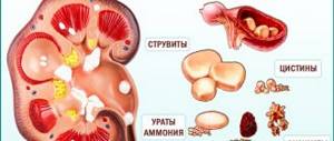
The essence of the work of modern lithotripsy devices is the impact on the calculus with a shock wave pulse. The stone is destroyed to the smallest particles, similar to sand. They do not interfere with physiological processes and are excreted in the urine. To find out which method will be effective in a particular case, the doctor evaluates the location, size, and hardness of the formation. Stones have different chemical natures: oxalates, phosphates and urates.
The longer the disease lasts, the larger the stone becomes.
Preliminary diagnostics are carried out. If the stones cannot be removed with medication, the doctor uses one of the lithotripsy techniques: remote or contact. They have advantages and disadvantages.
Remote
Remote lithotripsy of kidney and ureteral stones (DLT) is performed without direct contact with the patient’s body. The shock wave, which crushes the stones, penetrates the tissue. The patient does not experience any discomfort.
Lithotripters with different energies are used:
- piezoelectric,
- electrohydraulic,
- electromagnetic.
The doctor controls the focus on the stone using ultrasound, which allows you to remove all the stones in one procedure. The stones are destroyed gradually until they pass through the ureter. Based on the patient’s condition, operations can be performed under general anesthesia or local anesthesia.
The advantages of the method are the ability to destroy small stones up to 2 centimeters in diameter, non-invasiveness.
The lithotripsy method has disadvantages and limitations. When the stones are dense, more than 1000 units. according to the Hounsfield scale, they cannot be destroyed in one go, you have to repeat it or use another technique, resort to surgery.
If the size of the stone is more than 2 centimeters, the fragments may not pass through the ureter, clinging to its walls.
There is a risk of developing renal colic. Symptoms:
- severe pain in the lumbar region,
- nausea,
- vomit,
- the appearance of blood in the urine.
A dangerous complication is blockage of the lumen of the ureter. All the fragments accumulate and a stone path is formed. If the walls are damaged, internal bleeding may occur. Residual fragments that remain in the kidney are too large to exit into the ureter and can grow again, leading to relapse of the disease.
After lithotripsy, 90% of people experience postoperative hematuria. Damage occurs to the tissues and blood vessels of the kidneys and ureter. When using this method, the shock wave does not act accurately enough. It is not dangerous, but requires medical supervision. With a successful outcome of the operation and local anesthesia, the patient goes home after a few hours.
Contact
Contact or transurethral lithotripsy of kidney and ureteral stones is performed with a thin surgical instrument: a flexible urethroscope. It is inserted into the urinary canal. An impulse is passed through the tube and the stones are purposefully struck. An impulse uses different types of energy:
- pneumatic,
- laser,
- ultrasonic
Laser lithotripsy is effective.
Disadvantages of the method: invasive surgery using general anesthesia. Pneumatic lithotripsy can damage healthy tissue; ultrasound cannot destroy formations of medium and high density. If the method is chosen incorrectly, the procedure will have to be repeated.
Hard stones of more than 1000 units can be destroyed using a laser to the smallest particles, which are excreted with urine flow. After this, there is a smaller number of relapses of urolithiasis. Contact lithotripsy has a more targeted effect than non-contact lithotripsy, as a result, healthy tissues of the body are not damaged. The rehabilitation period is faster compared to percutaneous, lasting 1-2 days.
The percutaneous method involves the use of anesthesia. A puncture is made in the lumbar region, endoscopic instruments are inserted, and the remains of destroyed stones are cleaned out with their help. The rehabilitation period is about a week under the supervision of specialists. Painful sensations in the incision area, hematuria, which normally disappears after 24 hours.
This method is chosen in the following cases:
- stones larger than 2 cm have formed,
- coral stones completely fill the renal pelvis,
- the pelvis area is too narrowed,
- other methods are contraindicated.
Indications and contraindications
Let's consider under what circumstances certain procedures can be carried out to get rid of urolithiasis (urolithiasis):
Indications for DLT
- The size of a stone in the kidney or ureter is from 0.5 to 1.5 (in some cases up to 2) cm cm;
- Stone density up to 1300-1500HU
- Possibility of focusing the lithotripter on the stone (using an ultrasound or x-ray guidance system)
Contraindications to DLT
- Severe heart disease
- Acute inflammatory process in the urinary tract
- Kidney tumors;
- Pregnancy;
- Menstruation;
- Aortic aneurysm, in case of possible contact with it by a shock wave beam;
- Inability to focus the lithotripter due to anatomical features
Indications for CL
- Stones up to 3 cm in size;
- Stones of any density
Contraindications to CL
- Arrhythmia;
- Pregnancy;
- Weight over 130 kg;
- Curvature of the spine, bones;
- Pathological conditions (renal failure);
- The occurrence of neoplasms in the organ;
What is better contact or non-contact lithotripsy?
Contact is used in situations where non-contact would be ineffective. However, doctors and patients themselves prefer to use the “remote” method whenever possible.
Recommendations of the European Association of Urology for the treatment of urolithiasis
For a patient with an acute attack of renal colic, the first step is to relieve pain. Non-steroidal anti-inflammatory drugs are recommended:
- Metamizole,
- Paracetamol,
- Diclofenac,
- Indomethacin,
- Ibuprofen
The latter two drugs are also used to reduce the frequency of pain relapses after an attack. Long-acting alpha-blockers (for example, Tamsulosin) can be prescribed for the same purpose. When using drugs, you need to remember about contraindications and side effects.
In case of recurrent attacks of renal colic and low effectiveness of drug treatment, it is recommended to immediately drain the kidney using an internal stent or puncture nephrostomy, as well as removal (crushing) of the ureteral stone.
Dissolving urinary stones (or their fragments) can only be effective if they are composed of uric acid. Dissolution is achieved by alkalinizing the urine using chemicals (potassium citrate or sodium bicarbonate). Stone-dissolving therapy is carried out under mandatory monitoring of urine pH, the target value is 7.0 - 7.2. Excessive alkalization can lead to the formation of phosphate stones. In the process of treating urolithiasis, it is necessary to observe a urologist, monitor urine tests and ultrasound of the kidneys (or MSCT).
The effectiveness of extracorporeal lithotripsy depends on the lithotripter used, crushing modes, size, location and density of the stone, as well as on the characteristics of the patient (for example, obesity). When crushing large stones, pre-installation of an internal stent may reduce the risk of stone walk formation.