Visually, the renal pelvis is a type of funnel formed by a small and large bowl. The renal pelvis serves as a kind of reservoir for collecting fluid, which is then excreted. Being part of the general collector system of the body, the cups and pelvis are connected by a narrowed neck.
Important! When stones are removed, the narrow cervical junction becomes clogged with a stone and the cup, neck, and pelvis expand. The pathology is called calicoectasia and causes unpleasant painful sensations
The inner part of the renal pelvis is lined with mucous membrane, the walls are smooth muscle tissue of the longitudinal and transverse type. Due to the contraction of smooth muscle structures, urine moves through the urinary tract and is excreted. The peculiarity of the kidney structure is that the walls are impermeable to liquid, so urine cannot get outside the urinary system.
Structure and functions
The calyces of the medulla parenchyma are connected to the natural urinal by narrow formations - necks. The pelvis has the appearance of a funnel with an expanded side outward of the kidney, and the drainage is into the gate and ureter.
The storage structures of the renal parenchyma include:
- small cups - the total number varies from 6 to 12;
- large calyces - there are 2–4 of them in the human kidney;
- pelvis.
Starting with smaller formations, the calyxes merge with each other and form larger structures. The role of the pelvis is reduced to the accumulation and movement of formed urine through the ureters.
If there is difficulty in outflow through the urinary tract, pathological expansion occurs, then the size of the necks of the large cups increases. The process is called calicoectasia.
The renal pelvis is internally covered with a mucous membrane of epithelial cells. This type of epithelium is double-layered with basal and superficial layers. The cell type is called transitional. They can change depending on the degree of filling of the pelvis.
Histological examination of the transitional epithelium reveals cell nuclei similar to vesicles and grains inside the cytoplasm. Most often the cytoplasm is yellow because this is caused by the pigments characteristic of urine. The shape of the epithelium of the renal pelvis can look like cells:
- caudate,
- fusiform,
- pear-shaped,
- oval.
Determining exactly what type of epithelium is exfoliated into the urine is important for diagnosing the level of inflammation of the urinary organs. Typical cells are found in catarrhal pyelitis, when inflammation of the renal pelvis does not affect the deep layers.
In the case of purulent pyelitis, the epithelium undergoes a degenerative change, most often fatty degeneration
The wall contains smooth and transverse muscle bundles. This structure allows us to provide:
- reliable impermeability, complete isolation of collected urine, normally it cannot get outside the kidney;
- push the accumulated fluid into the ureters, causing peristaltic movements by contraction of the longitudinal and transverse muscles.
Further management of the patient
The detected enlarged renal pelvis requires dynamic monitoring. If the cause of its development is another disease (pyelonephritis, urolithiasis, cancer), then it is necessary to focus on its treatment. Usually, effective therapy (the use of antibiotics, uroseptics, lithotripsy) allows one to stabilize the size of the pelvis and achieve partial regression.
Surgical treatment is indicated if congenital pyelectasia is detected in a newborn, grade 3 dilatation (hydronephrosis), development of a staghorn stone, or complete obstruction of the ureter. Most modern intervention techniques are laparoscopic, which has made it possible to perform pelvisplasty with a much lower incidence of complications.
After surgery, patients are recommended to undergo regular ultrasound diagnostics of the urinary system (once a year). This makes it possible to prevent the re-development of pyelectasis, which occurs in approximately 5-7% of patients.
How to treat an enlarged renal pelvis?
Pyeloectosis is an expansion of the renal pelvis. Usually detected by ultrasound. IMG_5478
Urologists believe that this expansion occurs when there is an obstruction to the outflow of urine from the kidney. Sometimes pyelectosis can be transient (temporary) due to blockage of the ureter with a clot of mucus or pus and may go away after the clot is reabsorbed. But still, more often the detected pyeloectosis turns out to be permanent. Such pyeloectosis eventually disrupts the functioning of the kidneys, resulting in the development of a disease - hydronephrosis. Hydronephrosis is a more expanded concept than just pyelectosis, since it includes functional and anatomical changes in the entire kidney, and not just in the renal pelvis.
To understand what happens in the kidney during pyelectosis and hydronephrosis, you need to have an idea of the anatomy and features of the kidneys.
The kidneys are a paired organ. The length of the kidney in an adult is 12-13 cm, weight on average 150-200 g. The surface of the kidney is covered with a dense capsule, and under it there is a special tissue - the kidney parenchyma. This parenchyma contains many specific formations - glomeruli and tubules. It is in them that blood is filtered and urine is formed. The urine formed in the glomeruli and tubules first enters the so-called renal calyces, and from there into the renal pelvis. The renal pelvis is a thin-walled cavity. Its wall consists of three layers: the outer layer is made of connective tissue, the middle layer is made of muscle tissue, and the inner layer is made of epithelium.
The pelvis usually has a cone shape, its normal capacity is on average 5 ml. Tapering, the pelvis passes over the ureter. Due to contractions of the renal pelvis, urine enters the ureter, then from there it is collected in the bladder and released through the urethra from the body.
If there is any obstacle to the evacuation of urine from the kidney, retention and accumulation of urine occurs in the pelvis. The pelvis expands. This expansion is called pyeloectosis.
Obstacles to the outflow of urine can occur at the transition from the pelvis to the ureter, in the ureter itself, or in the bladder.
The dilated pelvis begins to disrupt the functioning of the entire kidney. Due to the accumulation of urine and pressure from the dilated pelvis, atrophy of the parenchyma occurs, destruction of the glomeruli and tubules. Not only urine excretion is disrupted, but also its formation. In such cases, they already talk about a disease such as hydronephrosis. Destruction of the kidney parenchyma is an irreversible process and can ultimately lead to renal failure.
The reasons that impair the secretion of urine from the pelvis can be congenital (primary hydronephrosis) and acquired (secondary hydronephrosis). Congenital causes are usually associated with developmental abnormalities in the kidney and ureter. These could be some kind of valves, “tongues” made of connective tissue or epithelium in the pelvis and ureter. Sometimes there are additional blood vessels that pass near the ureter and compress it. Pediatric urologists encounter congenital hydronephrosis not so rarely. Sometimes congenital hydronephrosis is detected even in the fetus during an ultrasound examination of a pregnant woman.
Acquired causes are usually associated with urolithiasis, tumors, cicatricial adhesions in any part of the urinary system. Stones block the lumen of the ureter, and tumors and adhesions can compress the ureter from the outside. In both cases, urine remains in the pelvis, which leads to its expansion.
Hydronephrosis, especially in the initial stage, can be asymptomatic and detected by chance on ultrasound. But more often, patients complain of dull, aching pain in the area of the corresponding kidney. Sometimes renal colic is observed, that is, attacks of exacerbation of pain. When an infection occurs, an increase in body temperature is observed.
As the process progresses, pain no longer occurs. Changes in urine analysis are observed.
With unilateral hydronephrosis, the patient may not have any complaints at all and feel quite satisfactory due to the fact that the work of the affected kidney is compensated by the second, healthy kidney. With bilateral hydronephrosis, the process is more severe and can lead to chronic renal failure.
The danger of hydronephrosis also lies in the fact that due to stagnation of urine, infection is often associated, which leads to pyelonephritis, and even purulent processes - pyonephrosis.
The diagnosis of hydronephrosis only based on patient complaints is almost impossible to make, since these complaints are nonspecific and can accompany other kidney diseases.
Therefore, to clarify the diagnosis, in addition to urine tests, additional examination, ultrasound, excretory urography or computed tomography is necessary.
What to do if hydronephrosis is detected?
You need to know that there are no conservative methods of treatment (tablets, injections) for hydronephrosis. Treatment is surgical. These may be operations to restore the outflow of urine from the pelvis, for example, removing a stone from the ureter, removing adhesions, additional blood vessels, or plastic surgery of the ureter. In advanced cases, especially with a purulent process in the kidney, the kidney is removed - nephrectomy. When choosing surgical treatment, doctors take into account many factors: the patient’s age, concomitant diseases, whether the process is unilateral or bilateral, the presence of pus. For example, if a young woman is planning a pregnancy, and one kidney has stopped functioning due to hydronephrosis, but the second kidney is healthy, then she is recommended to have a nephrectomy before pregnancy. In older patients (60-70 years old) with many concomitant diseases and a partially functioning kidney, the absence of acute pain, suppuration, surgical treatment is approached very carefully. Doctors believe that the earlier hydronephrosis is detected and the flow of urine from the kidney is restored, the better the prognosis for the patient. With unilateral hydronephrosis and a healthy second kidney, the prognosis is better than, for example, with bilateral hydronephrosis. Bilateral hydronephrosis quite early leads to chronic renal failure and a threat to the patient’s life.
With congenital hydronephrosis, doctors believe that the prognosis is better than with acquired hydronephrosis. Thus, the following conclusions can be drawn:
- Conservative treatment for hydronephrosis is ineffective;
- To correctly diagnose hydronephrosis, a comprehensive examination is necessary (urinalysis, ultrasound, expert urography, CT);
- If the cause that interferes with the flow of urine from the pelvis is not eliminated, you can lose a kidney;
- With unilateral hydronephrosis, the prognosis for the patient is favorable; with bilateral hydronephrosis, a threat to life may develop.
To date, no methods have been developed for the prevention of hydronephrosis.
It is necessary to monitor your health, pay attention to emerging kidney symptoms, in a timely manner, without delaying for “later”, visit specialists, carry out all examinations prescribed by the doctor - all this allows you to timely identify diseases and carry out the necessary treatment with the least risk. The kidneys are a paired organ with a complex structure that performs the functions of creating, storing and removing urine from the body. The pyelocaliceal system (PSS) plays an important role in its functioning: it collects urine formed in the nephrons and further evacuates it through the ureters into the bladder. There are several dozen diseases that occur with damage to the calyces and pelvis, and pyeloectasia of the kidneys is one of them.
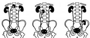
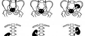
It is known that the normal size of the renal pelvis is 3-6 mm in children and adolescents and 10 mm in persons over 18 years of age. Pyeelectasis is a pathological expansion of the renal pelvis in adults and children. According to statistics, this syndrome is more typical for the stronger half of humanity. Due to the anatomical features of the urinary organs, pyeelectasis of the right kidney develops several times more often than the left.
What determines the size of the pelvis?
The size of the adult pelvis is no more than 10 mm. In women during pregnancy, the volume may increase to 18–27 mm, but this is considered a physiological norm and is due to the pressure of the uterus on the ureters and the obstructed outflow of urine.
If there is no connection with pregnancy, the following reasons should be considered:
- the likelihood of a tumor compressing the urinary tract;
- the presence of calculi (stones) in the ureters;
- structural anomalies (kinks or torsions).
The renal pelvis in a child is visible in the prenatal period at 17–20 weeks of pregnancy. Doctors can assume abnormal development or pathology using an ultrasound examination before birth and warn parents. An important difference is the absence of changes in size in children before and after urination.
The table shows the maximum normal dimensions of the fetal pelvis.
| Gestational age | Ultrasound size in mm |
| up to 32 weeks | 4 |
| 36 weeks | 7 |
A pediatrician will help determine how much the kidney has changed and whether anything needs to be done after examining and examining the newborn.
We will consider common kidney diseases affecting the pelvis area from the perspective of the most likely causes.
What affects the size of an organ?
The size of the renal pelvis may not always change for pathological reasons. In women carrying a fetus, dilation up to 27 mm is a physiologically acceptable norm. But it is still worth monitoring the condition of the expectant mother in labor and regularly conducting diagnostics. Other factors can also affect the size of the organ:
- probable neoplasms in the urinary organs;
- formed stones;
- an abnormal structure in which kinks and twists occur.
Inflammation
The inflammatory process in the pelvis is called pyelitis. It most often occurs in girls aged 2–5 years, pregnant women, and in men after surgical interventions on the prostate gland. Any stagnation of urine provokes infection. The dangerous pathogen is E. coli, which is always present in the body.
Other pathogens are actively involved in damage to the urinary tract. It is especially important to take this into account if a person has a chronic infection (tonsillitis, sinusitis, cholecystitis). Hypothermia can be an additional factor in the disease.
What to do and how to treat?
Treatment of such a disease largely depends on the cause that caused it. Since pathology is most often caused by a mechanical obstruction to the outflow of urine, it is necessary to conduct a series of examinations that will detect it - ultrasound , urography , cystoscopy . After this, surgical intervention is performed to eliminate the cause of the disease. After the operation, the patient needs a rehabilitation period, during which the normal outflow of urine and the size of the structures of the urinary system are restored.
If the disorder is caused by diseases of the organ parenchyma - cyst , tumor , hydronephrosis - then treatment is also surgical. The affected area is removed, after which a course of restorative therapy is carried out. In severe cases, removal of the entire organ is required. If both kidneys are affected, it may be necessary to remove both of them and place the patient on hemodialysis. In such situations, the patient is put on a waiting list for an organ transplant.
Stages of hydronephrosis
For congenital anomalies, surgery may be risky. To reduce the risk to the baby’s health, it is carried out after lengthy preparation. But it is surgical intervention that can give a small patient a chance for a relatively healthy life. A number of restrictions (on the amount of liquid and salt in food, physical activity, some medications and procedures) remain for life.
Conservative treatment of this pathology is ineffective and is used in cases where surgery is contraindicated or impossible, during the recovery period, as well as during preparation for intervention.
Pyeelectasia is a pathology that is practically asymptomatic in the first stages. If complaints or negative signs appear, after an in-depth examination, parents learn that the child has an enlarged renal pelvis.
How dangerous is the disease? Why does congenital pathology occur? How to treat pyeloectasia in children? The answers are in the article.
Fetal anomalies
Duplication of the renal pelvis is a rare anomaly. It is often combined with accessory ureters. If kidney function is not impaired, then the person does not feel any abnormalities. If detected in a child, it is not considered normal; treatment is suggested only if inflammation or other pathology occurs.
Double ureters, kidneys, pelvis occur in the prenatal period
Ectopic ureters - (disturbed location), when in girls the ureter is attached to the vagina, and in boys - to the urethra. Often combined with kidney duplication, it causes inflammation of the renal pelvis and its enlargement.
Symptoms of renal pelvis cancer and treatment measures
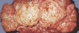
Renal pelvis cancer is less common than kidney or bladder cancer, but it requires special attention. One of the main symptoms that may indicate the development of cancer is the appearance of blood in the urine. Another symptom is lower back pain, which occurs due to blockage in the urinary system. This may be due to a blood clot or due to the development of a tumor. In addition, the person begins to lose weight sharply, he often feels sick and vomits.
If the doctor suspects the development of a tumor, he palpates the abdominal cavity, which allows him to identify a large tumor. Blood and urine tests are required, as well as an ultrasound examination of the said organ.
If an ultrasound shows that there is a tumor of the pelvis, the doctor prescribes a computed tomography scan, which allows a more accurate diagnosis, to determine at what stage of development the tumor is and whether it is possible to remove it.
In cases where the tumor is concentrated only in the pelvis or ureter and there are no metastases, it can be removed through surgery. This usually involves removing the entire kidney, ureter, and some of the bladder. There are cases, for example, if there is only one kidney, then only the tumor is removed, and healthy tissue remains. But this method of treatment is associated with the risk of relapse. If surgical treatment is not possible, chemotherapy is prescribed.
In any case, if you have an enlarged renal pelvis or other abnormalities, you should definitely consult a doctor who will prescribe all the necessary tests and examinations. Based on the results obtained, he will be able to make the correct diagnosis and prescribe effective treatment. The sooner this is done, the greater the chance that you will completely get rid of this disease.
What complications are expected with dilated pelvis?
The gradual development of the expansion process in an adult proceeds in parallel with the underlying disease. The consequences may be:
- hydronephrosis;
- urethrocele - at the confluence of the ureter, a spherical expansion forms on the wall of the bladder; it is usually located on the side of pyelectasis;
- vesicoureteral reflux - consists of the backflow of urine from the bladder into the ureters and further into the kidney, accompanied by infection and increasing pressure in the pelvis.
The causes of reflux are considered to be:
- impaired innervation of the bladder;
- mechanical obstacles to the proper flow of urine due to a neoplasm, a stone in the renal pelvis.
Pathologies and their manifestations
What symptoms are accompanied by inflammation?
A feeling of fullness in the suprapubic area is one of the symptoms of a possible pathology.
With an inflammatory reaction in the pyelocaliceal system (PSS), the patient may experience changes in the size of the organ. The deviation is called pyelitis, and is most often diagnosed in girls 2-5 years of age, pregnant women or men after prostate surgery. Due to inflammation, urine stagnates and the infection begins to multiply. The patient is concerned about the following manifestations:
- severe pain when going to the toilet;
- frequent urge to pee;
- feeling of fullness in the suprapubic area;
- changed color of urine.
What anomalies occur?
Rarely, the fetus has one double pelvis, which is often supplemented by a ureter. If urinary function is not impaired, the patient does not feel discomfort and treatment is not required. Also, common anomalies include ectopia, which occurs in girls and boys, and is characterized by the attachment of the ureter to the vagina or urethra.
Features of pyeloectasia
With pyelectasis, the pelvis increases in size and the outflow of urine is impaired.
The deviation is characterized by an enlargement of the pelvis, which occurs when the outflow of urine is disrupted, which is thrown back into the kidneys. Against this background, the fetus develops the following complications:
- abnormalities in the formation of urinary organs;
- blockage of the ureter;
- formation of the urethral valve.
In an adult, pyeelectasis can be caused by kidney stones or neoplasms in the pelvis. First, the latter becomes spherical in shape, due to which the parenchyma is pushed to the edge. When disrupted, the nephrons die, in their place fibrous tissue is formed. It is also possible to develop an infectious focus in the organ, as a result of which constant inflammatory processes develop.
Hydronephrosis
Hydronephrosis is a significant expansion of not only the renal pelvis, but also the calyces. The renal parenchyma gradually atrophies and becomes thinner, the boundary between the cortex and medulla disappears, and the main structural units of the kidneys, the nephrons, die.
Extensive sclerotic areas remain. The process can be one- or two-way. The outcome is kidney failure.
According to the mechanism of development, they are distinguished:
- acquired form;
- congenital.
Congenital hydronephrosis is detected in 1.4% of newborns. This is usually due to a hereditary predisposition.
The purchased one is formed:
- with a tumor;
- if kidney pathology is accompanied by vesicoureteral reflux;
- with urolithiasis.
Reasons for development
The form of pyelectasis depends on the cause that led to the abnormal expansion of the renal cavities. With the acquired organic form, there is a narrowing of the ureters as a result of injury or an inflammatory process. The reasons for the development of the pathological process can be:
- kidney stones;
- nephroptosis;
- tumors of the kidneys and urinary tract.
When these pathologies are detected, urine stagnates in the cavity of the renal pelvis, which causes expansion of the renal pelvis.
The congenital type of the disease is often detected during fetal development or in the first months of a child’s life. It occurs when the development of the walls of the upper sections of the ureters is disrupted. The dynamic congenital form occurs when:
- phimosis;
- urethral stricture;
- psychological disturbance of urine excretion.
In adults, it can develop against the background of blocking the lumen of the ureter with a calculus or mucous clot formed during inflammation. The pelvis enlarges with nephroptosis and wandering kidney. In adults, the start of the pathological process can be facilitated by the consumption of large amounts of fluid, when the kidneys become unable to perform their functions. In older age, the development of pathology is caused by impaired peristalsis of the ureters.
Pyeelectasia is often combined with a disease such as ureterocele, a narrowing of the area of the ureter that flows into the bladder. The presence of a cavity in the ureter is detected by ultrasound examination. At the same time, dilation of the renal pelvis is detected. Disruption of the posterior urethral valves occurs only in men; it is combined with bilateral pyelectasis and dilation of the urinary canals. With ectopia of the ureter, it ends in the urethra or vagina, accompanied by doubling of the kidney and an increase in the urinary tract.
Vesicoureteral reflux of urine into the kidney leads to pathological changes in the organ. An increase in size of the ureter leads to increased pressure in the bladder. The lower part of the urinary canal is narrowed, urine is thrown into the kidney. In adults, pyeelectasis is usually asymptomatic.
In some cases, there are signs of an underlying disease leading to dilation of the renal pelvis. Retention of urine in the kidneys leads to their sclerosis. This term refers to the death of tissues responsible for the production and excretion of metabolic end products. The disease is combined with renal failure, inflammation and atrophy of renal tissue.
Oncological processes in the pelvis
Tumors in the renal pelvis are rare localizations, if only an isolated structure is taken into account. Most often, the neoplasm affects the entire kidney, including the pelvicaliceal segments. The source of malignant growth is the epithelium covering the inner surface. Such tumors are called adenocarcinomas. According to the type of epithelium, they belong to transitional cells.
The tumor “masks” itself as an inflammatory disease for a long time. Severe symptoms appear only when they grow inside the wall of the pelvis.
What it is?
The renal pelvis is the “final” part of the kidney. It is a cavity into which urine flows through the renal tubules. The ureter originates in this same structure, which drains fluid into the bladder, where it accumulates.
The structure is located in the area of the so-called renal hilum - an area located on the side of the spine. This area received its name due to the fact that the renal artery, vein and ureter are located close to each other - the most important structures providing blood supply and function.
In its structure, this formation is a cavity in which urine collects. From the side of the parenchyma (main tissue), second-order tubules open into it, bringing urine that has undergone a series of successive changes. From the side of the gate, the ureter emerges from the pelvis. The function of this formation is to collect urine before it goes into the ureter.
Stone formation
Consequences of urolithiasis
The reason for the formation of stones is the intake of chemical and biological substances from food, which are broken down in the body into insoluble salts. These include:
- urates,
- carbonates,
- phosphates,
- oxalates.
A similar process occurs when metabolism is disturbed and it is impossible to bind and neutralize these components.
Salt sedimentation occurs in the pelvis, ureter, and bladder. Gradually, the stone in the renal pelvis reaches sufficient size. Due to this, the useful volume is reduced. The shape of the stones follows the structure of the kidney. They can be:
- triangular,
- oval,
- in the form of a cone,
- cylindrical.
Fixed stones are dangerous due to subsequent stagnation of urine and the development of hydronephrosis. Movable ones cause destruction of the wall, tissue rupture with the outpouring of urine into the peritoneal cavity.
Diagnostic measures
Before starting treatment for the pathology, it is necessary to establish the main cause of the expansion of the heart rate. A standard diagnostic plan for kidney disease includes:
- Collection of complaints and medical history. It is important for the doctor to outline the boundaries of possible problems and make a preliminary diagnosis.
- Inspection, palpation, percussion, determination of the tingling symptom. Allows you to diagnose enlarged kidneys, the presence of an inflammatory process or stones in them.
- Blood tests (general, biochemical). With their help, you can learn about existing health problems and possible somatic pathologies.
- Urine tests (general, according to Nechiporenko, according to Zimnitsky).
- Instrumental diagnostic methods - ultrasound, CT, MRI, excretory urography.
Instrumental tests are the main method for detecting pyelectasis. If the renal pelvis is dilated, this can be easily determined, as well as the exact size of the organ can be determined using visual diagnostic methods.
What symptoms should you pay attention to?
Developmental anomalies may be asymptomatic. They are detected accidentally during examination for chronic inflammation or when a neoplasm is suspected. Symptoms of damage to the pelvis are difficult to differentiate. Patients complain about:
- acute or dull arching pain in the lower back with irradiation to the perineum, pubic area,
- frequent urination with pain;
- bloating above the pubis and inability to urinate;
- change in the color of urine (cloudness due to an excess of leukocytes caused by inflammation, the presence of blood due to a tumor or after an attack of urolithiasis);
- temperature increase from low values to a sharp rise, depending on the nature of the inflammation.
Attacks of renal colic when a stone moves can lead to shock
Common symptoms include:
- malaise and weakness;
- nausea;
- weight loss;
- headache.
How dangerous is expansion?
The expansion of the cavity inside the organ itself can be dangerous only in advanced cases. A much greater risk to the patient’s health is posed by the causes that caused it.
If the outflow of urine is impaired, this causes not only its accumulation in the kidney cavity, but also partial absorption of unnecessary substances back. In such cases, the patient experiences severe pain in the lower back, his temperature rises, headaches, and a feeling of weakness and fatigue occur.
In especially severe cases, uremia - a high content of waste substances in the blood, which must be excreted in the urine. The reason for its development with increase is the overflow of the urinary tract, which inhibits the formation of urine and contributes to the accumulation of undesirable substances in the blood. Symptoms of uremia:
- Constant weakness, headache.
- Unpleasant odor of sweat (sweat smells like urine)
- Grayish skin color, peeling, deterioration of hair condition.
- Nausea, vomiting, loss of appetite.
- Urinary disorders.
The actual increase in formation, if it continues for quite a long time, can lead to disruption of the blood supply to soft tissues and gradual degradation of the parenchyma.
How is renal pelvis pathology detected?
There are no special diagnostic methods specifically for identifying diseases of the pelvis. A doctor has ample opportunities to research kidney diseases. The level and location of the lesion can be assessed by carefully interpreting the results. Patients are prescribed:
- general urinalysis with sediment examination;
- culture for pathological flora;
- Ultrasound of the kidneys;
- excretory urography with the introduction of a contrast agent;
- computed tomography.
An x-ray of the kidneys taken using the excretory method shows a “spot” on the left that is not filled with contrast; perhaps this is a tumor formation
Diagnostics
Often, enlargement of the renal pelvis is diagnosed in adults by chance during a medical examination. Unilateral pyeloectasia can be asymptomatic for a long time. With age, symptoms of pyelonephritis and urolithiasis increase.
Enlargement of the renal pelvis in the fetus and newborn is diagnosed using ultrasound.
- Ultrasound of a pregnant woman is performed 3-5 times throughout the entire gestation. Pyeelectasis in the fetus is diagnosed at the 2nd and 3rd screening - at 19-22 and 30-33 weeks, respectively.
- Ultrasound of a newborn child in the presence of such a pathology in utero is performed immediately after birth, then according to the scheme 3-6-9-12 months.
The main diagnostic methods that establish the dilation of the renal pelvis:
- Ultrasound with Dopplerography.
- Multislice computed tomography.
- Vaccine cystography.
- Radioisotope renography and nephroscintiography.
- Angiography.
- Excretory urography.
- Survey radiography.
- MRI.
Laboratory diagnostic methods:
- Urine tests: culture, general clinical studies, Rehberg tests, according to Zimnitsky, Nechiporenko.
- Biochemical blood tests, acid-base status.
The expansion of the renal pelvis corresponds to such results as leukocyturia, bacteriuria, increased creatinine, urea, decreased glomerular filtration rate, decreased hemoglobin, urate salts and oxalates are present in the urine sediment in large quantities, the relative density of urine decreases.
renal pelvis
The human kidney is an important organ that functions as a kind of filter for the body, and the renal pelvis and calyx are a single system functioning within this organ.
This component is a kind of sedimentation tank for secondary body fluid, which then enters the ureter for subsequent excretion. The renal pelvis is an area of the organ in which important processes of filtration and fluid storage occur.
Renal pelvis: description
The renal pelvis is a cavity whose main function is to collect urine, the formation of which occurs in the kidney. In appearance, it resembles a funnel formed by a small and large calyx, each of them has a narrowing - a neck, which is a kind of connecting element of the pelvis and the calyx system. Any violations in the form of blockages lead to an increase in this component.
The renal pelvis has a body: a muscular organ, internally covered with a mucous membrane, the walls of which are endowed with longitudinal and transverse smooth muscles. This structure provides contractile movements of the pelvis to move fluid through the urinary tract. One of the main features of the walls is their impermeability to all substances.
Parameters: norm and deviations
In medicine, there are generally accepted standards for the size of the pelvis for all age categories of people. From the fetus to the elderly, there are boundaries within which the parameters of the renal pelvis vary. Any deviations indicate the presence of a particular disease, the timely detection of which will help to begin treatment and avoid negative consequences and complications.
Adult sizes (+ during pregnancy)
The normal size of the renal pelvis in an adult should not exceed 10 mm. In women during pregnancy, the pelvis is enlarged, which is considered normal for this condition. In the first trimester, the size of both pelvis reaches 18 mm, and in the last stages - 27 mm. The main reasons for enlargement in the absence of pregnancy are:
- tumors;
- kinking or twisting of the urinary tract;
- stones in the ureters.
Normal in children
In children, the pelvis is smaller - 6 mm, less often - 7-8 mm. Exceeding this norm indicates a disease such as pyeloectasia, which practically does not manifest itself with visible signs. In newborns, this figure ranges from 7-10 mm, and any deviation beyond these limits requires consultation for the child with a specialized specialist.
Fetal parameters
Kidneys begin to form in the womb and this process continues after birth. Starting from 17-20 weeks, the doctor can examine the fetal urinary organs and give a tentative assessment of their condition. Their size returns to normal after six months of life. In view of this fact, the fetus does not have clear boundaries of the pelvis; there are approximate ones:
- 4 mm up to 32 weeks;
- 7 mm at 36 weeks;
- more than 10 mm is a signal for treating the disease after the birth of the baby.
Diseases of the renal pelvis can be congenital or acquired.
Diseases
Human kidney diseases have become natural for a number of reasons (for example, a sedentary lifestyle, an unbalanced diet), which lead to a disease that is subsequently firmly entrenched in the individual’s life.
Women are more often at risk, but the male half of humanity should not forget that even a seemingly harmless disease can lead to irreparable consequences for the body.
Kidney pathologies are divided into congenital and acquired.
Source: https://medic-online.net/2161/lohanka-pochki/