Urolithiasis is a common urological disease, accounting for about 30-40% of cases of the total number of renal pathologies. Statistics show that urolithiasis ranks second in frequency of occurrence, second only to infectious and inflammatory diseases.
Most often, urolithiasis is diagnosed in patients aged 20-55 years, among whom there are 3 times more men. However, in women, coral nephrolithiasis, which is the most dangerous type of pathology, is more often diagnosed. Statistics show that in most cases, uroliths affect only one kidney, but in 15-30% of cases, diagnosis can still indicate a bilateral course of the disease.
As medical practice shows, single stones can form in the kidneys, as well as multiple stones, the number of which can reach many dozens. Urologists more often detect small stones, the size of which is a few millimeters. However, in surgical practice it is necessary to identify large conglomerates, the size of which exceeds 10 cm, and their weight reaches 1 kg.
Any kidney stones provoke a malfunction of the organ, so doctors are focused on laser removal of kidney stones. Leading clinics in Russia successfully carry out diagnostics, including metabolic examination and medical imaging. Based on the diagnostic results, an individual treatment plan is developed, an operation is performed to remove kidney stones, after which, based on the results of a chemical analysis of the removed stone, measures to prevent further stone formation are determined.
What stones can pass on their own?
If there are stones (calculi) in the kidneys, a person experiences lower back pain, and blood may appear in the urine. However, not every case will require surgery.
According to various sources, in 70-75% of people stones up to 5 mm in size are able to pass on their own.
This process is accompanied by a set of symptoms called renal colic:
- severe, often unbearable pain in the lumbar region (right kidney);
- increased blood pressure;
- temperature increase.
The passage of the stone may take from several days to 2-3 weeks. If it is accompanied by long-term complications or malaise, it is better to consult a doctor immediately.
Diagnosis of urolithiasis
For all patients who suspect urolithiasis, doctors prescribe:
- ultrasonography. It can be used to determine the size and location of stones;
- survey urography of the kidneys. This research technique determines X-ray positive stones;
- intravenous urography, which will more accurately determine the size and location of stones, and also determine whether the outflow of urine is impaired;
- biochemistry and general blood test;
- general urine analysis;
- microscopy of urine sediment to clarify the structural features of stones;
- bacterial culture of urine.
Also, doctors may, if indicated, prescribe additional research in the form of:
- retrograde or antegrade pyelography;
- scintography;
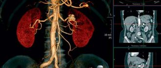
- computed tomography;
- biochemical study of urine.
Reasons for the passage of stones
The reasons for the formation of kidney stones are well known - they are associated with metabolic disorders and the formation of clots of poorly soluble salts. The passage of a stone provokes:
- mechanical pressure from neighboring organs (tumor of the kidney, ureter);
- complications after various operations;
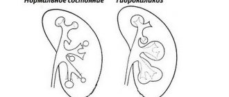
- inflammatory processes in the urinary tract due to diseases (periurethritis, hydronephrosis, kidney tuberculosis);
- blood clots in the veins of the kidney;
- congenital features of the genitourinary system.
Sometimes the process of stone passage is preceded by active physical activity, drinking large quantities of drinks and water.
Taking diuretic medications without a doctor’s recommendation also contributes to the movement of stones.
Open surgery
When stones are detected in the kidneys, abdominal surgery has recently been performed less and less often. But there are a number of indications when such an operation simply cannot be avoided:
- regular relapses;
- the stones are large and cannot be removed by any other method;
- inflammatory processes of purulent type.
Open surgery is performed under general anesthesia. During surgery, the body cavity is involved, and excision is performed through all tissue layers. A positive aspect is the presence of a calculus in the renal pelvis, which helps reduce the invasiveness of surgery. In addition, you can open the ureter and remove the stone from there.
Modern methods of performing such surgical intervention include laparoscopy. In this case, the stone is removed through a small incision. A camera is inserted into it to transmit the image to the big screen. Removal of stones using laparoscopy is performed only if there are special indications.
How a kidney stone passes: general symptoms
The main symptom is noticeably severe pain, which often becomes unbearable. Along with extraneous sensations, there are other symptoms that allow you to understand that the stone is coming out of the kidneys.
Pain and burning
Pain is the main symptom indicating the movement of kidney stones. The characteristics of pain are as follows:
- can be either dull or sharp (burning sensation);
- usually localized in the lumbar region;
- appears not only in the back area, but also under the ribs on the back side;
- lasts from 15 minutes to 1 hour;
- worsens after physical activity, with sudden movements and jolts.
Changes in urine
Certain changes also occur with urination and the composition of urine:
- little or no urine;
- frequent urge to pee;
- the appearance of blood impurities;
- white flakes in urine (loose, gelatinous texture);
- urine turns an unnatural color - reddish or dirty black.
In 65-70% of cases, similar symptoms appear in men. However, there are certain specificities in the signs of kidney stones in women. At the initial stage, the symptoms of the attack are similar. Subsequently, in men, pain symptoms spread to the scrotum and perineum, and in women - to the labia area.
Chills, temperature
A characteristic, but nonspecific symptom of kidney stones passing is an increase in temperature. It usually reaches from 37.2 to 38°C. But there are also cases when it rises a little higher. The person begins to shiver, as if he had a severe cold. If such symptoms persist for several days, it is recommended to consult a doctor.
Nervous system disorders
Signs of movement of kidney stones also appear from the nervous system:
- irritability;
- general fatigue, weakness;
- dizziness and heaviness in the head.
These symptoms are due to prolonged exposure to pain. Therefore, the first measure of assistance for colic is associated with mandatory pain relief.
Edema
Another sign of a stone leaving the kidney is swelling of the skin. This symptom is easily identified visually. Edema appears as a result of a malfunction of the kidneys. The stones that appear interfere with the normal excretion of urine, causing fluid to accumulate in the body.
High blood pressure
Failure of the kidneys leads to excess fluid accumulating in the body. It puts pressure on internal tissues, organs, and blood vessels. Therefore, an increase in blood pressure is observed (usually by 15 mm). Even if a person has always had normal blood pressure, with an exacerbation of colic, the upper blood pressure can reach 150-160 mm Hg. Art.
Digestive disorder
Even more interesting:
Echinococcal liver cyst surgery
Apple cider vinegar for dandruff reviews
Disruption of digestive processes during colic is not always observed. However, signs of a stone leaving the ureter or kidney are also characteristic of inflammation of the pancreas, stomach, and gall bladder. The accumulation of fluid can lead to digestive disorders, which will manifest itself in pain, diarrhea, and intestinal spasms.
Indications for surgery
Surgery to remove kidney stones is performed if there are a number of indications:
- Ureteral obstruction. This pathology requires immediate treatment, so the use of conservative methods of therapy is not effective.
- The development of renal failure or the presence of this pathology in the acute stage. If you ignore the symptoms inherent in this disease, there is a high probability of complications, including death.
- The presence of painful sensations that cannot be relieved with medications.
- Inflammation of purulent type.
- Presence of a kidney carbuncle. This term deciphers the area of purulent necrosis formed as a result of exposure to stones.
- The patient's desire to undergo surgery.
What to do if a kidney stone passes
Colic due to stones occurs suddenly and can occur on the road or at work, so it is better to immediately call an ambulance. Before doctors arrive, first aid measures should be provided.
First aid
One of the main questions is related to what can be done at home if stones pass. First of all, a person is shown complete rest. You should sit him in a comfortable position and ventilate the room. You also need to take care of how to relieve pain if a kidney stone passes. For this purpose, antispasmodics are given (No-Shpa, Baralgin, Papaverine, Revalgin).
Place a heating pad on your lower back or take a warming bath. However, this can only be done if the person is completely sure that the pain is associated precisely with the movement of stones, and not with other reasons (sciatica, appendicitis, etc.).
Medication assistance
Today, in pharmacy kiosks there is a fairly large number of medications that can cope with urolithiasis.
Today, in pharmacy kiosks there is a fairly large number of medications that can cope with urolithiasis. How to get rid of kidney stones? Before you start taking drug therapy, you should undergo a full examination, take the necessary tests and get advice from a specialist. Finding out the cause of the disease will help you find a suitable drug, and a specialist will tell you about the dosage and time of taking it. An important factor is also the type and shape of the stone; such nuances influence the choice of method for removing coral. So, how to remove kidney stones, as well as relieve pain and inflammation with the help of medications. Let's look at the most popular means:
- Canephron - the main effect of the drug is aimed at neutralizing inflammation, increasing the level of lactic acid production in the kidneys, as well as scavenging radicals;
- Blemaren - the main effect of the drug is aimed at plucking urine and completely dissolving nephrolithiasis formations;
- Potassium citrate - the main effect of the drug is aimed at completely removing stones.
For information! In order to understand how to remove a kidney stone, you need to undergo an ultrasound. It is this procedure that allows you to determine the size, type and shape of the stone. If there are large coral growths on the renal system, surgical intervention is performed.
Consequences of stones leaving the renal pelvis
The most common consequences of this phenomenon include:
- temperature about 37.2-38°C;
- general malaise;
- foreign sensations in the lower back;
- symptoms of the inflammatory process (pyelonephritis) - nausea, vomiting, pain when urinating, chills.
The movement of stones in the ureter is accompanied by pain both during exit from the kidney and after that. Therefore, at first it is better to consult a urologist in order to adequately assess your condition and, if necessary, undergo a course of treatment.
Prevention with herbs, juices and fruits
The most common vegetables and fruits that can be used to eliminate and prevent urolithiasis are lemon, cucumber, beets, and carrots. It is worth mixing them in equal proportions, diluting them with water 50/50 and taking them on an empty stomach 3 times a day. Course – 15 days.
Dried figs should be poured with milk, boiled for 5 minutes over low heat, removed, and allowed to brew. Serve hot.
Nettle and parsley are used to remove phosphates and urates, but in no case oxalates. You need to make teas from these herbs. It is advisable to periodically use parsley root. Add a little boiling water to the crushed raw materials and let it brew. Then take a small spoon on an empty stomach.
Not only teas and tinctures are prepared from nettle, but they are also added fresh to salads and other dishes. Nettle tincture can be mixed with other remedies. Among them are teas made from rosehip, elderberry, juniper, linden, and mint.
Important questions
People suffering from urolithiasis always have questions related to the duration of stone removal and the consequences of this process.
How long does it take for a stone to come out?
The period may take from several days to 2-3 weeks. But delays are possible due to the size of the stones. Therefore, the answer depends on the person’s condition.
What to do if a pebble comes out when urinating
In this case, it is better to immediately contact a urologist in order to adequately assess the condition of the kidneys and the genitourinary system as a whole. In most cases, stones do not exceed a centimeter in diameter. You should not throw them away - it is better to save them for demonstration to the doctor.
How long does the pain last after the stone passes?
Pain lasts for 1-1.5 hours and is usually relieved with antispasmodics. But as a result of complications against the background of inflammatory processes, pain and cramping may appear after this for several days.
Why did the temperature rise after the stone passed?
The phenomenon may be due to the fact that other stones, larger or smaller, remain in the ureter. Also, an increase in temperature occurs due to ongoing inflammatory processes in the kidney and ureter. In any case, you should not self-medicate - it is important to consult a urologist as soon as possible.
How to understand that a stone has passed
The main sign of stone movement is severe pain, that is, symptoms of renal colic. When the pebble is completely released into the bladder, the extraneous sensations stop and the state of health noticeably improves. The release of a calculus is independently determined by the fact of its appearance as a result of urination.
The movement of the stone is always associated with severe pain. First of all, you need emergency help - antispasmodic drugs. In the future, at the first opportunity, you should consult a doctor to understand how to speed up the passage of the stone, and whether surgery is necessary.
Kidney stones (uroliths) form gradually - sand appears first, and then larger stones. Small formations come out on their own without causing any discomfort. The movement of larger stones is accompanied by pain and other symptoms of varying severity, which in some cases may lead to the need for conservative therapy and even surgery. According to statistics, the disease develops more often in men, but in women it is more complicated due to the tendency to form stones of irregular shape and the structural features of the urinary system. If you have such a problem, you need to know how kidney stones come out in order to recognize the process in time and take the necessary measures.
Duration of surgery to remove stones in the ureter
The length of a patient's stay in a nephrology hospital depends on the patient's condition after treatment, the development of complications, and the type of intervention.
After percutaneous nephrolithotomy, the patient stays in the surgery department for 3 to 7 days. On the third day, the attending physician removes the drainage, assesses the condition of the postoperative wound, and conducts an ultrasound diagnosis of the kidneys and urinary tract. If no pathological changes are detected, the patient is discharged home. Doctors recommend that discharged patients follow a strict diet.
After urethroscopy, the patient can be free the next day. People with swelling and pain syndrome require inpatient monitoring.
Abdominal ureterolithotomy is more difficult to tolerate. Due to the high risk of complications (suture dehiscence, bacterial infection), the patient is discharged a week after stone removal. Sutures are removed and the catheter is removed 4-5 days after the procedure.
After discharge from the hospital, the patient is observed by a family doctor or nephrologist at the place of residence.
Which way does the stone come out?
The release of urolith can begin under the influence of external factors associated with vibration or increased physical activity. But most often it is provoked by taking diuretics or drugs that affect the condition of the kidneys.
The movement of the stone begins from the kidney into the ureter, from where it enters the bladder and then into the urethra. The narrowest and longest is the ureter - on average its dimensions are 0.8 cm in diameter and 40 cm in length. Therefore, the urolith’s passage of this particular segment of the “path” turns out to be the most difficult and most noticeable.
The severity of the symptoms of this process depends on the size of the stone:
- if it does not exceed 0.6 cm, then no sensations arise;
- with a size of 0.7–1 cm, the stone also usually passes due to stretching of the walls of the ureter, but can cause pain, and in the presence of sharp angles, blood discharge from the urethra;
- stones of a larger diameter can get stuck, blocking the flow of urine, which is fraught with dangerous consequences.
Note! Even small stones can have a rough surface and pointed protrusions. As they move along the ureter, they will damage the inner walls, causing severe pain and bleeding.
Removal of a section of a kidney
If kidney stones are detected, this type of surgery is performed only for very large stones. This surgical intervention makes it possible to preserve the internal organ, which is especially important if there is only one functioning kidney.
Resection is performed in the following situations:
- numerous stones localized at one pole of the organ;
- regular relapses of the disease;
- necrotic lesions;
- the last stages of urolithiasis.
Moreover, if the patient is in serious condition and doctors assume that surgical intervention may aggravate it, they refuse the operation.
Resection is performed under general anesthesia. The patient is placed on his healthy side, under which a bolster is placed. The surgeon makes an incision and then separates the underlying layers of tissue. A clamp is applied to the area of the kidney with the ureter to prevent bleeding, since this is where the maximum concentration of blood vessels is located.
Next, the affected area is excised. The edges are stitched. A drainage tube is removed from the kidney and the wound is sutured. The drainage tube should be in the kidney for 7-10 days after surgery. After the specified period and in the absence of complications, it is removed.
Signs of stone movement
There are several main symptoms that accompany the exit of urolith from the kidney and its movement along the ureter:
- Painful sensations of different nature, intensity and localization. The pain can be aching or paroxysmal, concentrated in the kidney area or encircling. Along with this, a strong internal burning sensation appears, at the peak of which a loss of consciousness is possible.
- A sharp jump in blood pressure caused by pinching of the renal artery by a stuck or slowly moving stone.
- The appearance of blood in the urine due to injury to the tissues of the ureter.
- Disorders of the digestive tract, characterized by prolonged vomiting without preceding nausea.
- Increase in the thermal state of the body to low-grade levels (fever, sometimes with chills).
- Swelling of the arms, legs, face.
- Nervous disorders - severe anxiety, irritability, disorientation in space and time.
Renal colic
Renal colic (colitis in the kidney) is unbearable pain in the lumbar region, cutting and stabbing type, preventing a person from moving, lying or sitting, tormenting constantly and not leaving him alone - an attack can last for hours. This is not an independent disease, but only a symptom, the causes of which can be very diverse, but all of them are somehow related to problems of the kidneys and urinary system. So, what is renal colic and why does it occur? What should a person do with renal colic - how to relieve pain?
Our readers successfully use Renon Duo to treat kidneys. Seeing how popular this product is, we decided to bring it to your attention. Read more here...
Renal colic has different causes, and the range of pathologies leading to this phenomenon is large. All causes have a common feature: blockage of one or another segment of the urinary tract occurs, and normal urodynamics are disrupted. Deterioration or cessation of fluid outflow causes a number of symptoms, including acute pain - colic. Blockage of the lumen can occur in any part of the urinary system, but the symptoms in any case will be very similar.
The folk way to cleanse the kidneys! Our grandmothers were treated using this recipe...
Cleaning your kidneys is easy! You need to add it during meals...
The main diseases leading to renal colic syndrome:
- Urolithiasis disease. It is the main cause of colic in the vast majority of cases. A urine stone blocks the pelvis or ureteral tube. If a patient comes to the doctor with acute pain in the lumbar region and suspected renal colic, the diagnosis begins with confirmation/exclusion of urolithiasis.
- Pyelonephritis. Pathogenic microorganisms, having entered the kidneys one way or another, cause infection in them. Common pathogens include Escherichia coli, strepto- and staphylococci and others. Under their influence, inflammation of the epithelium of organs begins, the appearance of purulent foci. Purulent discharge and exfoliated particles of inflamed tissue move along the urinary tract, gradually clogging them. It is important to understand that pyelonephritis often accompanies kidney stones and causes renal colic.
- Traumatic injuries. If the organs are damaged as a result of some mechanical impact, this will cause hematomas that compress the ducts or blood clots that clog the lumens. If there is damage to the left kidney, renal colic will occur on the left.
- Congenital anomalies and disorders. In some people, from birth, the ureter is incorrectly attached to the bladder, the kidneys are formed in the wrong position, etc. These problems themselves are not so critical, and their owner can live a long, happy life without knowing about them. But if the patient has suffered an injury or an infectious disease, these factors can be superimposed on congenital anomalies, disrupting the drainage of urine and causing colic.
- Tumors. The process of pathological tissue proliferation that has begun can, over time, compress the ureters and the kidneys themselves, causing urodynamic failures. This occurs if the tumor directly affects the urinary organs or nearby tissues.
- Renal tuberculosis. Tuberculosis itself is considered a pulmonary disease, but about a third of patients are carriers of its extrapulmonary form, and the kidneys are one of the possible sites of damage. The tissue of these organs often becomes a site of accumulation of mycobacteria, causing urinary disorders. Therefore, a doctor, when examining a patient with colic who has already been diagnosed with tuberculosis or is showing signs of it, must conduct a diagnosis for the presence/absence of tuberculosis pathogens. Characteristic symptoms: incessant cough, low-grade temperature, sudden weight loss of the patient.
An important nuance in determining the causes and providing further assistance to the patient is the need to confirm or refute the presence of stones in the ureter and pelvis. Having carried out first aid measures for the patient, you should first find out exactly this, and only then examine the person for other pathologies.
Symptom complex
Symptoms of renal colic in women and the stronger sex are generally similar. The main symptom of renal colic is acute pain that lasts for a long time. It also serves as the main marker (but not the only one) of diagnosis.
The pain syndrome can be supplemented by:
- vomiting (especially at the peak of negative sensations);
- urinary disorders.
These signs do not always appear, but are still quite common in patients with this serious condition.
As mentioned, people complain mainly of pain due to renal colic. The types of this sensation are different, directly with colic it is cutting, very strong - up to its complete unbearability. It occupies all the patient’s attention, displacing all feelings and thoughts; the person is unable to sit quietly, move, or even lie down; he becomes agitated and can literally “climb the wall.”
The sensations are localized in the lumbar area, with irradiation to:
- the front of the thigh;
- crotch area;
- external genitalia.
The pain becomes stronger if the doctor or the patient himself palpates the abdomen in the navel area, and it also intensifies if light tapping of the lower back is performed. Pain upon palpation is characteristic of the cause of the syndrome located in the ureter. Right-sided renal colic is characterized by pain on the right side, left-sided renal colic will cause acute discomfort on the opposite side. Symptoms of renal colic in men are felt somewhat more strongly, due to the peculiarities of the nervous system and physiological structure.
The condition is manifested not only by pain and spasm.
Other signs of renal colic in men and women:
- Impaired urination. This symptom occurs if the lumens of the urinary tract are narrowed or blocked. The patient feels a frequent urge to go to the toilet, little urine is released, and the process brings severe discomfort. It is accompanied by pain in the perineum and lower back, and a burning sensation. Injuries to the mucous membranes (for example, from the passage of stones) cause bleeding, turning the urine pinkish or red. There may also be other impurities, such as pus.
- Vomit. This phenomenon in patients with colic develops under the influence of two biological mechanisms. The human nervous system has a certain threshold of pain that can be tolerated, with which the brain’s compensatory mechanisms are able to cope. If this threshold is exceeded, autonomic disorders begin. The patient sweats, feels weak, and begins to feel sick and even vomit. Another mechanism is associated with the location of a nerve bundle - the solar plexus - in the painful area. The functioning of his nerves is disrupted, affecting the entire gastrointestinal tract.
Once nausea occurs, it is not a one-time occurrence. Its development is not affected by drinking or eating; the patient vomits completely spontaneously. The attack is not controlled by the drugs intended for this purpose.
Sometimes symptoms may go away on their own. This happens spontaneously and abruptly, and is associated with a change in the position of the stone, an increase in free space in the lumen of the urinary tract, and restoration of urodynamics. In certain cases, if the stone is small, it may even pass on its own. But this does not happen so often, and in most situations the patient still requires the help of a doctor.
Colic in children
Renal colic in children can be difficult to recognize due to age-related characteristics. A small child does not yet know how to accurately indicate where and what hurts, and the incompleteness of the nervous system leads to the localization of pain not in a specific place, but throughout the abdomen. Added to this are urinary disorders and symptoms of dyspepsia: nausea, flatulence, constipation or, conversely, diarrhea. This makes it difficult to make a diagnosis and misidentify other diseases.
In a child, it would be best to focus on the signs of dysuria. In combination with pain, it often speaks of nephrotic pathologies. In childhood renal colic, symptoms and treatment are similar to those in adults.
Diagnostics
A patient with an attack is taken to the hospital. Here, the doctor first collects anamnesis: asks the patient about his feelings, finds out whether he and his family members had similar problems before.
It is necessary to accurately determine the nature of the pain, its localization, where it goes, against which background it weakens or intensifies. By palpation of the abdomen, it becomes clear on which side the pain is stronger and in which area: this way you can determine the location of the blockage of the urinary ducts. The degree of tension of the abdominal muscles and the reaction to tapping in the projection of the kidneys are assessed.
In case of renal colic, clarification of the cause of the disorder and the current condition of the patient is carried out mainly in the laboratory.
It includes diagnostic procedures:
- blood tests - general and “biochemistry”;
- urine examination, including according to Nechiporenko;
- x-ray of urinary organs and excretory urography;
- tomographic examination;
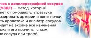
- ultrasound diagnostics of the kidneys;
- other procedures if necessary (laparoscopic diagnostics, etc.).
It is extremely important to examine the patient according to the principles of differential diagnosis.
Acute renal colic is accompanied by symptoms that are characteristic of very different pathologies, and the attending physician must be able to differentiate the attack from them. Acute pathologies of the abdominal cavity:
- appendicitis;
- perforated ulcers of the intestines and stomach;
- pancreatitis;
- cholecystitis;
- intestinal obstruction.
Female diseases of the pelvic organs and pathological processes:
- ectopic pregnancy;
- pipe ruptures;
- symptoms in women are similar to cystosis with its torsion and necrotization of the leg;
- inflammation of the ovaries/tubes, etc.
Sometimes in renal colic the symptoms resemble inflammatory processes of the urinary organs - urethritis, cystitis, etc.
The following diseases occur with colic-like symptoms.
Cardiovascular disorders (aortic aneurysm, heart attack).
Musculoskeletal problems: lumbar and thoracic osteochondrosis, neuralgia, hernia, etc.
Therapy
What to do with renal colic?
The strategy for helping people with colic involves two steps:
- Relieve pain (pain-relieving analgesics and antispasmodics are suitable for renal colic).
- Fix the problem that caused it.
It is extremely important to relieve pain and restore the flow of urine. Relieving renal colic and normalizing urodynamics will allow the patient to feel better, but will also help keep the kidney in working condition. When the primary goal is achieved, the doctor begins to eliminate the cause of the syndrome identified by the results of the examination. Treatment is carried out by a specialist whose profile the diagnosis relates to, after the exacerbation ends.
For renal colic, treatment is indicated in a hospital. The first step is to call an ambulance, after which the person can try to provide first aid while waiting for the team to arrive.
How to relieve an attack of renal colic:
- The lower back needs to be warmed up. It is best to take a warm bath with water between 38 and 40 degrees. The warmth of the water will dilate the blood vessels of the kidneys, help relieve spasms, and calm and relax the patient. A contraindication to this procedure will only be a suspicion of renal tuberculosis;
- take a painkiller. It is advisable to take a tablet of complex action - with an analgesic effect and the presence of an antispasmodic effect. These drugs include Baralgin and the like. You can take one of the non-steroidal anti-inflammatory drugs - Ketorolac, etc. No-Shpa, available in almost every first aid kit, helps a lot.
The bath and medication should be combined so that the effect is complex, in addition, we must remember that the tablets take about 30 minutes to start working. The effect of the product, superimposed on the warming, will allow the patient to wait for the doctor relatively comfortably. If you can’t take a bath for some reason, you can get by with a heating pad.
If these measures do not help reduce pain, the patient is given local anesthesia and subsequent emergency restoration of the dynamics of urine drainage. This, however, can only be done in a hospital.
Having arrived at the call, the emergency doctor will most likely suggest hospitalizing a patient with colic to continue treatment in a hospital. Despite the obvious necessity of this step, not everyone agrees to it when trying to be treated at home. Meanwhile, the lack of correct therapy (or treatment administered incorrectly to a patient with colic syndrome), even with sudden relief, can cause a relapse in the future, so it is highly advisable to carry out therapy on an inpatient basis.
There are some groups of patients who must be hospitalized both during exacerbation and during periods of relief:
- persons with symptoms of complications - a sharp drop in pressure, temperature of 38 degrees and above, confusion;
- persons with bilateral pain;
- people who have previously had one kidney removed (or were born with one organ).
Hospitalization of such patients is critical. If this is not done and normal urine output is not restored, the pathological process will become irreversible and the organs will lose their function. Possible death.
After providing first aid, you need to restore the flow of urine, this is done using:
- Medicines administered either orally or by intravenous injection. The drugs improve blood flow, promote the dissolution of stones and their removal. When administered as injections, they tend to work more effectively and quickly.
- Modern surgical methods include through the opening of the urethra or insertion of instruments into a small puncture in the skin.
Surgical treatment of renal colic is carried out using the following methods:
- percutaneous nephrostomy. This is an emergency method for restoring urodynamics in patients in urgent conditions, when endoscopic techniques are ineffective or impossible to use. A special drainage is inserted into the pelvic cavity through an opening in the skin;
- ureteral stenting. The doctor places a drainage in the pelvis, creating a bypass path for urine movement and relieving symptoms;
- endoscopic extraction of urolith. This is the most effective method of “natural” treatment, when the stone is removed through the urethra. It allows the patient to be healed quickly and with minimal injury.
Possible complications
With timely assistance, the prognosis for renal colic is favorable, patients quickly return to normal life. In the opposite case, however, complications of renal colic are possible - if help is late or therapy is carried out incorrectly. The severity of the consequences depends on how long the stagnation of urine lasts and the general condition of the person.
The most common complications:
- states of shock;
- pyelonephritis;
- sepsis;
- atrophy and death of the kidney, sclerosis of its tissues.
There are known cases of death of people who tried to treat renal colic on their own using traditional methods. Preventing the development of complications is simple - you just need to contact a qualified medical specialist for help.
Diseases of the urinary system often occur with severe symptoms. One of the most dangerous conditions with painful manifestations is renal colic.
How to relieve pain during an attack? What signs require urgent medical attention? Why does pain develop? The answers are in the article.
- What is renal colic
- Clinical picture
- General rules and effective methods of treatment
- How to relieve pain
- Medications
- ethnoscience
- Diet and nutrition rules
- Renal colic during pregnancy
- Useful tips
What is renal colic
A dangerous condition, accompanied by severe pain, develops suddenly. A person cannot sit and move calmly, severe pain increases rapidly.
The main factor causing painful symptoms is a violation of the outflow of accumulated urine from the problem kidney. Compression of the tubules or blockage of the ducts is caused by stones: phosphates, oxalates, urates or tumor formation. Acute symptoms often develop after lifting weights, high loads in the gym, long bicycle rides, or after riding a motorcycle.
The main causes of renal colic:
- urolithiasis in women and men;
- inflammatory kidney diseases;
- development of a tumor compressing the kidney;
- gynecological diseases;
- kidney tuberculosis;
- exacerbation of chronic pyelonephritis;
- severe pathologies of the retroperitoneal region.
Renal colic ICD code – 10 – N23.
What to do if the kidney on the left side hurts? Find out effective treatment options.
For a list of antibiotics for the treatment of chronic pyelonephritis in women, see this address.
Clinical picture
Renal colic in men and women has similar symptoms:
- during an attack, a person rushes about, looking for a more comfortable position in which discomfort is not felt so much;
- pain occurs sharply in the kidneys and spreads across the upper abdomen;
- unpleasant sensations quickly cover the ureter, bladder, are heard in the area of the ribs, and spread to the legs;
- often a sharp pain pierces the back, iliac region, and groin area;
- in the absence of timely assistance, painful shock is possible;
- the attack is accompanied by nausea and vomiting;
- One of the difficulties that arises during the relief of painful symptoms is problems with taking medications orally. Vomiting causes rapid dehydration;
- against the background of pain due to renal colic, frequent urges to empty the bladder appear and gross hematuria develops;
- Blood clots are often visible in the urine;
- blood pressure rises, pain and dizziness;
- with pyelonephritis (an inflammatory disease), the temperature often reaches 38–39 degrees;
- sometimes the patient feels the urge to defecate.
Peculiarities:
- symptoms of renal colic in men. The pain often affects the genital area;
- symptoms of renal colic in women. Pain sensations occur not only in the abdomen, lower back, and bladder, but also appear in the perineum and radiate to the thigh. One of the reasons is kidney prolapse, kinking of the ureter against the background of sudden weight loss or physiological characteristics of the body: a small retroperitoneal layer of fat or the absence of a layer that supports important organs in the correct position.
The symptoms of an attack in the kidney area are considered one of the most severe and painful, along with toothache. The shooting continues for 10 hours or more, sometimes lasting two or three days with short breaks. It is important to call an ambulance in time and, if necessary, hospitalize the patient in order to avoid serious complications due to dehydration, intoxication if the outflow of urine is impaired.
General rules and effective methods of treatment
The first rule is to seek emergency medical help. Sometimes negative symptoms indicate the development of other pathologies that have similar manifestations. Wrong actions and delay in calling an ambulance in some cases pose a risk to the victim’s life.
What is an attack of renal colic often confused with:
- ectopic pregnancy;
- attack of radiculitis;
- lumbago – lumbago in the lower back;
- acute appendicitis;
- biliary colic;
- perforated ulcer in the stomach;
- acute inflammation of the pancreas.
How to relieve pain
Emergency care for renal colic:
- immediately call a medical team;
- give painkillers and antispasmodics;
- if you are 100% sure that an attack develops against the background of urolithiasis in men and women, you need to place the victim in a bath with hot water or apply a heating pad;
- in other cases, the use of heat is unacceptable: high temperature causes active progression of inflammation or other pathological process in the peritoneal area.
Medications
Oral administration due to vomiting and nausea is difficult; taking a tablet is quite difficult. To quickly relieve acute symptoms and allow active components to enter the bloodstream, injections of analgesics, non-steroidal anti-inflammatory drugs (NSAIDs) and compounds that relieve spasms of smooth muscles are prescribed. Intramuscular and intravenous administration should not be carried out for longer than three days; then painkillers and suppositories and tablets are prescribed.
Injections:
- Baralgin.
- Metamizole.
- Ketoprofen.
- Pitophenone hydrochloride.
- Ketorolac.
- Diclofenac.
Pain relieving suppositories:
- Ketanov.
- Diclofenac.
- Spazdolzin.
- Baralgin.
Tablets for the treatment of renal colic:
- Paracetamol.
- No-shpa.
- Spasmalgon.
- Platyfillin.
- Drotaverine.
- Baralgin.
Important nuance:
- before the doctors arrive, painkillers are given in minimal quantities: too high a dose of analgesics “blurs” the picture of the attack, making it difficult for doctors to figure out what factor caused the excruciating pain;
- It is difficult to endure pain during lumbago, but addiction to analgesics can negatively affect the diagnosis and development of a treatment regimen. You should not frequently take painkillers in the hope of self-cure: only a specialist can determine the cause of the problem;
- It is important to know: advanced cases of self-medication often provoke renal failure.
Learn about the diagnosis of urolithiasis in men and treatment options for the disease.
Read about the causes of kidney failure and the treatment of pathology in males at this address.
Follow the link https://vseopochkah.com/bolezni/mochekamennaya/y-zhenshin.html and read the information about the treatment of urolithiasis in women with the help of medications.
ethnoscience
During an attack, herbs and homemade ointments help little: without the use of NSAIDs, antispasmodics and analgesics, acute pain cannot be relieved. It is important not only to relieve discomfort, but also to understand why the attack develops and to treat the underlying disease.
Folk remedies for renal colic are ineffective; herbal decoctions to improve urine flow, relieve inflammation, and normalize the functioning of nephrons are prescribed after eliminating dangerous signs. In the treatment of urolithiasis, pyelonephritis, and other kidney pathologies, medicinal plants are beneficial, but during an attack you should not rely on corn silk or nettles: you need to call a medical team and use medications.
How to remove stones
Measures to eliminate kidney stones are prescribed depending on the characteristics, severity of the pathological process and the individual characteristics of the patient.
Options for stone expulsion tactics
There are several ways to treat this disease:
- Conservative therapy. If stones are detected in a timely manner, drug treatment is carried out aimed at dissolving them. If the size of the formations does not exceed the diameter of the ureter and urethra, drugs are prescribed to cause their release.
- Special diet. Simultaneously with taking medications, the patient is transferred to a special diet and increased drinking regimen, which must be combined with the use of diuretics.
- Traditional medicine. Home recipes are used in addition to drug treatment, but can also be an independent method if only sand or very small stones are found in the kidneys.
- Crushing uroliths using hardware techniques. If the stone is large, it is crushed using non-contact or urethral (contact) lithotripsy. The resulting small particles can be removed naturally or using an endoscope.
- Surgical intervention. Most often, stones are removed using minimally invasive endoscopy. In particularly difficult cases, open abdominal surgery is performed.
In most cases, the stones are first crushed and then removed using medication or endoscopic extraction.
Hardware crushing methods
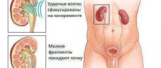
Three types of lithotripsy are used to crush stones:
- shock wave - the most common method, which is based on the impact of shock waves directed from the outside, without penetration of instruments into the tissue;
- urethral - the instrument is inserted through the urethra, and then the stone is crushed by ultrasound, pneumatics or laser;
- percutal - performed laparscopically (through a hole in the soft tissues with a diameter of no more than 0.5 cm) with mechanical crushing of the stone.
All of these procedures are performed under general anesthesia.
Surgical extraction
Surgery is performed in several ways:
- endoscopic treatment is the most minimally invasive, since it is performed through the urethra and completely eliminates tissue trauma, but for large stones it requires preliminary crushing using the above methods;
- laparoscopic removal - is carried out in cases where the problem cannot be solved endoscopically, through a puncture of 0.5 cm in size, and for large stones it also requires preliminary crushing;
- open (cavitary) operations - are carried out in extreme cases by making an incision in all surrounding tissues to access the stone and remove it.
Due to the availability and accessibility of modern minimally invasive methods of getting rid of uroliths in the kidneys, surgical intervention is used extremely rarely.
Indications and contraindications for lithotripsy
When performing this operation to remove kidney stones, it is possible to get rid of only stones up to 2 centimeters in size, and determining the location does not cause difficulties. If urolithiasis has reached the fifth stage, then this method of treatment is not only useless, but even dangerous.
This operation to crush kidney stones is not recommended in the following situations:
- gestation period;
- injury to the musculoskeletal system, which makes it impossible to take the position on the couch required for the operation;
- the patient’s body weight is more than 130 kilograms, height is above 2 meters or below 1 meter;
- problems with blood clotting.