Poor quality food and excessive alcohol consumption lead to the formation of sand and kidney stones. Experts say that if you follow basic preventive measures, you can avoid urolithiasis. If stones appear in your life, do not wait until the disease worsens, contact a urologist who will prescribe diagnostics and effective therapy. In this article we will talk about what to do if you have kidney stones. We will analyze the types of stones, their sizes, causes of formation, and also find out in which case it is necessary to immediately go to the hospital.
Types of kidney stones
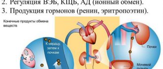
The mineralogical classification indicator for kidney stones is considered an international standard in urology. To provide proper medical care, and most importantly, the purpose of effective treatment, the urologist needs to know not only the reason for the formation of corals, but also their shape and size. There are four main mineralogical types of urinary stones:
- An inorganic compound of calcium salts, resulting in the formation of oxalates and phosphates. These formations are common and are diagnosed in more than 70% of patients;
- Infectious formation, appearing as a result of previous diseases of the urinary system, is diagnosed in 20% of patients;
- Urate formation or uric acid stone is diagnosed in 10% of patients;
- Xanthine or cystine formation, diagnosed extremely rarely, in only 5% of patients, is caused by hereditary pathologies or a violation of amino acid processes in the body.
For information! Almost 50% of patients suffering from stones have mixed types.
Kidney formations are:
- multiple and single;
- bilateral and unilateral;
- flat, round, with spikes and edges;
the size can be from 3 mm to the size of the kidney itself (staghorn stone that forms the cliche of the collecting system).
mama.tomsk.ru
Tentorium is not a dietary supplement, it is a living food product containing various beekeeping products: honey, propolis, pollen, royal jelly, wax, etc. The KTR program is designed to get rid of stones and does not drive stones anywhere. The stones dissolve, if taken correctly, there will be no emergency situations.
Karma wrote: At least because Tentorium products are not a medicine, but a dietary supplement. And its use can help prevent the appearance of stones, relieve inflammation of the gallbladder, improve its contractions, and change the composition of bile. And if there are stones, it’s too late to drink Borjomi when the kidneys have fallen off. Small stones can come out on their own, but I wouldn’t specifically drive them away with a nasty broom; who knows, maybe they’re not that small. And an emergency operation to remove the gallbladder due to a violation of the outflow of bile is an extremely unpleasant thing.
Yeah, I’m cracking a tangerine right now - it’s also like a living food product, but it doesn’t dissolve anything (and thank God) I don’t think that honey is any different from it in any way. In general, it becomes sad when in general a good product begins to be attributed with all sorts of miraculous properties for no apparent reason, only trust is lost in both the product and the seller
Sue wrote: oh, not pleasant at all. and then at 27 you can’t wear a swimsuit.
Why is it so sad? Scars, of course, mainly decorate men, but if a girl has a good figure, scars, in my opinion, are the twenty-fifth thing people notice. And the diet after cholecystotomy only contributes to the preservation of forms. As a last resort, you can remove the scar; it still has no benefit to the body.
marfa Important person / Forum authority Messages: 36161 Registered: February 27, 2008, 18:32 From: Tomsk, Kashtak
Not everything is so bad, stones can be removed endoscopically, and you can wear a swimsuit all year round. And the tentorium is, of course, nonsense in terms of dissolving stones.
It’s sad, but a person usually finds out about the existence of a gallbladder when he has pain in the right hypochondrium or in the upper abdomen. The pain may radiate to the back, right arm, or collarbone. This means that, firstly, stones have formed in the gall bladder, and, secondly, they have started to move, causing inflammation of the bladder - cholecystitis. A situation that requires immediate medical attention.
The fact is that, having set off on their own, the stones can completely block the outflow of bile and cause inflammation of the gallbladder - cholecystitis, inflammation of the pancreas - pancreatitis or obstructive jaundice.
“Gallstones form for two reasons: due to an imbalance of bile components and due to its stagnation,” says the head of the abdominal department of the Institute of Surgery. Vishnevsky RAMS, Doctor of Medical Sciences Vyacheslav Egorov. — The occurrence of cholelithiasis can be provoked by a sedentary lifestyle, in which, as a rule, metabolic processes in the body slow down.
But the main risk group is those who eat irregularly, as well as those who like fatty foods with high cholesterol content. For these people, each feast is accompanied by a change in the composition of bile, and the likelihood of stone formation in such cases increases many times over.
Depending on the components, gallstones can be cholesterol, pigment, if they are formed from the coloring substance of bile - bilirubin, and calcareous, if calcium salts predominate in them. Most often, mixed stones ranging in size from 0.1 mm to 3–5 cm are found.
It is difficult even for an experienced doctor to make a diagnosis of gallstone disease by eye. This will require additional studies - ultrasound of the abdominal organs; in the most complex cases, X-ray studies with the introduction of a contrast agent into the bile ducts may be required. Currently, there is a study that allows the doctor to see the stones with his own eyes - choledochoscopy. These procedures allow the doctor to assess the size of the stones, their location, predict the further development of the disease and prescribe treatment.
Doctors are relentless: only a surgeon can get rid of gallstones! However, if there are no symptoms of the disease and the gallstones are “silent”, they can be left alone. The most important medical instruction for patients with gallstone disease is to adhere to proper nutrition and a strict diet. Spicy, fatty, fried and smoked foods are strictly prohibited.
Sometimes they try to dissolve small cholesterol stones with the help of medications - chenodeoxycholic acid and ursofalk. The treatment is long-term - the course lasts at least a year, it is expensive and, unfortunately, does not always lead to the desired results. By the way, in case of cholelithiasis, choleretic herbal remedies are strictly contraindicated. They can contribute to the migration of stones, and this is fraught with the most dangerous complications.
You can try to destroy small single stones with a shock wave. During this procedure, the stones are crushed into small pieces (up to 1-2 mm in size), which independently exit the body. This procedure is painless, well tolerated by patients and can be performed on an outpatient basis.
Often patients with gallstone disease undergo surgery for emergency reasons, when removal of the gallbladder - cholecystectomy - is vital.
The gold standard for gallstone disease is laparoscopic surgery , in which the gallbladder is removed through small punctures in the anterior abdominal wall. The patient is usually discharged a day or two after the operation, and he quickly returns to his usual rhythm of life.
Many people are concerned about the question: is a full life possible without a gallbladder?
Doctors say yes. The purpose of the gallbladder is to store bile until food is consumed. It was vitally necessary only for primitive people, who did not sit down at the table every day. Modern man has no need to eat in reserve.
Integrate Pravda.Ru into your information flow if you want to receive prompt comments and news:
Add Pravda.Ru to your sources in Yandex.News or News.Google
We will also be glad to see you in our communities on VKontakte, Facebook, Twitter, Odnoklassniki.
Chemical composition of renal formations
Absolutely all formations in the renal system have a different chemical and heterogeneous composition. If a urologist calls the formation urate, phosphate or oxalate, he means the predominant chemical components of the growth. In modern medicine, there is a concept that the formation of corals occurs due to changes or disturbances in the relationship between salt and urine colloids. According to their chemical composition, stones are divided into:
- urate - formed as a result of uric acid salt;
- oxalate - formed due to increased levels of oxalic acid in the body;
- phosphate - formed due to apatite or calcium phosphate;
- carbonate - formed as a result of calcium salt and carbonic acid;
- struvite - formed due to ammonium phosphate.
For information! Absolutely all stones measuring from 5 to 10 mm can be passed painlessly and independently with urine. If there is pain and cutting in the lower abdomen and lower back, urologists prescribe diuretics and antispasmodics.
It is worth noting that urologists also distinguish between stones of organic origin. These include:
- amino acid formation or xanthine and cystine;
- cholesterol formation, small black stones, as a rule, crumble easily, they cannot always be detected during the diagnosis period;
- protein formation, a small clot of fibrin that contains salt and bacteria, they are easy to detect during diagnosis.
How do stones pass out?
The process of stones passing through the biliary system is not always accompanied by pain. If the stones are small in size, they can pass out unnoticed along with the bile, and this will only be detected during an ultrasound examination.
Large stones can cause significant discomfort. They injure the mucous membrane of the biliary tract, and sometimes completely block their lumen, leading to the development of the following symptoms:
- sharp paroxysmal pain in the right hypochondrium, which intensifies when eating fatty foods or when pressing in this area;
- discoloration of stool;
- darkening of urine;
- the appearance of a yellow tint to the skin, mucous membranes and sclera of the eyes;
- dyspeptic disorders (nausea, feeling of bitterness in the mouth);
- increase in body temperature (with the development of inflammatory complications).
The duration of the process of stone release also varies. A gallstone can pass out in as little as 10 minutes or as quickly as several days.
Stone sizes
Kidney formations can reach completely different sizes. This may be one large stone in the kidney, but in urological practice multiple stone formations are also quite common. Their sizes can vary, from 1 mm to 8 mm. For example, a 4 mm kidney stone may be present and not manifest itself in any way, i.e. the person does not feel discomfort or pain. And there are such growths that in a short period of time can grow and fill the entire calyx and pelvis, and in size they can reach up to 20 mm. If there are small formations, for example five millimeters, removal can be done independently with the help of medications.
Removing stones using ultrasound
If the crystal does not come out on its own, or when there is a possibility of injuring the genitourinary system, doctors resort to ultrasonic breaking of stones. Many people are afraid of the procedure, but in vain. There is nothing complicated or painful about it. The waves pass through the body and, encountering a dense object (stone), crush it. The whole process takes no more than an hour. In this case, the patient is in the operating room, he lies on a special water cushion. In modern clinics, the patient is placed in a reservoir of liquid. This way the crushing process goes faster, and the waves travel better. For those with low blood pressure, doctors suggest administering a painkiller. By removing kidney stones with ultrasound, you can be confident in the effectiveness of the method. The crystals break down into small pieces and turn into sand, which is released when you urinate.
Sources used:
- https://m.baby.ru/community/view/73449/forum/post/348191624/
- https://opochk.ru/idet-kamen-iz-pochek-chto-delat-i-kak-oblegchit-bol/
- https://myhube.ru/signs-of-a-stone-moving-in-the-kidney-renal-stone-disease-occurs-frequently/
- https://fleynaro.ru/delat-esli-tronulsya-kamen-poc/
- https://lecheniepochki.ru/kamni-v-pochkax/kamni-v-pochkax-chto-delat.html
- https://xn—-itbjbjcjri0ff.xn--p1ai/chto-delat-kogda-kamen-v-pochke-shevelitsya/
How to remove stones?
People suffering from kidney diseases often wonder how to remove a kidney stone? Urologists say that not all stones can be removed on their own. For example, a 6 mm kidney stone can be eliminated with the help of special medications. A 6 mm stone can cause the following symptoms in a patient:
- cramps and pain in the abdomen and when urinating;
- the appearance of red cells in the blood and urine;
- increase the temperature;
- inflame the mucous membrane.
If the stone formation has a heterogeneous and too dense chemical structure, and the size of the kidney stone is 8 - 20 mm, then such formations should be removed surgically. Crushing stones up to 20 mm in size is performed without cutting the skin or kidney directly. A special instrument is inserted through the urethra and corals are crushed using laser and ultrasonic waves. If stone formations reach more than 20 mm, the operation is performed using the open method. This operation is quite traumatic and has a large number of complications, because During the intervention, an incision is made into the skin and kidney in order to remove the corals. Experts do not recommend self-medication; if you feel discomfort and pain, consult a doctor for advice and a full diagnosis.
Main symptoms
Symptoms of kidney stones can be divided into the following categories:
- Kidney stone 5 mm what to do
- Spastic conditions;
- Urinary syndrome;
- Dysmetabolic disorders.
Spastic conditions due to urolithiasis lead to impaired urine flow. Frequent urge to urinate, lower back pain and tension in the abdominal wall - symptoms often occur against the background of contraction of small arterial vessels.
Localization of pain with urolithiasis
During physical activity, the movement of a stone can provoke not only a spasm of the urinary tract, but also pain in the lower back.
Irritation of the kidney tissue and ureters causes persistent narrowing of the ureter.
The urethra responds to any type of mechanical impact with a reflex contraction of smooth muscles. Persistent spasms can lead to severe obstruction and stagnation of urine in the kidneys. In such a situation, renal colic develops.
Passage of kidney stones - symptoms of renal colic are manifested by acute pain in the lower back. Its duration ranges from several hours to a day. With pathology, a person takes a forced position, because he is afraid that a careless movement will cause severe pain.
The peculiarity of pain syndrome in kidney diseases, in contrast to intervertebral hernia, is that it is localized on one side (right or left).
There is a high probability of pain radiating to the perineum and groin area (if the stone is located in the lower third of the ureter).
- How to remove urinary stone from toilet
Urinary syndrome occurs in 50% of patients with kidney stones. With it, blood, a small number of leukocytes and protein appear in the urine. If the calculus damages the mucous membrane of the ureters or pelvis, total gross hematuria is formed. With it, an abundant amount of red blood cells can be traced in the urine.
Gross hematuria
If urolithiasis is combined with inflammation, then leukocytes (leukocyturia) appear in laboratory urine tests. This symptom is not pathognomic for stone formation, but with large and abundant stones it is often found in people.
Prevention of coral formation
To avoid worsening the disease, experts offer simple recommendations that can alleviate the condition and help the stones pass painlessly:
- try to drink 2 liters of purified water daily, the liquid will help cleanse the kidneys of sand and salts;
- consume more diuretic foods, such as watermelon or teas;
- spend more time outdoors, take walks;
- avoid hypothermia of the lumbar region, especially in the cold season the back should be covered;
- always use protection during sexual intercourse, this will help avoid the transmission of genitourinary infections, but if the act was without additional protection, you must immediately go to the toilet, this will help avoid infection in the kidneys;
- follow a diet, exclude seafood, salted, smoked foods, alcohol, smoking, all foods that contain high levels of oxalic acid;
- use diuretic herbal decoctions of St. John's wort and corn silk;
- consume more cranberry juices.
Can your kidneys hurt?
Due to various diseases and abnormalities, the kidneys really hurt. The sensations associated with kidney disease may resemble those experienced by a person suffering from osteochondrosis or radiculitis. Discomfort in the back spreads to the lower half, is felt in the area of the kidneys, extends to the thigh, groin area.
Abnormal mobility of the kidney, in which it descends, is called nephroptosis. Stress is blamed for surges in blood pressure during nephroptosis, dull nagging pain is often attributed to osteochondrosis, and recurring inflammation is attributed to colds.
The lower back may hurt due to the kidneys on one or both sides. In most cases, the cause of pain is inflammation - pyelo-, glomerulonephritis, displacement of stones due to impaired salt metabolism.
Causes of kidney stones
The main cause of urolithiasis is various metabolic disorders in the body: changes in the chemical composition of the blood, water-salt imbalance. The result is clogging of the renal pelvis and ureter with salt crystals, from which stones gradually form.
There are several factors that provoke stone formation:
- constant consumption of water with a high salt content;
- abuse of sour, salty, spicy, fried foods;
- regular dehydration of the body;
- features of climatic conditions;
- sedentary lifestyle;
- infections of the kidneys and genitourinary system;
- genetic factor;
- imbalance of vitamins in the body;
- alcohol abuse;
- abuse of diuretics (diuretics);
- uncontrolled use of medications;
- injuries.
The chemical composition of stones is determined by specific reasons. Calcium is formed due to excess calcium in the blood and urine, urate - due to increased acidity of urine. Excessive intake of oxalic acid can cause the formation of “deposits” with ammonium, magnesium or phosphate composition.
Causes of the disease
The most common causes of the disease are:
- inflammatory process of the gallbladder;
- the content of bile in increased amounts of cholesterol, bile pigment and calcium serves to convert bile into water-insoluble bilirubin;
- genetic factor is one of the reasons for the formation of stones. The child is likely to develop gallstone disease if one of the parents has had it;
- treatment with drugs such as octreotide, cyclosporine, clofibrate;
- long intervals between meals and fasting foreshadow the formation of stones;
- Crohn's disease, cirrhosis of the liver and Caroli syndrome can lead to the formation of gallstones;
- when the lower part of the intestine is removed, stones form in the bile;
- drinking alcohol in large quantities causes the formation of stagnation of bile, as a result of which bilirubin crystallizes and stones appear.
One of the main reasons for the appearance of cholelithiasis is the intake of large amounts of fatty foods with cholesterol. Due to impaired nutrition, obesity develops, which is a secondary risk factor for the development of the disease. In addition, incorrect functioning of internal organs and liver function failure can negatively affect bile and subsequently lead to the formation of stones.
Stones appear when the walls of the gallbladder contract poorly. The causes of poor bladder contraction may include problems such as flatulence and dyskinesia. Surgeries on the gallbladder can also affect its proper functioning. Another reason may be pregnancy, during which the gallbladder becomes stressed and its functioning is disrupted. Cysts, tumors, adhesions, swelling of the bladder walls, congenital defects - all of this can serve as a factor contributing to stone formation.
Infections that enter the bladder through the lymphatic or bloodstream may be one of the causes of cholelithiasis. Inflammation of the gallbladder and its canals is caused by any infection that gets into it. In the future, this leads to cholangitis and cholecystitis, and subsequently to the development of cholelithiasis. In medicine, stone formation is divided into two types:
- primary – the formation of stones occurs slowly and without any signs for a long time;
- secondary – the cause of stones is congestion in the gallbladder.
Bile contains various components and, therefore, the stones that form have a distinctive composition. Stones are classified into the following types:
- round cholesterol stones with a diameter of 16-18 mm;
- limestones containing large amounts of lime;
- mixed stones - have a layered structure, and sometimes have a cholesterol shell and a pigmented center, ranging in size from 0.1 to 5 cm;
- pigment stones - appear due to an increase in water-soluble bilirubin. They contain substances from calcium salts and bilirubin. In addition, there is a possibility that small bilirubin stones may appear in the bladder, which are located in the ducts and sac.
What can be done?
The exact treatment regimen for kidney stones is selected by the doctor individually, taking into account the characteristics of the disease, age and general health of the patient.
Measures that you can take yourself without fear of side effects are determined by the previous points:
- take measures to purify drinking water from salts and harmful impurities;
- The use of mineral water and its quantity should be agreed with your doctor;
- minimize the consumption of fried, spicy, salty, sour;
- drink the amount of fluid recommended by your doctor;
- before a trip that involves a sudden change in climate, consult a doctor;
- walk and swim more;
- promptly treat all infectious diseases;
- take vitamin complexes only as prescribed by a doctor;
- stop drinking alcoholic beverages, including beer and tonics;
- take diuretics only as prescribed by a doctor;
- do not self-medicate even with “harmless” medications, dietary supplements, freely sold in pharmacies;
- control physical activity and conduct sports training only under the guidance of an experienced trainer and with medical permission.
Following simple rules of a healthy lifestyle will help reduce the risk of exacerbation and at least slow down the process of stone formation. This will not remove kidney stones, but it will help reduce some of the symptoms of the disease.
Eliminating pain
When pain in the lumbar spine spreads to the legs, qualified medical help is needed . A set of measures has been developed for this purpose. Basically, these are medical procedures and special medications. Naturally, before treatment, a thorough examination is carried out to identify the cause of the pathology. Then symptomatic therapy will be prescribed to stop the progression of the disease.
The choice of treatment regimen depends on various factors:
- Degrees of severity.
- Main pathology.
- The presence of inflammation and acute pain.
- Gender and age.
- Period of pregnancy or lactation.
Therapy will have the following goals:
- Identifying and eliminating the cause of the disease.
- Relieving the slightest discomfort.
- Complete restoration of mobility.
- Eliminate pain and stop the inflammatory process.
If a disease of internal organs is detected, then therapy is selected strictly individually.
First aid
Self-medication is not encouraged, but sometimes situations arise in which it is necessary to eliminate acute pain, at least for a short time. The most effective way is considered to be the fetal position .
If the pain radiates to one leg, then you need to lie on the opposite side and pull the sore leg towards your stomach. When the pain syndrome spreads to both legs, you need to lie on your back, pulling both legs to your stomach. Do not roll over onto your stomach.
Medicines
Treatment includes several groups of drugs:
Analgesics. For acute pain, you cannot do without Paracetamol, Sedalgin and Nise. These painkillers are very strong, but their effects are short-lived.
Chondroprotectors. These products restore cartilage tissue. The most popular:
- Chondroxide tablets.
- Teraflex Advance capsules.
- Chondroxide gel or ointment.
- Chondroitin ointment AKOS.
Anti-inflammatory drugs. Diclofenac and Ketotifen are usually used. They are considered the most effective means. Non-steroidal medications are excellent at relieving swelling and inflammation. The pain also goes away quickly.
Corticosteroids. The medications contain hormonal substances. Mostly. Prednisone and Diprospan are used.
Myoresistants. Relieves muscle spasms and eliminates pain by relaxing muscles.
Medicines are prescribed only by a doctor; independent treatment should be excluded. It is necessary to strictly adhere to the dosage and follow the instructions included with the drug.
Physiotherapy
If lumbar pain radiates to the legs, then a complex of physiotherapy procedures is often used. They promote faster pain relief, significantly improve blood flow and restore metabolism.
If there are no contraindications, then it is recommended:
- Electrophoresis.
- Mud baths.
- Acupuncture.
- Darsonvalization.
- Applications and therapeutic compresses.
- Manual therapy.
All procedures are carried out in courses that are repeated after 3 weeks.
Surgery
If medications and physiotherapy do not help, they resort to surgery.
Indications for this method are:
- Paralysis.
- Paresis of the limbs.
- Complications affecting the functioning of internal organs.
- Unbearable pain that is not relieved by analgesics.
But first, traditional methods are used, since during the operation there is a risk of complications with negative consequences.
Folk remedies
In addition to conservative treatment, alternative methods are often used.
There are effective traditional medicine recipes:
- Pepper tincture . Add 100 g of black pepper to 300 g of vodka. The mixture is infused in a glass container for two weeks. The lower back is rubbed with the tincture 2 times every 7 days.
- St. John's wort tincture . Grind a tablespoon of the plant. Add to boiling water and leave for 10 minutes. The tincture is drunk daily, one spoonful for a month.
- Horse chestnut . Ghee is mixed with mashed chestnut and camphor oil is added. All ingredients are in equal proportions. Apply the resulting ointment to a piece of bread and apply to the lower back.
- Ointment made from lard and hops . Grind hop cones (100 g) and mix with lard (150 g). After a week, the ointment can be applied to the sore spot of the lower back.
- Salt bath . Add sea salt (200 g), lemon juice (50 g) and a few drops of lavender oil to warm water. The bath takes 20 minutes.
Although the recipes are time-tested and quite effective, you should still consult a doctor first.
Video: “Advice from a specialist on eliminating pain in the lumbar spine”
Exercise therapy and massage
Pain in the lower back that radiates to the legs can be eliminated with the help of special exercises. They restore metabolism in the painful area.
A well-designed complex will allow:
- Ensure normal blood supply.
- Provide adequate nutrition to the problem area.
- Increase the elasticity of the muscles that support the spine.
Exercise therapy is selected only by the attending physician, since improper execution of movements can aggravate the condition, even leading to surgery.
But while doing the exercises you must follow certain rules:
- The first training is carried out under the supervision of a doctor.
- Therapeutic exercises are strictly prohibited during exacerbation of the disease.
- The recommended complex cannot be changed.
- If pain occurs, training stops immediately.
- Exercises are performed smoothly and slowly.
- There is always a 30 second break between sets.
- It is useful to hang on the horizontal bar.
It is advisable to supplement physical exercise with massage. Various types are used: classic, vacuum, spot, honey, jar.
But for some diseases massage is prohibited:
- In the presence of active tuberculosis.
- For oncology.
- If there are cardiac pathologies.
- For diseases of lymphatic diseases.
- If you have varicose veins.
The combination of therapeutic exercises and massage sessions is necessarily used in complex therapy.
Kidney stones - what are the symptoms?
The process of formation of salt deposits in the kidney can occur for a long time without external manifestations or be accompanied by symptoms similar to those of other diseases. Therefore, a sudden attack and diagnosis of a “stone” is often a complete surprise for the patient.
Doctors strongly recommend undergoing examination for the following phenomena:
- frequent urination in small portions;
- back pain, radiating to the lower abdomen, and in men - to the genital area;
- intermittent stream of urine;
- discomfort, burning when urinating;
- urine is cloudy or contains blood;
- nausea or vomiting.
Attacks of aching pain in the lumbar region can be triggered by physical activity, long walking, or an uncomfortable position during a long trip.
The movement of the stone may be accompanied by:
- dizziness;
- a drop or increase in blood pressure;
- bloating;
- stool retention;
- temperature rise.
The cessation of pain or disappearance of symptoms is not a reason to postpone a visit to the clinic until later. Delayed consultation with a doctor significantly worsens the prognosis of the disease.
Exercising when sick
If cholelithiasis is detected, you should limit yourself in physical activity. This also implies the exclusion of household physical activity. Physical activity during the course of the disease can help:
- rapid formation of bilirubin (this is dangerous for people with bile stagnation);
- movement of stones (if there are stones, then physical activity will move them out of place);
- likelihood of complications of cholelithiasis;
- complications after surgery.
According to the doctor's indications, daily 30-60 minute walks, gymnastics without stress on the abdominal area and sudden movements, swimming without diving are allowed. These loads are considered to prevent the occurrence of gallstones and also promote recovery after surgery. Professional sports involving heavy loads are prohibited for patients with cholelithiasis.
What should you not do if you have kidney stones?
The main thing is that you should not attempt self-treatment.
Kidney stones can only be removed through surgical intervention in a clinic.
If you have kidney stones, you should limit the use and, during an exacerbation, stop using:
- leafy vegetables and herbs;
- strong meat, fish and mushroom broths;
- legumes;
- chocolate, peanut butter, confectionery and fruit preserves;
- strong tea and coffee;
- carbonated drinks;
- dairy products;
- pepper, horseradish and mustard;
- sour berries and fruits.
The treatment complex for kidney stones may include traditional medicine, but the possibility of their use should be discussed with your doctor.
Urolithiasis is a pathology characterized by the deposition of solid organic formations in the kidneys.
Concretions in the form of sand and stones in the renal pelvis are often the result of an irresponsible attitude towards one’s health.
Doctors believe that in most situations they can be prevented by performing simple prevention.
But what to do if kidney stones have already formed? Waiting for them to “resolve” on their own or come out naturally is not the best solution. Left unchecked, urolithiasis can lead to dangerous consequences, including renal failure.
Prevention of exacerbation of cholelithiasis
After a course of treatment in a hospital, rehabilitation therapy is prescribed. It includes various drugs that improve liver function, for example, Essentiale and other hepatoprotectors. It is important to prevent a possible attack in the future. A sedentary lifestyle, obesity, diabetes are risk factors.
You should stick to a strict diet for as long as possible; table No. 5 is recommended. It is necessary to refuse semi-finished and instant products. Food should be fresh and balanced towards increasing the proportion of protein and decreasing fat. Sweets are allowed only of natural origin: honey, dried fruits, berries. Physical exercise and quitting smoking, as well as avoiding stress (for example, changing occupations) greatly contribute to recovery.
Causes and mechanism of kidney stones
To understand what to do with kidney stones, you need to understand the mechanism of their formation.
The main cause of urolithiasis is considered to be a violation of water-mineral metabolism, when an excessive concentration of salts is observed in the urine.
Why this happens - either the body produces salts too actively, and they do not have time to be excreted through the kidneys in a timely manner, or the problem is related to poor urination - it is difficult to say without examining a specific patient.
A person may well have a natural tendency to form stones. This happens with improper development of the kidneys in the prenatal period, with a family predisposition to pathologies of the urinary tract, and congenital metabolic diseases.
- unhealthy diet (“too much” with proteins, salt, calcium);
- insufficient volume of fluid intake;
- excessive mineral content in drinking water (so-called “hard” water);
- sedentary life and bad habits;
- chronic inflammatory processes in the body.
Of course, all this contributes to stone formation not on its own, but against the background of pathological conditions associated with infections of the genitourinary organs, endocrine diseases, and physiological processes (for example, pregnancy in women).
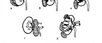
Urogram of the kidneys of patients with different types and localization of stones
When, due to such disorders, problems with the outflow of urine begin, the prerequisites for the deposition of stones are created in the renal pelvis. Mineral salts in stagnant urine settle, fall out in crystals and form pebbles of different shapes and sizes.
An excess of uric, oxalic, phosphoric, and carbonic acids, synthesized in the body and entering it from the outside, leads to the formation of stones from their salts (usually calcium). The tactics of their treatment depend on the composition of the formed stones.
Diagnostics
Before figuring out how to treat kidney stones in women or men, it is necessary to get a proper diagnosis. Modern diagnostic methods will help us with this:
- ultrasound examination of the diseased organ;
- laboratory testing of urine and blood;
- urography (survey and/or excretory).
As an additional examination, the following is prescribed:
- multislice computed tomography - this method allows you to determine the size and type;
- nephroscintigraphy - determines the level of functional disorders in the kidneys;
- determination of sensitivity to antibiotics - the level of development of the inflammatory process is determined.
Naturally, the patient is examined and questioned - it is necessary to find out the possible reasons that provoked metabolic disorders before the formation of kidney stones. Such a diagnosis is basic - based on the results obtained, a conclusion can be drawn and treatment can be prescribed.
Dissolving kidney stones
There are many myths about dissolving kidney stones:
- It is believed that you can “cleanse” kidney stones by drinking liters of water and taking diuretics.
- Urate stones can be dissolved by acids, that is, you need to consume as much acidic drinks (lemon, cranberry juice) and medications (aspirin, asparkam) as possible.
- To combat oxalate stones, you need to “alkalize” your urine.
The use of these dissolution methods has no medical justification.
Drinking plenty of fluids actually helps flush the kidneys and serves as a prevention of nephrolithiasis by reducing the concentration of salts in the urine.
But if the stones have already formed, it is impossible to dissolve them in this way.
Sour drinks can generally be harmful: cause stomach problems and further stimulate stone formation, and “alkalization” is only suitable for urate stones.
Only a doctor can distinguish these stones from other types: the level of urea in the tests will be increased, the pH of the urine will be shifted to the acidic side, the stone will be visualized with ultrasound, but not with x-ray.
Once you are sure that urolithiasis is of uric acid origin, you can try a chemical effect on the kidneys.
The task is to make the urine reaction slightly acidic or alkaline, to reduce the level of urea in the body, for which drugs are used that disrupt its synthesis: Blemarin, Allopurinol.
For stones of mixed composition, litholytic therapy is no longer so effective, and experts highly doubt that any stones can be dissolved with the help of pharmaceutical and folk remedies.
For medications to work, they must be used for a long time - from 2 months to six months, combined with a diet and sufficient drinking.
Important information
If you suspect kidney inflammation, you should undergo appropriate examination. You already know how to determine whether your kidneys or back hurt. However, inflammatory processes can change the structure of the internal organ. Thus, the membranes of the choroid plexuses of the kidney can be affected, and the tissue itself can be deformed. With pyelonephritis, on the contrary, the organ often increases in size. Of course, the patient will not be able to determine the condition of the internal organ by touch. An ultrasound examination will provide the necessary and accurate information. Remember that in 80 percent of cases, kidney disease occurs without pronounced pain symptoms.
Removing kidney stones
If the stone is insoluble, it can be removed from the kidney naturally (without surgery) only after the crushing procedure.
Many patients make a big mistake when trying to treat urolithiasis on their own, arranging all sorts of cleansing and rinsing.
If you have kidney stones, any diuretics should be used with great caution, and it is better not to do this at all without the direct recommendation of a doctor - there is a danger of moving the formation into the ureter. What does this mean?
- When a stone moves through the urinary ducts, they become injured, causing pain and bleeding.
- The lumen of the ureter is blocked, as a result of which the passage of urine is difficult.
- Impaired urodynamics lead to renal colic, a prolonged attack of acute pain that may require medical intervention.
- A stone stuck in the ureter can only be removed surgically.
If the stone “goes”, it doesn’t matter whether it is soluble or not - there is no time left for conservative treatment. In this situation, it is important to have a clear understanding of the size of the stone.
Shock wave crushing of kidney stones
If you have not been to a doctor and have not had an ultrasound, it is better not to risk it and if signs of renal colic appear, go straight to the hospital. If you are sure that the stones have a small diameter (2-5 mm), you can try to endure the pain and remove them with urine:
- Take a combined antispasmodic (such as Spazmalgon) to relieve pain and relieve spasm of the ureter.
- Soak in a hot bath for half an hour.
- Lie in bed with your back elevated and place a warm heating pad on your sore kidney.
- During this time, constantly drink water or tea with lemon.
Heat and an antispasmodic help expand the ducts, and a large amount of fluid helps push the stone out. If there are very small pebbles in the pelvis, similar to sand, they can be forcibly washed out of the kidneys in a similar way, adding to the procedure physical activity that promotes the advancement of stones (jumping, running, outdoor games) and diuretics (Phytolysin, folk remedies) with plenty of drinking. But when expelling stones in this way, you need to be prepared for the appearance of renal colic.
In this article, read about the characteristics of urolithiasis in women. How and why stones form.
Malfunctions of the urinary system
Malfunctions of the urinary system due to urolithiasis manifest themselves in different ways (which depends on the properties of the stone and the severity of urolithiasis). With any type of stones, the following symptoms form:
- Sclerosis of the kidney parenchyma;
- Damage to microcirculation in tubules and glomeruli;
- Inflammation and narrowing of the urinary tract.
Malfunctions of the kidneys can be manifested by the following conditions:
- Pain syndrome in the lower back (Pasternatsky's symptom is positive);
- Dysuria – urinary disorders;
- Hematuria – blood in the urine;
- Sand in urine and bladder.
The size of the stones significantly affects the urination pattern. Large stones block the urethra and lead to dilation of the ureter. Sand is not characterized by specific symptoms. Small stones cause itching and burning during urination.
Inflammatory diseases (glomerulonephritis, pyelonephritis) lead not only to the formation of infiltrates. When they occur, vasoactive substances are released into the blood, narrowing the lumen of blood vessels.
As a result, with glomerulonephritis, an increase in blood pressure is observed.
- If a kidney stone is 4 mm, what to do?
When a stone is located in the lower part of the ureter, an unproductive urge to urinate is formed. With them, Fr. Tapping on the back in the area where the kidneys are located increases the intensity of painful colic in the lower back (if there is a stone in the ureter).
Mechanical damage to the renal tissue leads to narrowing of the ureter and the release of psychoactive substances that promote spasm of the renal tubules and glomeruli.
The types of failures described above occur in most people with urolithiasis.
Folk remedies for getting rid of kidney stones
Treatment with traditional methods for kidney problems is very common.
Urologists quite often themselves prescribe herbal remedies to patients, as well as home procedures as auxiliary measures in the conservative treatment of nephrolithiasis.
Traditional medicine experts say that many natural medicines help get rid of kidney stones, and it’s not for nothing that almost all pharmaceutical preparations for treating kidneys are made on the basis of herbs. Of course, the fact that natural ingredients can completely dissolve and remove stones is an exaggeration.
However, the diuretic, anti-inflammatory, antiseptic, and antispasmodic properties of some plants may well be useful in the treatment of urolithiasis. In addition, many infusions and decoctions can affect the acid-base balance of urine, helping to soften salt formations.
Here are some popular folk recipes:
- Drink a glass of compote made from dogwood berries and wild pear 4 times a day.
- Prepare an infusion of parsley and take it throughout the day: pour a teaspoon of chopped root and leaves into a glass of boiling water and leave for 3 hours.
- Drink a decoction of rose hips as tea for 3 months, then take a break.
- Take the roots of ripened sunflowers and chop them finely. Boil a glass of crushed root in 3 liters of water for about 5 minutes, cool. Drink the resulting medicine within 3 days.
In cases where neither decoctions nor pharmaceutical preparations can help, surgery is not necessary. You can try to crush the stones.
Drug treatment
We will tell you about all the medications that will help relieve lower back pain and eliminate numbness in the legs. All medications must be prescribed by a doctor, because they can seriously harm the body.
Analgesics
These are universal painkillers that will help cope with any pain. With a short course of treatment, analgesics are practically harmless to the patient. The downside is that they only relieve moderate pain.
- Baralgin M – from 310 rubles;
Baralgin M is a good analgesic and antispasmodic - Aspirin – from 18 rubles;
- Paracetamol – from 19 rub.
For severe pain shock, doctors prescribe narcotic analgesics. They are sold strictly according to prescription. They can be used with other painkillers. These are medications such as Tramadol, Vicodin, Morphine, Fentanyl, Promedol.
Nonsteroidal anti-inflammatory drugs (NSAIDs)
These remedies are often prescribed by doctors for pain due to a variety of diseases of the spine and joints. NSAIDs have many contraindications and side effects. However, with a short course of therapy they do more good than harm.
Attention! A common side effect from taking NSAIDs is stomach pain. Therefore, along with painkillers, take Omeprazole or other medications that protect the gastrointestinal mucosa.
Doubling the dose of nonsteroidal drugs does not enhance the analgesic effect. List of effective NSAIDs:
- Diclofenac (analogues: Dolaren, Voltaren, Naklofen and Diklovit) – from 25 rubles;
- Nise (analogs: Nimika, Nimesulide and Nimesil) – from 219 rubles;
- Artrosan (analogues: Movalis and Artrosan). New generation NSAIDs are well tolerated by patients. Costs from 215 rubles;
Artrosan is an effective NSAID for aching lower back pain - Arcoxia. Has a high analgesic effect. Costs from 415 rubles;
- Ketonal (analogs: Artrosilene, Dexalgin, Flamax and Bystrumcaps). The active substance ketoprofen helps relieve moderate pain, as well as fever. Costs from 105 rubles;
- Ibuprofen. A similar active ingredient, which is included in this NSAID, reduces pain, aching joints, and fever. Costs from 35 rubles;
Ibuprofen is a relatively safe drug with a short course of treatment - Lornoxicam. An effective analgesic that even reduces the symptoms of rheumatism. Cannot be used by people with stomach problems. Costs from 210 rubles;
- Ketorolac (analogues: Ketanov and Ketorol). Another potent NSAID. Relieves severe pain. Take with caution for gastrointestinal diseases. Price – from 50 rubles;
- Piroxicam. Helps with moderate pain. Price from 45 rubles;
- Dilaxa (Celecoxib). This is a new generation non-steroidal drug. Does not harm the stomach, even relieves chronic pain. Costs from 250 rub.
Muscle relaxants
This is an important component of conservative therapy. Muscle relaxants help relax the lower back muscles and also significantly increase the effect of NSAIDs. Therefore, most often they are prescribed as a complex, and sometimes B vitamins are added.
Muscle relaxants should be prescribed by your doctor, since uncontrolled use can cause serious side effects.
- Mydocalm - from 405 rubles;
- Sirdalud - from 265 rubles;
Sirdalud is an inexpensive and effective muscle relaxant - Tizanidine – from 175 rubles;
- Tizalud - from 180 rubles;
- Baklosan – from 315 rub.
Hormonal steroid drugs
Corticosteroids contain a human hormone called cortisol, which is produced by the adrenal glands. Thanks to this hormone, drugs effectively relieve severe pain and inflammation, even if NSAIDs were powerless.
Steroid medications should be prescribed by a doctor because they can cause severe side effects.
- Kenalog – from 370 rub.;
Kenalog is a strong hormonal drug that should only be prescribed by a qualified specialist. - Methylprednisolone – from 220 rubles;
- Cortisone – from 920 rub.;
- Dexamethasone – from 60 rubles;
- Diprospan - from 230 rub.
For severe pain and spasms, novocaine blockades are prescribed, which contain the above-mentioned hormonal drugs.
Candles
They are otherwise called rectal suppositories. They are inserted into the anus (preferably at night). Provides the same pain relief as tablets. Suppositories have fewer side effects and do not harm the gastrointestinal tract.
- Diclovit - from 165 rubles;
Diclovit suppositories help well with lower back pain - Ketoprofen – from 155 rubles;
- Artrosilene - from 200 rubles;
- Voltaren - from 315 rub.
B vitamins
Beneficial properties of these drugs:
- Improving nerve conduction, restoring damaged nerve endings.
- Normalization of metabolism.
- Reducing pain.
They are often prescribed concomitantly with NSAIDs and muscle relaxants.
- Milgamma - from 315 rubles;
- Neurobion - from 300 rubles;
- Combilipen - from 185 rubles;
Combilipen is a good analogue of Milgamma - Trigamma – from 195 rub.
Homeopathy
Contains plant substances. They activate the body’s own forces in the fight against the disease.
- Goal T - from 465 rubles;
- Traumeel S – from 156 rubles;
- Disc compositum - from 1389 rub.
Combination drugs
Complex products contain several active substances.
- Katadolon Forte. Allows you to replace NSAIDs and muscle relaxants. Relatively safe for patients. Costs from 950 rubles;
Katadolon Forte - helped many patients with severe lower back pain - Neurodiclovit. NSAIDs and vitamin B. Price from 325 rubles;
- Panoxen. Contains diclofenac and paracetamol. Costs from 165 rub.
Medicines for numbness in the legs
- Amlodipine (analogs: Corvadil, Normodipin, Norvasc, Cardilopin). Effectively dilates blood vessels and arteries.
- Nifedipine (analogues: Cordaflex, Cordafen, Corinfar). It works very quickly.
- Vazonit (analogues: Pentoxifylline, Trental, Trenpental). Normalizes blood circulation, helps saturate tissues with oxygen, thins the blood.
Ointments
A good addition to tablets and injections. Helps get rid of pain and inflammation even faster. Can be used 3 times a day for 1-2 weeks.
Warming ointments:
- Finalgon - from 380 rubles;
- Nicoflex - from 365 rubles;
- Kapsikam - from 375 rubles;
- Apizartron - from 330 rubles;
- Turpentine ointment – from 56 rub.
Pain relieving ointments:
- Ketoprofen – from 110 rubles;
Ketoprofen is an effective inexpensive ointment - Nise – from 280 rub.;
- Bystrumgel - from 265 rubles;
- Amelotex - from 195 rubles;
- Fastum gel – from RUR 276;
- Viprosal B – from 380 rub.
Plasters
An even more convenient and effective form of local remedies. They just need to be placed on the painful area and wait for the healing effect. They have almost no side effects or contraindications.
- Versatis. Contains lidocaine, relieves minor pain and inflammation. Valid for 12 hours, course of treatment is 5 days. Costs from 905 rub.
- Voltaren. Contains diclofenac sodium. Reduces moderate pain and inflammation. Valid for 24 hours. Doesn't stick well to the skin. Costs from 265 rub.
Crushing stones
Kidney stones that cannot pass on their own could previously only be removed through surgery. Today's medicine allows you to do without surgery, offering several methods of crushing stones:
- Percutaneous lithotripsy is a procedure performed through an endoscope with an ultrasound tip inserted into the renal pelvis. Allows you to break a large formation into smaller fragments and then remove them with urine.
- Laser crushing helps to vaporize stones located in both the kidneys and ureters. A laser endoscope is inserted into the urinary organs through the urethra, where it affects the stones with holmium-based light beams.
- External lipotripsy – destruction of large stones using the shock wave method. This method is more traumatic, but still preferable to surgery.
As a rule, these procedures are not free and are expensive types of treatment. In order not to bring their use to extreme necessity, you need to focus on proper nutrition and active prevention of urolithiasis.
Lithotripsy - is this treatment worth trying?
Recently, doctors have been using laser methods to remove crystals. Lithotripsy of kidney stones is performed under general gentle anesthesia. By evening a person can go home. The essence of the method is that the stones are not crushed, but melted. This is done using a special device that is inserted into the genitourinary canals. The doctor can watch what is happening on a special screen. The advantage of the method is that even small crystals are removed. And large remnants are immediately washed out with liquid.
A holmium laser is used for the operation. It gently and gently melts stones, turning them into sand. This method is considered more effective than the different cost of the procedure, which provides for a price ranging from 16 to 60 thousand rubles, depending on the size and structure of the crystal itself. Only a doctor can advise and prescribe it.
Proper nutrition for kidney stones
As you know, diet is one of the main factors that causes the growth of kidney stones.
Patients with nephrolithiasis need to adhere to a diet aimed at eliminating substances that provoke stone formation.
The goal of a therapeutic diet is to reduce excessive levels of acids in the body, achieve the urine pH necessary to soften stones, and ensure good urodynamics.
General recommendations for nutrition for ICD are as follows:
- drink 8 – 10 glasses of water per day, one of them at night;
- avoid heavy fatty foods;
- minimize salt intake;
- give up alcohol;
- if you are overweight, bring your body weight back to normal through physical activity and proper diet;
- take decoctions of diuretic herbs.
For each type of stones there is a list of prohibited products:
- For urate stones, it is important to reduce the level of uric acid in the blood; for this you need to follow a purine-excluding diet. Avoid offal, fatty meat, fish, strong broths, smoked and canned foods, legumes, and dairy. Sour drinks are recommended: cranberry or lingonberry juice, lemon juice.
- Oxalate stones are formed from oxalic acid, so foods containing it should be removed from the menu. These are potatoes, spinach, rhubarb, lettuce, oranges, nightshades.
- Phosphate stones grow in alkaline urine conditions. With this type of ICD, you need to eat differently: reduce vegetables and fruits, remove dairy, and, on the contrary, add meat and fish. If you have phosphates, it is not advisable to drink a lot of liquid.
- If the stones contain mainly calcium salts, cheeses, cottage cheese, yoghurts and other dairy and fermented milk products should be excluded.
Pulsation in the liver area
- 1 Symptoms that indicate liver disease
- 2 Causes of throbbing pain in the liver area
- 3 Diagnosis of violations
- 4 Therapy and recovery
Have you been struggling with LIVER PAIN for many years without success?
Head of the Institute of Liver Diseases: “You will be amazed at how easy it is to heal your liver just by taking it every day.
Unpleasant and throbbing pain in the liver area is a sign of enlargement of the organ itself and tension of its walls, since pain receptors are located in the tissues of the capsule, and not inside the organ. The more the liver enlarges, the more intense the pulsation and pain become. In addition, the liver pulsates due to a possible cause of external pressure from the gallbladder, due to inflammation of the appendix or the presence of intercostal neuralgia.
Impulses that a person can feel from the area where the liver is located sometimes signal the development of pathology in the organ.
Symptoms that indicate liver disease
The liver is located on the right side under the diaphragm and is involved in 500 activities. Therefore, when a “problem” arises, our gland can react by pulsating. There are diseases that are carefully masked at an early stage, so pulsation, a feeling of “moving,” spasms in the liver area will help to recognize disorders in time. Why does it feel like something is moving or twitching? There are two cases:
- Echinococcosis is the reproduction and parasitism of a tapeworm (echinococcus), causing the feeling that something is moving in the liver.
- Impaired intestinal motility. When the intestines swell, each nearby organ begins to “move.” Including the liver. The intestines may become bloated due to increased gas production due to eating unhealthy foods.
In addition to the above symptoms, you can add a few more that will require medical intervention:
- Spasms in right side. A symptom indicating several problems, it is important to correctly determine the nature of the pain. Sharp attacks of pain, alternating with subsidence, are a symptom of biliary colic, when the bile duct is blocked by a stone, a ruptured cyst, an abscess, an attack of pancreatitis, an anomaly or appendicitis. Acute pain is a sign of biliary dyskinesia, and a typical sign of cholelithiasis and hepatic colic. Aching pain is typical for a gland abnormality or a disease that has been developing asymptomatically for a long time. Throbbing pain is the pulsation of the liver itself, which, when working synchronously with the heart, indicates problems with the heart valve.
- Hyperthermia, simultaneously with pain under the ribs.
- Frequent vomiting with bile.
- The skin and eyes become yellow.
- Spider veins appear throughout the body.
Return to contents
Causes of throbbing pain in the liver area
Throbbing pain in the liver most often occurs due to inflammation, stretch marks or hepatitis.
Externally, the liver is covered with a protective membrane, which has an interesting structure in the form of protective connective tissue. This tissue contains pain receptors. Receptors react with throbbing pain in the following cases:
- long-term stagnation processes in our gland;
- organ growth, which provokes
- stretching of capsular tissue;
- inflammation of the gallbladder or chronic cholecystitis;
- symptoms that relate to cardiac cirrhosis and hepatitis.
Synchronous (transfer) pulsation - pulsation of the organ simultaneously with the work of the heart, warns of disruption of the heart valves, namely the aortic and tricuspid.
Note that in order to notice a pulsating organ, it does not need to be palpated (felt). Pulsating spasms in the right hypochondrium often indicate a rare disease - hepatic artery aneurysm. You shouldn’t think long about the causes of throbbing pain and it’s better not to put off seeing a therapist.
Return to contents
Diagnosis of disorders
Liver treatment takes place in a hospital, but before this the patient will be prescribed a series of tests to establish the correct diagnosis:
- blood chemistry;
- bile analysis;
- undergo an ultrasound examination of the abdominal cavity;
- sometimes duodenal intubation.
Return to contents
Therapy and recovery
Although the liver is vulnerable to disease, it recovers easily. If the disease has not yet taken on a chronic form, you need to take care of a healthy lifestyle:
- stick to normal Restoring a sore liver will require a healthy diet, physical activity, and eliminating bad habits.
diet (do not starve the body);
- eliminate the consumption of alcoholic beverages;
- avoid fatty, spicy foods;
- move more often;
- get enough sleep;
- protect yourself from stress.
- low fat and cholesterol intake, increase protein and carbohydrate intake;
- eat dairy products more often;
- introduce light soups with cereals into the diet;
- boiled meat and fish will provide the very proteins that the liver needs;
- more vegetables and fruits;
- Avoid confectionery products, spicy and salty foods, canned food, coffee.
If it was not possible to avoid a more serious disease, the doctor will use drug therapy, which, although effective, will still leave a negative mark. After all, not all components of medications are “sparing” to our gland. To eliminate pain in the liver area, some painkillers will be prescribed. To ensure the effectiveness of drug treatment, the patient must follow a strict diet, which will reduce the feeling of heaviness and “stirring” in the right side. The basics of this diet are:
Preventive actions
Methods for preventing back pain in the kidney area caused by diseases of the spine and urinary tract are similar.
These include:
- active lifestyle, regular exercise;
- giving up bad habits (smoking, drinking alcohol);
- avoiding hypothermia, drafts;
- control your own weight;
- drinking enough clean water (from 2 liters per day);
- timely treatment of pathologies of the kidneys and spine;
- balanced diet.
The causes of lower back discomfort are varied: from urinary tract injury to osteochondrosis or pregnancy. To find the root cause of the discomfort, you need to be diagnosed by a doctor, and then follow all his instructions.
(1
ratings, average:
5.00
out of 5)
If the cause of pain is incorrectly determined and another organ is treated, the condition of the kidneys will worsen. They will additionally process drugs they do not need and work under increased load. Kidneys or back hurt, how to determine. After all, pain from the spine occurs in the lumbar region of the back 15 times more often.
Collapse
A significant part of toxins is eliminated from the body in urine through the kidneys. With poor nutrition and lifestyle, the organ receives additional stress. How to understand that your kidneys are hurting in order to take action in time and prevent it from becoming chronic or cancer. Is the disease always accompanied by severe pain?
Sharp pain due to kidney inflammation
The kidneys can become inflamed during hypothermia and send signals. Most diseases occur without obvious symptoms. They can be identified by the appearance of swelling and changes in the color of urine. The occurrence of pain indicates the neglect of the disease and its transition to the acute phase.
The most common kidney diseases include:
- pyelonephritis;
- glomerulonephritis;
- hydronephrosis;
- nephroptosis;
- urolithiasis disease;
- renal failure.
The most acute pain is caused by urolithiasis at the moment when the stone blocks the ducts. The only thing stronger than this is labor pain. In men with a low pain threshold, they lead to shock and loss of consciousness.
With urolithiasis, pain can become unbearable
Nephroptosis - prolapse of one or both kidneys, and hydronephrosis - expansion of the pelvis, signal deviations first with weak, aching, periodically fading pain.
Pyelonephritis and glomerulonephritis occur as a result of infection of the body with viruses, as a secondary sign of damage. The pain gradually intensifies and becomes severe.
Kidney failure is the result of an advanced disease resulting from viral infection, colds and inheritance. The pain syndrome intensifies as the destruction of nodular tissues progresses.
How to find out that the kidneys are hurting, and not other organs. Only a doctor can answer this question accurately. Since pain in the lumbar region can occur with other pathologies:
- gastritis of the antrum;
- duodenal ulcer;
- inflammation of the pancreas;
- obstruction of the small intestine;
- inflammation of the appendix;
- diseases of the genitourinary system.
Pain of a different etiology may occur in the lower back
In case of diseases of organs located in the pelvic area, pain may radiate to the lumbar region. To determine the source of the disease, other symptoms and signs must be taken into account. Among the listed diseases, discomfort in the lumbar region occurs in 4% of cases.
How to distinguish lower back pain from kidney pain. Pinched nerves in the vertebrae and disease in the back muscles can radiate to the sides, not just in the center.
- The pain is sharp, piercing and shooting.
- The aching and nagging pain when turning the body sharply intensifies and is spasmodic in nature.
- When the body position changes, the intensity drops, or vice versa, the backache prevents you from straightening up.
- In a relaxed lying position, the pain gradually goes away.
Localized pain caused by bruises of the discs, vertebral body and the formation of a hernia can radiate to the lower extremities - legs, pull. The main focus is clearly felt in the center of the lower back. Radiculitis and constant presence in a tense position, leading to deterioration of posture, localizes pain above the lower back, at the level of the lower ribs.
The pain intensifies with movement and is felt below the waist line, in the area of the tailbone, if its cause is a disease of the spine:
- radiculitis;
- hernia;
- osteoporosis;
- scoliosis;
- a consequence of heavy loads, especially heavy lifting.
Often back pain is associated with pinched nerves or ligament damage.
When resting in an upright position and during night sleep, the pain goes away, but constrained movements and an attempt to stand up provoke the return of discomfort.
Hypothermia can cause inflammation in various organs. How to understand that your kidneys hurt, not your back. Only by location, nature of pain and additional signs. The kidneys are very sensitive to cold and can become inflamed during colds and hypothermia, especially the feet and lower back.
First of all, you need to decide where the kidneys hurt. They are located in the area of the lower ribs, above the waistline - lower back. Moreover, the right one is slightly lower than the left one. Aching or sharp is determined by the disease. The following symptoms are typical for kidney pain:
- severe pain in the upper lumbar region;
- does not subside when changing position, turning and relaxing while lying down;
- does not appear immediately, at first a weak one appears periodically;
- analgesics do not relieve it.
You can quickly verify that the cause of the pain lies in kidney disease. First you need to turn to the sides, if nothing has changed, lightly hit the lower back with the back of your palm. Kidney pain radiates inwards in waves.
How to understand that your kidneys hurt, symptoms and signs of pathology and inflammation. If there is an organ disease, there are additional symptoms that will help determine the cause of the pain:
- swelling;
- change in urine color;
- the appearance of a protein deposit;
- sand;
- blood in urine;
- decrease in the volume of urine excreted;
- fatigue;
- increased blood pressure;
- temperature.
Change in urine color indicates kidney problems
When the disease is advanced, the purification of the blood from toxins worsens, and a rash or acme may appear. If the problems are caused by the genitourinary system, then the nature of the pain may change when bending, squatting and pulling the legs towards the stomach - squeezing and releasing the bladder and ureters. or appear there.
With poor urine outflow or a disease of the genitourinary system, excess urine accumulates in the pelvis, causing them to stretch. Pain during hydronephrosis appears gradually, is aching in nature, and intensifies over time. The root cause is prostate disease, bladder inflammation and prostatitis.
With bacterial infection and inflammation, pyelonephritis occurs. In the initial stage there are no pain symptoms. Then, as inflammation develops and complications caused by blood intoxication due to taking medications to treat the primary disease, a relapse occurs. Pain in the kidneys and fever appear. Inflammation can spread to the ureters, causing cystitis with pain in the lower abdomen.
Abdominal pain and fever may occur
Swelling may appear without any other external symptoms of kidney disease. In the morning there are swollen bags under the eyes, a flabby face, swollen fingers. In the afternoon, excess water accumulates in the lower body and is most noticeable on the legs. If the swollen limbs in the area of the bone are pressed with a finger and released, a depression remains, leveling out very slowly. less than usual is released per day. The body swells especially strongly after a heavy dinner with pickles, herring, hot spices and alcohol. They increase the feeling of thirst and retain fluid in the tissues. Working under increased load, the kidneys cannot cope with removing water from the body.
Lower back pain can occur with nephroptosis - prolapse of one or both kidneys. The chronic form is often hereditary. The acute form occurs, in most cases, with heavy loads and heavy lifting. Weakening of the retaining muscles may be a consequence of rickets, a sedentary lifestyle, or a previous illness. It differs from radiculitis and pinched nerve in less acuteness and does not go away with changes in body position.