Posted by Andro-gynecology clinic at 2018-06-12. Published: Women, Diseases
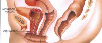
The Androgynecological Clinic provides conservative and surgical treatment of cystocele - prolapse of the anterior wall of the vagina and bladder.
Back to contents
Incompetence of the pelvic floor muscles. Cysto-rectocele
The prolapse or loss of a woman's internal genital organs is commonly called cysto-rectocele. This term refers to a violation of the position of the uterus and vaginal walls relative to the vaginal opening. Strictly speaking, pathologies associated with cysto-rectocele should be considered as a type of pelvic floor hernia.
Sometimes a synonym is used for terminology - genital prolapse. For isolated prolapse of the anterior wall, the term cystocele should be used, and for the posterior wall, rectocele.
As a rule, the disease occurs during reproductive age, developing at a relatively high speed. Of course, as the pathology develops, the functions of certain pelvic organs worsen. Unfortunately, cysto-rectocele causes not only physical suffering; there are often cases when the development of the disease led to complete disability. Failure of the pelvic floor muscles is always accompanied by an increase in intra-abdominal pressure.
There are four main reasons for the occurrence of this pathology: 1. Failure in the functioning of the genital organs, namely their synthesis. In addition, too much or too little estrogen also leads to the development of the disease; 2. Failure of connective tissues, which thereby form insufficiency of woven structures; 3. Injuries and other physical damage to the pelvic floor; 4. Various chronic diseases that to one degree or another affect intra-abdominal pressure.
As a result, under the influence of one or more of the above factors, failure of the ligamentous apparatus begins to develop. As a result, intra-abdominal pressure increases and pushes out the pelvic floor organs.
The main symptoms of this pathology are the sensation of a foreign body in the vagina. In addition, patients are always haunted by nagging pain in the lower abdomen. Of course, cysto-rectocele affects the entire urinary system. And all this happens against the background of severe constipation.
Diagnosis of cysto-rectocele should be comprehensive and include the following tests: - vaginal smear; - vaginal culture; - colposcopy; — Ultrasound of all pelvic organs; — oncocytology of the cervix.
After determining the stage of the disease, doctors will formulate a further course of treatment. If the initial stage is present, the patient will be offered physical therapy, consisting of exercises that are designed to restore normal functionality of the pelvic floor muscles.
In other cases, either drug treatment or surgery is used. Typically, the goal of medication is to restore normal estrogen levels. As for surgical intervention, its goal is not so much to eliminate the incorrect position of organs, but to correct and restore the functioning of adjacent organs: the bladder and rectum.
The Republican Center for Human Reproduction and Family Planning has excellent doctors who have extensive experience in dealing with these diseases. Come to us for diagnostics and we will answer all your questions.
Home » Gynecology » List of publications on gynecology » Incompetence of the pelvic floor muscles. Cysto-rectocele
What treatment can be prescribed by a doctor?
Treatment for cystocele depends on how severe the disease is and what associated disorders are present.
In mild cases,
when there are no manifestations or they are not particularly pronounced, treatment may not be necessary.
It is enough to observe and perform special gymnastics (Kegel technique)
for the pelvic muscles.
You can fight the disease, depending on its degree, by
- medications,
- ointments,
- surgery,
- massage,
- gymnastics
Conservative therapy
Among other things, there are also conservative methods for treating cystocele of the bladder.
At the same time, gymnastics, which is performed for cystocele, shows good results. It consists of increasing the muscle tone of the pelvis
.
Used in the initial stages of the disease
. Kegel exercises are aimed at strengthening the walls of the vagina and involve the use of a method of squeezing and relaxing the vaginal muscles.
This technique is not dangerous for a woman and can be used anywhere, since it is absolutely invisible to others. In addition, these exercises are excellent helpers for combating stress urinary incontinence.
Rehabilitation gymnastics includes several exercises for the whole body and is selected for each patient separately.
Depending on the severity of the disorder, certain exercises are prescribed.
It is important that they are discussed with the treating doctor, since self-medication can cause harm to weakened muscles of the pelvic area.
Yoga is often used to treat bladder
. The practice of special yoga positions not only greatly helps in neutralizing the disease, but also has a positive effect on the body as a whole, relieving a woman of pain, discomfort, and excessive irritability.
Women also use herbs
– snakeweed and centaury, a decoction of viburnum, which helps to increase the tone of the uterine muscles.
Irrigation with the compositions of these herbs is also carried out.
.
The disease should be treated immediately because there is a risk of infection. This happens due to the fact that the drooping walls, extending beyond the boundaries of the vaginal slit, can be rubbed by underwear
.
Therefore, redness and abrasions form. Among other things, the urinary organ descends and, therefore, does not empty completely.
Infections can develop in the remaining urine. If vaginal prolapse
, then cracks and inflammation may occur, which also threatens complications.
Surgical intervention
Surgeries to treat a disease have several goals
:
- Reducing the manifestations of urinary incontinence;
- Improving life and its sexual component;
- Bringing the pelvic organs to their normal position;
- Prevention of new problems with the pelvic diaphragm.
Anterior colporrhaphy
This operation involves suturing the vaginal walls in case of prolapse. Used to correct the central defect of the pelvic diaphragm
.
During the operation, under anesthesia, a speculum is inserted into the patient's vagina. After making an incision in the vaginal wall, the location of the defect is determined. The vaginal wall is then sutured.
Lateral defects of the pelvic floor are eliminated through access through the vagina or through the peritoneum. The procedure involves an incision and approach similar to anterior colporrhaphy.
At the next stage, the defect in the pelvic fascia is removed, then it is sutured to the arch of the tendon, and the incision in the vaginal wall is also sutured in layers.
Transabdominal access
also used if the disease factor is a lateral defect. In this case, the incision is made slightly above the pubic hairline.
Having determined the location of the lateral defect, doctors suture the fascia to the tendon arches. Sometimes the procedure can be combined with retropubic colposuspension
.
The operation is prescribed for the treatment of urinary incontinence
, and its essence is to suspend the urethra using the vaginal walls.
Other surgical treatments may be combined with paravaginal correction
.
Laparoscopy
Laparoscopy has advantages over conventional surgery
on the peritoneum: due to the reduction in trauma, the pain syndrome is significantly reduced, and it also takes less time to restore the patient’s strength and performance.
In addition to cystocele, prolapse of other organs of the urinary system also occurs. They are very unpleasant, so at the slightest manifestation of the disease, treatment should be started immediately. Leukoplakia of the bladder is a precancerous condition. you can learn about the most effective treatment methods.
Other treatments
Vaginal pessary
These are rings made of plastic or rubber that are inserted to support the bladder at the top and back. In some cases, the doctor may recommend using a large tampon or vaginal diaphragm instead of a pessary.
Many women who use pessaries use them as a temporary alternative to surgery.
. But some people can use these products for many years.
Hormone therapy
A doctor may recommend the use of estrogen—in pill or cream form— if a woman is going through menopause
, since after its onset the production of these hormones that strengthen muscle fibers decreases.
But estrogen therapy has its own nuances.
Women with certain forms of cancer should not take estrogen and should discuss any concerns with their doctor.
Manchester operation in gynecology: reviews
Manchester operation in gynecology, reviews of which show that it really helps in the treatment of prolapse, is a combination of cervical amputation, anterior colporrhaphy, strengthening of the cervical stump with additional ligaments and plastic surgery after all these procedures. Although this surgery is considered very effective, it is not performed as often as other treatments. Indications for its implementation are complete or incomplete prolapse of the uterus or vagina. This is especially true with cystocele or cervical elongation. My periods continue to come after the Manchester operation.
Manchester operation: patient reviews and preparation
To perform the operation, standard conditions of a surgical hospital are required. Before the actual surgical intervention, there is a long preparatory process. During it it is necessary to carry out such tests as:
- Extended colposcopy;
- Standard general clinical examination;
- A smear from the vaginal part of the cervix and cervical canal;
- Analysis for atypical cells;
- STI;
- Analysis for the presence of infections.
Manchester operation: video and implementation
Before the operation, the woman is put under anesthesia. The procedure itself is carried out in a gynecological chair. Then the cervical canal is dilated using a Hegar dilator. First, the minimum allowed number is used, after which it goes up to the tenth. Using bullet forceps, the uterus is pulled closer to the vagina, after which it is fixed in the closest possible position.
Using a scalpel, the mucous membrane is cut in a circle. The dissection is made at the level of the posterior and anterior vaginal vault. Then they retreat another two centimeters towards the urethra and another incision is made. After this, part of the mucous membrane is simply removed. At the same time, the wall of the bladder is separated from the wall of the uterus if they were in contact, which often happens with prolonged prolapse.
Then the uterine ligaments are strengthened. They are stitched together crosswise and attached to the uterine wall located in front. After such fixation, the membrane that covers the bladder and uterus needs to be sutured. Only after this the vaginal wall is sutured. A stump is formed from what is left of the uterus. Next, using several types of sutures, the vaginal vault merges with the stump of the cervix. Before sewing, specialists check the patency of the cervical canal with a special probe.
If bleeding occurs in the vessels during the operation, they are pressed using a coagulator. The biggest bleeding results from careless actions with the uterine artery, which is a fairly large blood channel passing through the cardinal ligament. If the need arises, bladder plastic surgery is also performed at the same time. First, its membrane is cut, after which it is sutured. The cut edges overlap each other. With this procedure you can get rid of cystocele. The Manchester gynecological operation on video shows the main nuances of this process.
At the final stage of the operation, perineal plastic surgery is performed. The muscle layer of the pelvic floor is also corrected. To do this, the back wall of the vagina is incised, after which part of the tissue and perineum is removed. The next step is to match the edges of the muscles and their fascia. The wound is sutured. The uterus can be removed from bullet sutures only after all sutures have been placed. The surface of the wound is treated with an antiseptic.
How to cure cystocele without surgery
Conservative treatment of bladder prolapse begins with drug therapy. These are preparations with estrogen that will maintain the condition of tissues and strengthen them. Patients are also prescribed ointments and suppositories with estrogen, which are used vaginally.
Today, treatment with a pessary is also actively used. This is the name of a special silicone ring that is installed at the beginning of the vagina (closer to the entrance). It helps to support the walls from sagging and maintains the position of the muscles under load. Installing and removing the ring is not difficult, so the woman controls it herself. The pessary is removed at night.
Exercising is an essential part of conservative therapy for cystocele. The training method developed by the American Arnold Kegel helps women around the world strengthen their intimate muscles. The beauty of exercise is that it can be done anywhere and anytime.
To do this, you simply need to contract the vaginal muscles, relaxing and tightening them. Those. first you need to imagine that you want to urinate and relax. Then try to stop the “stream” by contracting the muscles and maintaining this position for as long as possible.
By the way! You can also perform Kegel exercises while urinating, trying to hold your urine several times. If you do this regularly, you can avoid surgery.
Treatment of cystocele with folk remedies involves the use of sitz baths with datura herb, the use of tincture of astragalus root, milkweed or horsetail infusion, quince or birch bark tea, and mint decoction. All prescriptions are selected individually and agreed with the attending physician.
Manchester operation in gynecology: postoperative period and its features
- And patients experience early activation, almost on the second day after surgery, but the lower limbs must be bandaged;
- On the first day, moderate infusion therapy is used;
- From the first day, cuff pneumatic compression is used, which lasts until the patient’s immediate discharge;
- Prevention of thromboembolic complications is carried out;
- At the same time, you need to take medications to increase the body’s regenerative abilities;
- Sutures after surgery are processed daily;
- On the third day after surgery, daily douching begins;
- The average postoperative period in the hospital is one week.
Manchester operation: consequences and complications
- During the operation, artificial holes may appear in the bladder, which cause peritonitis;
- Excessive excision of tissues of internal organs, which happens when the vagina is too close or far away, causes discomfort during sexual intercourse in the future;
- Perforation can also appear in the rectum, which happens due to carelessness during plastic surgery of the posterior wall (if this happens, the intestine has to be sutured);
- Bleeding after surgery may be caused by injury to the uterine artery, which will then need to be sutured;
- The appearance of infection, which happens when the effect of antibacterial drugs is weak.
Surgery for cystocele
The operation is performed when the vaginal walls are significantly stretched and the bladder literally falls out, as well as when conservative treatment is ineffective. The principle of the intervention is not only to return the bladder to its anatomical position and condition, but also to create new muscle fascia that will reliably hold the internal genitourinary organs. For this purpose, bioinert prostheses are used.
By the way! Surgery for cystocele is low-traumatic, but it is technically very complex. Some hospitals do not do it, and in a commercial clinic, surgical treatment will cost 30 thousand rubles or more. Often after surgery it is necessary to undergo vaginoplasty so that the woman can lead a normal sex life.
Manchester operation in gynecology: when can you sit?
Many patients have a question about when they can sit after Manchester gynecological surgery. Doctors can answer this question in different ways, since it all depends on the specifics of the operation and the patient’s health condition. Some allow you to sit down immediately, while others are not recommended to do this even after a few days.
Factors associated with the disease
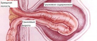
Rectocele (prolapse) is a protrusion of the rectum towards the vagina. This condition occurs when the pelvic floor muscles are severely stretched. It may be accompanied by unpleasant symptoms during bowel movements and cause various complications. Very often, rectocele develops after childbirth when the perineal ligamentous apparatus is weakened.
Rectocele
Factors associated with the disease
During pregnancy, women may experience such a pathological change in the wall of the rectum. This is due to a violation of the supporting function of the pelvic muscles. Rectocele reduces the ability to work and worsens the quality of life. The vaginal wall becomes fragile, thins, and its shape is disrupted. In women, the disease may be accompanied by complications such as uterine prolapse, hemorrhoids or protrusion of the small intestine.
To prevent the possible development of pathology, it is necessary to know the symptoms of intestinal rectocele, what it is, what its complications and treatment are. In men, this pathology develops much less frequently. In them, the protrusion occurs towards the tailbone.
Symptoms of prolapse (prolapse) of the uterus
Many women experience symptoms of uterine prolapse, especially during the postmenopausal period.
According to statistics, this pathology is detected in 10% of women under 30, 40% between the ages of 30 and 40, and 50% of women over 50 years of age. Despite the fact that the problem is accompanied by quite noticeable, unpleasant symptoms, women often put off visiting a doctor, thereby aggravating their condition. Uterine prolapse is an incorrect position of an organ associated with its displacement. Displacement occurs when the muscles responsible for holding the uterus weaken and cannot fully perform their function. Uterine prolapse (uterocele) and prolapse are the most common problems related to female genital prolapse.
In this case, signs of uterine prolapse are displacement from the correct position, and signs of uterine prolapse are its exit from the genital slit. If displacement occurs, the uterus will not be visible from the outside, even if the woman pushes, but this condition precedes prolapse, so if it is detected, you should immediately go to the doctor. Today, this problem can be easily treated, so women who experience unpleasant symptoms of prolapse of the vaginal walls should not worry. A visit to a good specialist will be the first step on the path to recovery.
Prolapse of the uterine walls can occur for various reasons. This is often encountered by those who have given birth several times, as well as those who experience significant physical activity in everyday life.
Causes of rectocele
The reasons for the development of this disease are:
Surgeon
- Multiple pregnancy, frequent and difficult labor, polyhydramnios.
- Heavy physical activity, repeated regularly.
- Prolonged increase in intra-abdominal pressure (constipation, paroxysmal cough).
- Chronic gynecological, proctological diseases.
- Natural age-related weakening of the pelvic floor and perineal muscles.
- Congenital pathology or genetic predisposition.
When answering the question of what rectocele is, it should be remembered that women with a history of more than three births are at risk. Also, a high risk of development occurs in persons with obesity of 3-4 degrees, with low physical activity.
Classification of rectocele
Protrusion occurs:
- Anterior, towards the vagina.
- Posteriorly, towards the anal-coccygeal ligament.
The initial form of this disease may not manifest itself in any way and may become a diagnostic finding during a routine examination.
| Symptom. | 1 (mild) degree. | 2nd degree (moderate). | 3rd degree (severe). |
| Complaints. | Can not be. | Bursting pain in the lower intestines. | Pulling pain in the lower abdomen, in the rectal area, difficulty urinating. With grade 3 (severe) rectocele, there is a bulging of the vaginal walls beyond the genital opening. |
| Defecation. | Not broken. | Periodic constipation, feeling of incomplete emptying. | Difficulty during bowel movements, the need for manual pressure on the vaginal wall for quality bowel movements. |
| The size of the protrusion. | Palpation examination reveals a bulge of up to 2 cm. | Reaches 4 centimeters. | Convex, with possible fecal stagnation, more than 4 cm. |
The last stage of the disease is characterized by the development of complications:
- the appearance of fistulas between the rectum and vagina or bladder;
- necrosis of surrounding tissues;
- anal fissure;
- inflammation of the subcutaneous tissue;
- fecal incontinence.
Stage 2 development of rectocele may be accompanied by the sensation of a foreign body in the vagina. Rectocele stage 1 (initial) is usually asymptomatic.
Degrees of cystocele
The process of bladder displacement depends on a combination of many factors. There is no one-size-fits-all scenario for the development of cystocele.
For example, in some patients, only the anterior part of the vaginal wall with an enlarged urethra (urethrocele) is displaced, as a result of which the bladder remains in its proper place.
The nature of cystocele is significantly influenced by individual anatomical features, the patient’s age, obstetric history, and the presence of concomitant non-gynecological pathology.
Depending on the topography of the bladder, three degrees of cystocele are distinguished:
— Cystocele of the first degree is the most favorable, mild degree of displacement of the bladder, most often does not cause discomfort to the woman and is detected during a gynecological examination after straining.
— Moderate, second-degree cystocele is detected during examination without the woman having to strain the anterior abdominal wall. A protrusion of the anterior vaginal wall is detected, which does not extend beyond the boundaries of the genital slit.
— The third degree of cystocele is the most severe. The protrusion of the vaginal wall overcomes the border of the genital fissure (partially or completely), and is detected even in a state of complete physical rest.
The degree of cystocele characterizes the stage of development of pathological displacement of the bladder, and also serves as a decisive criterion in choosing a method of therapy.
Symptoms of rectocele
This pathology is chronic, which characterizes it as a gradually developing disease.
The main signs of rectocele are:
Pain in the butt
- Reducing the frequency of bowel movements to once a week, which contributes to trauma to the intestinal walls.
- Feelings of inadequate emptying, which provokes the patient to constantly use a cleansing enema or laxatives. This leads to the body becoming accustomed to constant stimulation, and the person is unable to empty his bowels on his own.
- Feeling of a foreign body in the vagina. May get worse while standing or moving, and get better when lying down.
- Pain and discomfort during sexual intercourse in women.
- The urge is accompanied by pain and discomfort in the anal area, which indicates secondary damage to the mucous membrane.
- Reducing the amount of feces excreted.
- The appearance of streaks of blood in the stool.
- Making extra effort to defecate.
- It is possible to develop incontinence of urine, feces, and gases, which is often observed in old age.
- Prolapse of other organs also occurs: the uterus, bladder.
In the final stages, in order to fully go to the toilet, the patient has to press on the perineum, squeeze the vaginal wall, or put pressure on the buttock area.
When initial signs of rectocele appear, you should contact a surgeon or proctologist for treatment and examination.
Clinical picture of the disease
With cystocele, which is in the earliest stages of development, the symptoms corresponding to the pathology are practically absent, or are so weakly expressed that women do not attach any significance to them. This is the insidiousness of the disease, since it can be cured using a non-surgical method only at the initial stage of development. That is why experts recommend that all women who are at menopausal age or have suffered a difficult birth should be more attentive to their health. An urgent visit to a gynecologist is required if the following alarming signs appear:
- Vaginal discomfort, complemented by pain in the sacral region.
- Unpleasant sensations during sexual intercourse.
- Violation of the process of urination (increased urge and their adoption of an urgent nature).
- Urinary incontinence that occurs during a coughing or laughing attack.
The expansion of the vaginal opening and canal due to weakening of the vaginal muscles facilitates the penetration of pathogenic microorganisms into the pelvic organs, causing the development of infectious diseases. To avoid this, it is necessary to contact a specialist in a timely manner, who, after a full gynecological examination, will be able to make an appropriate diagnosis and prescribe adequate treatment.
Diagnosis of rectocele
To make a diagnosis, the following measures are carried out:
How is the disease diagnosed?
- Patient interview. He develops specific complaints (frequent constipation, false urge to defecate, feeling of insufficient bowel movement), which indicates the possible development of a rectocele.
- An increase in intra-abdominal pressure occurs, and prolapse of the intestinal wall into the vagina is noted. This indicates the development of grade 2, 3 rectocele.
- External inspection. In patients, one can see anal fissures and hemorrhoidal protrusions, which is characteristic of the progression of the disease. Symptoms of fecal incontinence may also be observed: maceration of the mucous membrane, the appearance of skin ulceration.
- Gynecological and digital examination. When palpating the rectum, internal hemorrhoids, polyps or protrusion of the intestinal wall can be detected. In this way, the size of the pocket and the presence of feces in it are determined.
- Vaginal examination. During the examination, the patient is asked to tense up.
- Instrumental methods: sigmoidoscopy, irrigography, defecography (x-ray examination of the defecation process). Carried out to clarify the diagnosis. These measures will help identify protrusion of the intestinal wall, which is often found with this pathology.
- Electromyography. Determines the functional state of the pelvic floor muscles and sphincter.
Be sure to take a blood and stool test, which is necessary for any illness. There you can see the severity of the inflammatory process or the possible presence of red blood cells in the stool.
Treatment of rectocele
The choice of treatment method depends on the stage of the disease. At the initial stage, the following methods are used:
What can you eat
- Diet. The diet should consist of fresh vegetables, fruits, cereals, and bran bread. You can diversify your diet with fermented milk products and broths, which will lead to looser stools.
- Water mode. It is recommended to drink at least 2 liters of liquid per day, excluding first courses.
- Dosed physical activity. Race walking or swimming, and the use of special exercises will strengthen the muscles of the perineum, which will speed up recovery. They are based on simulating urinary retention, with maximum tension and relaxation of the perineal muscles. They must be repeated several times a day.
- Conservative drug treatment. Taking laxatives, preferably with an osmotic effect (Duphalac, Forlax), which increases the effect of treatment. Antispasmodics (Motilak, No-Spa), probiotics (Bifido, Lactobacterin), probiotics (Bifidum, Lactobacterin, Acipol, Linex) will improve digestion and normalize microflora.
- Physiotherapeutic procedures.
The lack of effect from conservative therapy indicates that it is necessary to move on to the next stage of treatment: surgery. Surgery for rectocele is indicated for stages 2 and 3.
Excision of the thinned walls of the rectum is performed, followed by suturing. They also use suturing of the muscles that hold the rectum and the back wall of the vagina.
A more pronounced effect is achieved by plastic surgery of the rectovaginal septum. Its implementation indicates further normalization of fecal excretion. In the presence of concomitant pathology (polyps, anal fissures, protrusion to the bladder), a combined operation is performed to simultaneously eliminate these problems.
The rehabilitation period lasts up to 2 months. During this time, it is necessary to follow a diet, limit sexual intercourse, and normalize bowel movements. The combination of these options will lead to bowel movements returning to normal faster.
Upon completion of the recovery period, the functions of the intestines and genital organs are completely restored. You can additionally use baths with chamomile, calendula or potassium permanganate, which will speed up tissue regeneration.
For rectocele, the use of folk remedies has a good effect:
ethnoscience
- Pour boiling water over 8 prunes and leave overnight. In the morning, eat the steamed fruits and drink the liquid. This method has a laxative effect, which helps prevent the development of constipation.
- Prepare a tablespoon of sena grass and 100 g of pitted prunes. Chop the herbs and grind the prunes in a blender. Pour a glass of boiling water over this mixture and leave for two to three hours. Before use, the resulting composition must be mixed and taken two spoons three times a day.
- Pour 4 tablespoons of buckwheat into two glasses of kefir. Leave until morning and eat for breakfast.
This normalizes the intestinal flora, which will have a beneficial effect on its function. The absence of heat treatment will improve digestion and soften stool.
Prevention measures
It is quite simple to prevent changes in the position of the bladder; follow the useful recommendations of doctors:
- do gymnastics regularly, pregnancy is no exception (in the absence of contraindications);
- before the birth process begins, discuss with your obstetrician gentle methods of childbirth;
- promptly treat persistent coughs and prevent chronic constipation;
- when lifting minor weights, distribute the load correctly, avoid lifting large loads;
- Beware of stressful situations, asthenia, lose weight evenly, sudden changes in weight adversely affect the entire body;
- Monitor your weight and constantly stay in shape.
The main prevention of cystocele is to take a serious and careful attitude towards your health. If any unpleasant symptoms appear, consult a specialist; do not delay going to the doctor in the hope of solving the problem.
Next video. The TV show “Live Healthy” about what cystocele is in women and the features of treating the pathology: