Urologist Urology Bladder diseases
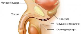
The structure of the ureter (μd10) is a pathological abnormal narrowing, which is caused by the replacement of healthy mucous membrane with scar tissue. As a result, there is difficulty urinating from the urinary canal and then from the kidneys.
This leads to the following complications:
- Cystitis (chronic infection);
- Chronic prostatitis;
- Urolithiasis disease;
- Inflammation of the epididymis, testicle (orchiepididymitis);
- Development of hydronephrosis (the kidney striate system expands);
- The outflow of urine from the kidney is impaired;
- Kidney failure.
- Kidney cyst.
The length of the urinary canal of an adult reaches 34 cm. Its length depends on the location of the kidney and on the person’s height. The arterial branches of the urinary canal from the ovarian and renal arteries extend into the upper section. It branches off from the internal iliac arteries into the lower section.
Normally, narrowings have the ability to expand if necessary. This happens due to the elastic wall, but when fibrosclerotic changes develop in the walls, atrophy of the muscle elements occurs, which soon change to scar tissue. The consequence of this is ureteral stricture (micd10), which is manifested by symptoms of pathologies: urolithiasis, kidney cyst, pyelonephritis (kidney inflammation), chronic renal failure, hydronephrosis.
Ureteral stricture (μd10) can be located in different parts of the urinary tract and have different lengths (from 0.5 cm). More common is acquired pathology of the urinary canal, which develops as a result of injuries, radiation injuries, and tuberculosis. Congenital narrowing can be caused by compression of the ureter by abnormal vessels.
Due to the development of narrowing, the diameter of the excretory duct decreases significantly. This process can be accompanied by either partial disruption or complete blockage of the urinary canal. From prolonged stagnation of urine, elongation, stretching, and tortuosity of the urinary tract is observed, which leads to expansion of the renal pelvis (hydronephrosis), ultimately to renal failure and a renal cyst.
Ureteral stricture occurs:
- double-sided
- one-sided,
- multiple,
- single,
- false,
- true.
Depending on these types, the reasons for the development of narrowings, their origin, and location are identified. The disease is usually observed in the juxtavesical and pyelourethal areas.
Contents of the article:
- 1 Causes of occurrence
- 2 Symptoms
- 3 Diagnostics
- 4 Treatment
Description of the pathology
The ureter is a hollow tubular organ that connects the kidney to the bladder (in most mammals).
It starts from a narrowed area of the renal pelvis, where urine formed in the kidney flows. Its outlet end ends in the wall of the bladder.
For a healthy person, narrowing of the ureter of an anatomical or physiological nature is considered an acceptable norm. This phenomenon occurs due to the elastic properties of its wall. However, if stenosis or stricture occurs, the changes begin to take on a fibrosclerotic form. As a result of this pathological process, a violation of the submucosal membrane, as well as the muscular and outer walls of the ureter, occurs. Some muscle elements die off and are replaced by scar tissue, which is unable to perform any functions because it is atrophied.
Organ dysfunction
The lumen of the urinary duct at the site of ureteral stricture decreases, which disrupts the normal functioning of the organ. Urine cannot be completely eliminated from the body and begins to accumulate in the bladder, eventually causing increased pressure on the ureter. Subsequently, it stretches and elongates. In some cases, it comes to curvature of the ureter. In the absence of proper treatment, the pathology affects the kidneys.
Ureteral stricture can develop in any area of the organ. Most often, the pathology is localized in the space between the bladder and ureter. In addition, there are cases of identifying a stricture between the ureter and the pelvis.
Diagnosis of urethritis
Diagnosis of the ureter includes a number of procedures:
- general blood analysis;
- blood chemistry;
- urography (an X-ray method that detects disturbances in the outflow of urinary fluid and the presence of urinary fluid);
- (procedure for checking the inflamed ureter for swelling);
- ureteroscopy (detection of swelling and damage to the ureter);
- catheterization of the ureter (detecting the presence of pus and cloudiness in the urinary fluid);
- (ureters and kidneys are examined)
Types of strictures
Narrowing of the ureter can take different forms depending on the area of localization of the pathology, as well as on the nature of the disease. First of all, a distinction is made between acquired and congenital stenosis. The latter appears during the intrauterine development of the unborn child.
The pathological process may occur due to thickening of the walls in certain places. Congenital ureteral strictures appear as a consequence of certain disorders during fetal development, namely:
- An inflection that occurs due to the curved shape of the ureter.
- The appearance of a membrane in the ureteral valve, which provokes the accumulation of urine in the bladder.
- Ureterocele. This is a disease that is characterized by a narrowing of the lumen in the lower part, while the ureter expands, and in some cases falls into the cavity of the bladder.
- Squeezing of blood vessels.
- The formation of diverticula, which provoke protrusion of the lower part of the ureter.
An acquired form of narrowing of the ureter can occur under the influence of various factors, depending on the person’s state of health. Depending on the area where the stricture was localized, right-sided and left-sided stenosis are distinguished. It also happens that both sides of the ureter are affected. Also, stenosis can be localized both in the upper part of the ureter and in its lower part, where the transition to the renal pelvis occurs. If the pathological process develops in the middle section, then both the upper and lower parts of the organ are affected.
Kinds
Inflammation of the ureter (ureteritis) in most cases is a secondary urological disease, which is provoked by infection.
In medicine, the following types of disease are distinguished:
| Name | Description |
| Septic | A pathological condition that is characterized by an inflammatory process on the surface of the mucous membrane of the ureter. Septic urethritis most often occurs as a result of pathogenic microorganisms entering the urinary system. |
| Aseptic | The urinary ducts become inflamed as a result of mechanical damage, tumor processes, impaired innervation or due to trauma. |
Inflammation of the ureter in women is called ureteritis.
There is also primary ureteritis, when the cause of the inflammatory process is injury to the ureter. Secondary develops against the background of concomitant diseases. In each case, the woman develops characteristic symptoms that will help the urologist establish a preliminary diagnosis and prescribe an examination.
Provoking factors
Factors that can cause the development of ureteral stricture are:
- Formation of kidney stones. This falls under the category of internal injuries. Urolithiasis causes inflammation, and the mucous membranes are easily damaged by the stones, leading to scarring.
- External injuries to the lumbar spine. As a result, a hematoma appears behind the peritoneum, which later becomes the basis for a stricture.
- Injury sustained during surgery.
- Radiation therapy and radiation injury.
- Tuberculosis, inflammation in the ureter.
The causes of ureteral stricture should be determined by a doctor.
In addition, in some cases, pathology appears as a consequence of gunshot or knife wounds. Also, self-treatment of sexually transmitted diseases can lead to injury to the ureter. Men are more susceptible to injury and overuse and are more likely to develop strictures. If any of the listed factors are excluded, the doctor makes a conclusion about the congenital form of the disease.
The ICD-10 code for ureteral stricture is N13.5.
Causes of inflection
Considering that often the reason that led to the bend can be called nephroptosis, you should figure out what is to blame:
- rapid loss of body weight, due to which the size of the fat capsule decreases;
- heredity, which entails increased tissue extensibility;
- lower back and abdominal injuries;
- sudden lifting of heavy objects;
- pregnancy, due to which there is a stretching of the abdominal muscles, which provokes a decrease in intra-abdominal pressure;
- various factors that affect the stretching of the ligamentous apparatus of organs, for example, jumping;
The condition, when the kidney puts pressure on the ureter, is more susceptible to females than males. This is due to the fact that the connective tissue of women is prone to stretching much more, and the female sex also has much weaker abdominal wall muscles. In addition, women become pregnant and give birth, unlike men, and during pregnancy the abdominal wall tends to stretch, thereby reducing intra-abdominal pressure.
Return to contents
Symptoms
As a rule, symptoms and severe pain accompany bilateral stenosis. Unilateral stenosis, on the contrary, mostly occurs in a latent form. For this reason, it is almost impossible to diagnose the disease at an early stage of its development. With bilateral damage, the following symptoms are observed:
- Increased pressure in the arteries.
- Pain syndrome in the lumbar region.
- Nausea and vomiting.
- Convulsive syndrome.
- Passage of a small amount of urine.
- Pain during urination.
- An increase in body temperature, which indicates an inflammatory process in the body.
- Presence of blood in the urine.
The symptoms of ureteral stricture are very unpleasant.
In the absence of proper treatment, the pathological process can progress and spread to adjacent organs, including the kidneys. Due to incomplete excretion of urine from the body, the risk of stagnation increases, which will ultimately lead to urolithiasis, pyelonephritis, hydronephrosis, as well as chronic kidney failure. It is important to promptly identify the pathology and receive qualified medical care.
Why is it important to treat ureterocele?
First of all, due to ureterocele, hydronephrosis develops - a disease in which, due to problems with the outflow of urine, the calyces and pelvis of the kidney expand, and a large amount of urine collects in these expansions.
Hydronephrosis leads to the development of pyelonephritis - inflammation of the kidney due to urine accumulating in it, which is a breeding ground for pathogenic microorganisms. Another consequence of hydronephrosis can be kidney stones: salts from the urine, as a result of stagnation, precipitate and form calculi (“stones”).
In addition, it is possible to develop renal arterial hypertension - increased blood pressure in the kidney that is not eliminated by medications with a hypotensive effect. Another consequence of ureterocele may be kidney atrophy, in which the nephrons are replaced by connective tissue that is not capable of filtering blood and producing urine.
The result of the process is chronic renal failure, which is a threat to the health of the entire body and human life.
Diagnostic methods
To get a complete clinical picture, it is necessary to schedule a detailed examination of the patient. Diagnostic procedures include blood and urine tests, and ultrasound of the genitourinary system. In addition, the patient is prescribed computed tomography and magnetic resonance imaging. Endoscopic examination is contraindicated if the patient has an inflammatory process in the vagina, uterus, urethra or prostate gland.
The most informative and common method of research for ureteral stricture is urethrography. The procedure is an X-ray examination using contrast. This technique makes it possible to identify those areas in which there is stagnation, as well as localize the presence and position of narrowed areas. The contrast is injected directly into the urethra or intravenously.
Ureteral stricture (ureteral narrowing, ureteral stenosis, ureterohydronephrosis)
- 13 Jul 2017Non-standard treatment of gene mutations in men with infertility Israeli doctors have identified a condition in the male body when, with a sufficient amount of androgens, their identification does not occur. As a result...
- 13 Jul 2017Achievement of Israeli medicine in the field of urologyModern urology in Israel makes it possible to treat diseases with minimal pain. This is where the most advanced technologies are used, which…
- 13 Jul 2017Recently, a detailed study of the treatment of stage 1 prostate cancer was carried out in the UK. Its results showed that therapeutic measures did not…
- 12 Jul 2017Prostate cancer treatment: lutetium irradiation Prostate cancer is the most common form of cancer in men. Medical statistics show that every seventh man...
- 13 Jul 2017The 4Kscore test is a guarantee of early diagnosis of prostate cancerIn the USA, Israel and some European countries from April 1, 2014 for men who need to consult a urologist if they suspect…
- 13 Jul 2017Prostate surgery: a cancer diagnosis is not a death sentenceIsraeli medicine is known all over the world for its level. The country's research activities in the field of medicine are amazing. Latest news...
- 13 Jul 2017Preventive removal of ovaries - expert opinion Ovarian and breast cancer are very dangerous for every woman. However, today there are many ways to both diagnose the disease in time and…
- 12 Jul 2017The IVF procedure using the IMSI method is the end of male infertility. The desire of a couple to have a child is natural, but not every one of them can afford such a “luxury” naturally. Today, every fifth couple with...
Source: https://israel-clinics.guru/diseases/striktura_mochetochnika_suzhenije_mochetochnika_stenoz_mochetochnika_ureterohydronefroz_/
Preparation for urography
Urography is considered an effective, safe diagnostic method. The study is prescribed if there is a suspicion of kidney pathologies, bladder diseases, problems with filtration and urine output.
The basic rules for preparing for urography will be the following:
- 3 days before the procedure, the patient must refuse food that causes excessive gas formation.
- A test must be carried out to detect an allergy to a radiocontrast substance.
- Meals should be taken no later than 8 hours before the test; you should not drink too much liquid during the day.
- You can't eat in the morning.
- In the office, you need to remove metal products and jewelry, and, as directed by the doctor, empty your bladder.
- If there is anxiety shortly before urography, you can take a sedative (calming) drug.
Treatment of inflection
Treatment of conditions when the kidney puts pressure on the ureter directly depends on what causes contributed to the appearance. Taking into account the fact that the pressure resulted in the prolapse of the kidney, this particular anomaly needs to be cured. Therapy for kidney prolapse is divided into conservative and surgical.
Return to contents
Traditional tactics
The consequences of kinking of the ureter can be smoothed out with the help of exercise therapy, normalization of exercise and nutrition.
Specifics of conservative treatment:
- sock bandage;
- specialized physical education, which consists in strengthening the abdominal muscles;
- increased and balanced food intake, which is aimed at thickening fatty tissue;
- avoiding carrying heavy objects.
Specialists can also prescribe drug treatment when the kidney puts pressure on the ureter, however, this happens in cases where the bend is accompanied by complications such as pyelonephritis, kidney stones, colic and high blood pressure.
Return to contents
Kink surgery
Surgical intervention is resorted to in the later stages of the curvature and in the presence of complications. During the operation, doctors place the kidney in the right place and secure it. Surgery helps restore blood supply to the kidney, remove the bend and restore the flow of urine. After the operation, the patient must strictly adhere to bed rest for at least 2 weeks so that the kidney can strengthen in the required position.
- Megaureter in children under one year of age: treatment methods, indications for surgery
Return to contents
Therapy
After a thorough examination and clarification of the diagnosis, the patient is prescribed the necessary treatment. The main goal of therapy is to normalize urine excretion. The treatment regimen is selected based on the results of the studies. It is also important to consider the general condition of the kidneys and genitourinary system. Another important factor in determining treatment is the size of the stricture.
Ureteral stricture cannot be treated at home or with traditional medicine. Contrary to popular belief, you should not heat the affected area, as this may make the pain more intense.
One of the effective treatment methods is plastic surgery in urology centers. This is a rather complex procedure with a long rehabilitation period, so it is prescribed only as a last resort. The operation is not suitable for every patient, as it has a number of contraindications.
Another treatment method is bougienage of the ureter. The procedure involves using a metal rod that is inserted into the ureter and dilates it. The procedure is very painful, and the effect is short-lived. Bougienage is rarely used.
Plastic replacement method
Urology centers also use the plastic replacement method. This method is suitable for the treatment of small strictures, the size of which does not exceed 20 mm. The operation involves making an incision and replacing the scars with tissue from the patient. In addition, optical urethrotomy using a cystoscope is used. Any intervention to treat stenosis must be agreed with the attending physician and supervised by a qualified surgeon.
The pathology is quite serious and cannot be treated with medications or traditional methods. If surgery is not performed, complications may occur that affect the kidneys and other organs.
Prevention and prognosis
Stenosis develops rapidly, especially when it is preceded by injury. A hematoma forms in the affected area, which must be detected and drained. If first aid is carried out correctly, the formation of strictures is excluded. Any, even minor, injury to the lower back requires contacting a specialist for examination and examination. It is important to avoid injury to the pelvic area when playing sports. It is important to use special protective shields that can soften the blow.
The sooner surgery is performed after detection of strictures, the better for the patient and the less likely it is to develop complications. In addition, this will shorten the recovery period, and the operation itself will not be so painful. An important point in proper rehabilitation is compliance with all instructions of the attending physician.
Complications
If the above conditions are not met, complications arise that can affect the functioning of the genitourinary system and other organs. The operation can also have consequences if the patient’s tissues have not grown together correctly or have not taken root at all.
Lack of treatment can lead to the development of pathologies such as cysts or kidney failure, as well as hydronephrosis, when the renal pelvis expands. In some cases, cystitis and kidney stones appear against the background of strictures.