Position of red blood cells in a general urinalysis
A general urine test (UCA) is a laboratory study of the organoleptic properties and biochemical composition of a waste product excreted by the kidneys. Organoleptic characteristics and norms for adults:
| Volume | Color | Smell | Transparency | Density | Acidity |
| ≈ 75% of the liquid drunk | light yellow (straw) | without harsh shades | close to absolute | 1.01–1.02 g/l | 4–7 pH |
Components in the urine of a healthy person
| Determined in minimal quantities | Not defined |
| red blood cells (Bld) | acetone, otherwise ketone bodies (Ket) |
| leukocytes (Leu) | granular and waxy cylinders |
| glucose (Glu) | bacteria |
| protein (Pro) | bilirubin (Bil) |
| hyaline casts | yeast fungi |
| urobilinogen (Uro) | salt |
Urine examination allows us to identify pathological changes in the body to further determine the exact cause of the disorders. The analysis is prescribed for diagnosis, monitoring of therapy and for prevention purposes. The results are assessed by comparing the obtained indicators with the reference values accepted in laboratory microscopy.
The results are recorded in the study form. Undetected elements are indicated by a dash, Russian “absent” or English “neg” (negative). When identifying components that should not be in urine, the term “trace intact” is used. Another result accepted in laboratory examination is the “presence of traces”, which is recorded in the final conclusion as “trace lysed”.
The structural units of the kidneys, the nephrons, are responsible for the formation and excretion of urine. They consist of a renal corpuscle, where the liquid portion of the blood is filtered, and a system of tubules, in which resorption (absorption) and excretion (excretion) of substances occur. Only micromolecular material leaks through the capillary walls. Red blood cells are too large in size and, in the absence of pathologies of the renal apparatus, practically cannot penetrate into urine.
Normal values
The Bld measurement value is a digital indicator (how many red blood cells are counted) per microliter (µl). In literal equivalent, the decoding looks like ca x ery/ul, (where x is the number of Bld, ery/ul is red blood cells per microliter). The term “in sight” is used for counting.
Depending on the laboratory performing the urine test, the designation rbc/ul may be used. The norm of red blood cells in urine is graded according to age (in children and adults) and gender (in women and men). Standard values of red blood cells by age and gender:
| Infants up to one year old | Children and teenagers under 16 years of age | Adults | |
| women | men | ||
| complete absence | 0–2/µl | no more than 3/µl | 1–2/µl |
A slight increase in the number of red blood cells in the urine is allowed in women during the perinatal period. The upper limit of the norm for pregnant women is Bld ca 5 ery/ul. A high concentration of red blood cells, otherwise the presence of blood in urine can be visually suspected by the changed color of the biofluid. However, it is impossible to determine this without laboratory examination. A temporary change in the color of urine is caused by eating asparagus, beets, blackberries, and taking laxative medications.
An incorrect procedure for collecting urine in the first seven days of the follicular phase of the menstrual cycle (the period of bleeding) can distort the results of the analysis in women. It is recommended to postpone the study until the cessation of menstruation. If an urgent analysis is required, before collecting urine, it is necessary to perform thorough hygiene of the genitals and insert a tampon into the vagina.
Deviations from the norm
Exceeding the standard values of red blood cells in the urine is called hematuria, which is classified:
- by the number of detected Blds (micro- and macrohematuria);
- for the reasons for the occurrence of deviations (extrarenal, renal, physiological).
Microhematuria is characterized by an increase in analysis parameters to a maximum of Bld ca 20 ery/ul. In this case, the color of the biofluid has no external changes. Red blood cell counts are performed by microscopy of urine sediment. Macrohematuria is a deviation of values above Bld ca 25 ery/ul, which is accompanied by pronounced changes in the color of urine.
A multiplication of red blood cells one hundred times the natural norm (ca 200 ery/ul) indicates that the bladder is actually filled with blood. This condition is dangerous to health and life; emergency hospitalization is indicated for the patient. Blood levels in the urine exceeding 250/μl are grounds for suspected oncology of the urinary system.
Renal hematuria is a clinical sign of kidney disease. Additional confirmation is the detection in the urine of an increased content of protein (proteinuria) and casts (granular, waxy, erythrocyte, epithelial).
Extrarenal hematuria indicates the development of pathologies in the organs of the genitourinary system and hematological abnormalities. Physiological hematuria occurs under the influence of external factors or due to the fault of the patient himself. As a rule, the manifestation is short-term and disappears after the cause is eliminated.
Additionally
In a newborn child, the reasons for the presence of blood in the urine may be congenital anomalies of the urinary organs, genetic pathology of the circulatory system (hemorrhagic disease of the newborn). During the perinatal period, a slight increase in the number of red blood cells is associated with a doubled load on the renal apparatus of the expectant mother in the third trimester.
If, according to the results of several tests, more than 10 ery/ul are consistently detected, then pregnancy nephropathy is diagnosed. The pathology is characterized by proteinuria, cylindruria (presence of cylinders in urine), persistent increase in blood pressure (blood pressure), swelling (hydropsis of pregnant women), dysania (sleep disorder). To avoid premature birth, a woman needs hospital treatment.
If the result of the initial test is unsatisfactory, the doctor should prescribe a repeat test. If an elevated level of red blood cells is confirmed, the patient is prescribed additional laboratory tests and hardware diagnostic procedures.
Hematuria in a child
Expert opinion Elena Anatolyevna Kovaleva Doctor-Laboratory Technician. 14 years of experience in clinical diagnostic services. The symptom is detected much less frequently in children than in adults, and its cause is predominantly kidney disease, often glomerulonephritis.
The causes of hematuria in children can be classified according to the frequency of their manifestation depending on the age of the child:
- Hematuria in infants often occurs with thrombosis of the renal vein due to acute infections, with malformations of the genitourinary system, and hemorrhagic disease of the newborn.
- In preschool children, the appearance of blood in the urine is often caused by injuries and damage to the external genitalia, urinary tract infection, glomerulonephritis, kidney tumor or abnormalities of their development.
- In school-age children, red blood cells in the urine can be detected in cases of kidney injuries and tumors, inflammation of the urinary tract (cystitis, urethritis), glomerulonephritis, pyelonephritis, thrombopathy, systemic lupus erythematosus.
You need to know that it is impossible to diagnose the disease based on hematuria alone. Additionally, a wide range of studies are carried out: ultrasound of the kidneys and urinary tract, microscopy analysis of urine sediment, three-glass samples, blood biochemistry and related diagnostic techniques to determine possible pathologies.
Renal causes of increased urinary Bld concentrations
Pathologies of the renal apparatus causing renal hematuria:
- Immunoinflammatory damage to the glomeruli of the kidneys (glomeruli), otherwise glomerulonephritis. It has an independent character or develops as a complication of previous infectious viral diseases. Untimely treatment threatens the development of acute renal failure.
- Acute or chronic inflammation of the renal tubular system (pyelonephritis). Has a bacterial etiology (origin).
- Nephrolithiasis (kidney stone disease). Characterized by the formation of calculi of various organic nature in the kidneys.
- CRF (chronic renal failure). Gradual loss of kidney function. It progresses as a concomitant complication of pathologies of the renal apparatus and endocrine system. It has four stages of development, the primary of which can be diagnosed with Bld ca10 ery/ul indicators.
- Enlargement of the renal calyces and pelvis due to impaired urine outflow (hydronephrosis). In a chronic progressive course, it leads to the death of renal tubules and nephrons.
- Extrapulmonary infection with Koch's bacillus (nephrotuberculosis). Most often, it is a consequence of advanced tuberculosis of the bones and lungs.
- Benign renal neoplasms (adenoma, cyst, hemangioma, fibroma, lipoma).
- Malignant tumor (kidney cancer). Multifactorial disease. The only treatment is nephrectomy (removal of the kidney) in combination with chemotherapy.
Blood spots and blood clots can be observed with mechanical damage (trauma, bruises, etc.) to the kidneys. The release of blood into the urine means a violation of the integrity of blood vessels and organ tissues.
What does the quantity 10 rbc/ul mean?
The determination of erythrocytes in urine in the amount of 10 cells per field of view can be observed in chronic renal failure, nephrotic syndrome, and pyelonephritis. Also, a small amount of them can be detected during inflammation of the reproductive system in women (endometritis, cervical erosion, vulvitis). You can also differentiate kidney disease from inflammatory diseases of the genital organs through a general clinical urine test, paying attention to the following criteria.
In chronic renal failure, leukocytes are also found in small quantities. The main sign indicating chronic renal failure is a decrease in urine density (“SG” on the form) to 1.010. Traces of mucus and single cylinders are also found.
Nephrotic syndrome is indicated by a protein content of up to 40 g/l, up to 20 leukocytes and a large number of cylinders.
Pyelonephritis is characterized by a large number of leukocytes up to 100, a low protein content - up to 2 g/l, and the presence of casts, bacteria and mucus.
This number of red blood cells in the urine test form is also indicated by the “+” symbol.
Extrarenal causes of blood in urine
Extrarenal hematuria accompanies the following diseases:
- genetic bleeding disorder (hemophilia);
- acute inflammation of the bladder walls (cystitis);
- rupture of the vessels of the urethra (urethra) and bladder due to injury;
- STIs (sexually transmitted infections);
- malignant tumor of the mucous membrane or wall of the bladder (cancer);
- inflammation of the walls of the urethra (urethritis);
- hypertension in stage III;
- genital injuries;
- urolithiasis (stones in the bladder or urinary ducts).
Diseases of the genitourinary system by gender:
| Men | Women |
| acute or chronic inflammation of the prostate gland (prostatitis) | violation of the integrity of the cervical epithelium (erosion) |
| BPH | uterine bleeding of various etiologies (endometriosis, adenomyosis, etc.) |
| malignant prostate tumor | postpartum pathologies |
| penile cancer | inflammation of the vaginal mucosa (colpitis) |
| malignant and benign neoplasms of the genital organs (fibroids, cervical or uterine cancer, etc.) |
What does the 250 rbc/ul indicator mean?
Red blood cells in the urine in an amount of 250 indicate gross hematuria, that is, we can say that a person’s bladder is emptied with blood, its content is so high. Urine is red or brown in color. This condition is dangerous to human health. Gross hematuria often indicates malignant kidney tumors. It can also be observed with renal infarction and urolithiasis after attacks, with acute glomerulonephritis.
The patient must be subject to additional examination: microscopy of urine sediment, culture test, three-glass or two-glass samples, ultrasound and MRI of the kidneys and bladder, blood biochemistry, kidney biopsy.
The author of the article is Elena Anatolyevna Kovaleva, Laboratory Assistant. 14 years of experience in clinical diagnostic services. 121 articles written Excellent article 3
Physiological hematuria
Non-pathological reasons for increased levels of red blood cells in urine include:
- Staying in conditions of elevated ambient temperature. This could be working in hot workshops, a hot microclimate in a bathhouse (sauna).
- Distress (constant neuropsychological tension provokes an increase in the fragility of small vessels).
- Passion for alcoholic drinks. Alcohol causes constriction of renal vessels and increases capillary permeability.
- Sports or other physical overload. Capillaries and larger renal vessels can rupture from excessive stress.
Detection of Bld in urine can be caused by incorrect use of certain medications: antithrombotic drugs (anticoagulants), phospholipids, drugs that improve blood microcirculation (Pentoxifylline and its analogues), drugs used to treat oncological tumors (cytostatics). In women, drug-induced hematuria can be caused by inappropriate hormonal contraceptives.
Clarifying the diagnosis
To determine the exact cause of the increase in the concentration of red blood cells in a general urine test, the patient undergoes additional diagnostics. Based on a comparative table of indicators, the doctor prescribes laboratory tests and hardware procedures.
| Bld values | 4–10 ery/ul | 11–50 ery/ul | 50–150 ery/ul | 200–250 ery/ul |
| Pathologies | inflammatory processes in the urinary and genital organs | primary stage of chronic renal failure | chronic and severe infectious pathologies | benign or malignant neoplasms |
The list of auxiliary laboratory tests includes:
- biochemistry and general clinical analysis (CCA) of blood;
- microscopy of three portions of urine obtained during one emptying of the bladder (three-glass sample);
- urine sample according to Nechiporenko (analysis of sediment after centrifugation of biological fluid);
- urine tests using the Amburger and Kakovsky-Addis method;
- collection and examination of tissue fragments from the kidneys or urinary organs (biopsy).
Hardware diagnostic procedures include ultrasound of the abdominal cavity and kidneys, examination with a tomograph (MRI and CT). Hematuria is not a diagnosis. Blood in the urine is a consequence of the development of diseases of the renal apparatus and organs of the genitourinary system. The doctor’s task is to identify a specific pathology for the most effective treatment in the future.
Results
Bld in the general urine test form indicates the content of erythrocytes (red blood cells). The number indicated opposite the abbreviation corresponds to the number of cells detected during the study. Ideally, the presence of blood in urine should not be detected. The maximum permissible Bld value for men is 2 ery/ul (erythrocytes per microliter), for women - 3 ery/ul (in the perinatal period up to 5 ery/ul).
Exceeding the reference values is defined as hematuria: up to 20 ery/ul - microhematuria, over 25 ery/ul - macrohematuria. An increase in indicators to 200–250 suggests the development of oncopathologies of the kidneys, bladder or genital organs (uterus, prostate, penis).
Depending on the reasons that led to the penetration of blood into the urine, renal hematuria is determined, associated with diseases of the renal apparatus, extrarenal, characteristic of diseases of the genital area and urinary system, as well as physiological, provoked by external influences or an unhealthy lifestyle. To make an accurate diagnosis, the patient is prescribed a complete examination of the genitourinary system.
A person often has to take tests to diagnose his body, but he often does not understand what is hidden under the medical concepts of BLD in a urine test. What information do red blood cells provide, and why are they dangerous to humans? What is this – physiology or pathology? All details are below.
Red blood cell norm
Normally, the color of urine should be from light yellow to yellow, and red blood cells in its sediment should not be detected. However, it should be remembered that its color may change under the influence of various dyes; for example, after eating beets, its tint may be pinkish. On general clinical test forms, blood in the urine is indicated by the abbreviations “KRO” or “BLD.” Normally, opposite these designations there is a “NEG” symbol, which means a negative result.
Expert opinion Elena Anatolyevna Kovaleva Doctor-Laboratory Technician. 14 years of experience in clinical diagnostic services. In some cases, in healthy people, up to 3 red blood cells may appear in the urine in the field of view after heavy physical work, after prolonged strength exercises, or while standing for a long time. In this case, the patient is re-referred for a general analysis.
The determination of red blood cells in quantities exceeding the norm is recorded with the symbols “+”, “++”, “+++”, which means their number or numerical values.
What does urine analysis include?
During a urine test, physical parameters are assessed and the presence of sediment and organic matter is determined. Attention is also paid to transparency, smell, color. Optimal urine should be yellow, shades may vary. If it is brown or almost black, then there is a suspicion that hemolytic anemia is observed in the body, the development of malignant tumors or poisoning occurs. A red tint may indicate inflammatory processes, injuries, or renal infarction. Pink – about defects in hemoglobin production. The BLD CA norm in a urine test will be presented below.
If a person suffers from diabetes, then his urine is pale or colorless. A milky color occurs when there is a high concentration of phosphates, fats or pus. In addition, the shade may be affected by the foods consumed before analysis.
Normally, urine is clear, but due to salt deposits it gradually becomes cloudy. The smell, although specific, is not too strong. If the aroma of ammonia is felt, then there is reason to judge inflammation. The smell of apples is typical for diabetics. The urine smells sharp in those who eat sweet-smelling foods or take strong medications. It's quite normal. But what does BLD mean in a urine test?
Acidity should be 7.0. If the urine is slightly acidic, then this indicator is lower. Increased acidity is observed with kidney pathologies, stones, and fever.
A very significant indicator is density, which is denoted by the abbreviation SG. It reflects the concentration renal function. Optimal indicators range from 1003 to 1028, but small fluctuations are acceptable. In men, the specific gravity of urine is higher. If deviations are observed in one direction or another, this indicates pathological processes or diseases.
Let's look at how a BLD trace urine test is performed.
General urine analysis with sediment microscopy: indications, how they are performed, norm, explanation
Among general clinical tests, a general urine test is prescribed by doctors more often than others. The fact is that it is very informative, simple and cheap, and at the same time reflects fairly complete data on the state of human health.
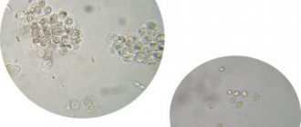
Urine analysis with sediment microscopy
Urine, or urine, is a product of the kidneys, the final component of metabolic processes. Urine contains water, as well as hormones, electrolytes dissolved in it, dead cells of the mucous membrane of the urinary tract, salts, mucus, leukocytes, etc. A general urine test (UCA) provides a set of information about the physical and chemical parameters of urine and the presence of various metabolites in it.
A general urine test allows you to evaluate the activity of the kidneys, bladder and other organs of the system; these are its most important, but not exhaustive purposes. The study will also help to identify disturbances in the activity of internal organs not related to the urinary system.
Microscopy of urine sediment is one of the routine diagnostic methods, which is used both for screening various diseases and for monitoring the course of diseases and the results of therapy.
The data that will be obtained after completing the study is as follows:
- General analysis (urine examination using dry chemistry) - urine specific gravity, hue, transparency, acid-base index, protein, sugar, nitrites, hemoglobin, ketone bodies, bilirubin, urobilinogen.
- Sediment microscopy (qualitative and quantitative assessment of a number of insoluble components) - leukocytes, casts, erythrocytes, epithelial cells, salts, bacteria.
Indications
For patients with various pathologies of the urinary and other systems, the analysis is prescribed according to the doctor’s recommendations; for healthy people, for preventive purposes, it should be done once every 6-12 months.
After a streptococcal infection, TAM is taken 7-14 days after recovery. The test results should only be interpreted by a doctor in order to correctly diagnose or exclude it.
Other indications for analysis:
- Screening studies, medical examinations.
- Monitoring the development of complications.
- Evaluation of treatment effectiveness.
- In a comprehensive examination of the body.
- For the purpose of differential diagnosis.
- To identify metabolic diseases and electrolyte imbalances.
- For the diagnosis of infections, inflammatory pathologies.
- For the purpose of monitoring the clinical condition of the patient after surgery.
- To analyze the condition of the kidneys when taking nephrotoxic drugs.
Most often, it is recommended to take an OAM if there are symptoms of inflammation in the urinary system - changes in the daily volume of urine, lower back pain, swelling, changes in smell, color of urine, increased temperature, etc.
Photo shows microscopy of urine sediment
How do they do it?
A container for collecting analysis should only be purchased at a pharmacy; home containers and previously used containers are absolutely not suitable! Disposable sterile jars prevent foreign substances from entering the material and distorting the results.
The day before, you should not consume coloring foods - beets, carrots, wine, blueberries and other natural dyes. Determining the correct color of urine is very important, because, for example, a dark shade of urine is sometimes a sign of liver inflammation.
The day before taking an analysis with sediment microscopy, it is not advisable to take medications - some of them distort the results, for example, Aspirin turns urine pinkish.
If the patient is taking antibiotics, antiseptics, or uroseptics, this must be reported to the doctor.
Also, the day before sample collection, you should avoid:
- Drinking alcohol;
- Consuming large amounts of fluid;
- Conducting sexual activity;
- Perform cystoscopy.
Before collecting urine, you should wash yourself without using soap or disinfectants. After using the toilet, you need to collect the morning portion of urine (the first after sleep) in a container. The container should be delivered to the laboratory within 24 hours, stored at a temperature of +2+24 degrees.
Urine examination is carried out as follows:
- The physical indicators of urine are assessed by appearance and with the help of the necessary equipment.
- A pipette is lowered to the bottom of the container, which has stood for 2 hours.
- Collect 10 ml of urine, which is centrifuged for 5-7 minutes.
- The composition of the sediment droplet is analyzed, obtaining all the basic data.
Decoding
OAM standards are given in the table:
Parameters Standard Units of measurement
| Hue | Yellow, straw yellow | – |
| Transparency | Transparent | – |
| Specific gravity | 1,010-1,025 | – |
| pH | 5,00-7,00 | pH |
| Protein | No or traces | g/l |
| Ketone bodies | No | mmol/l |
| Glucose | No | mmol/l |
| Bilirubin | No | mmol/l |
| Hemoglobin | No | Ery/uL |
| Nitrites | No | – |
| Urobilinogen | No | mmol/l |
Deciphering deviations in a general urine analysis with sediment microscopy:
Parameters Norms Units of measurement
| Leukocytes | Up to 3 for men, up to 5 for women | In sight |
| Red blood cells | Until 3 | In sight |
| Epithelium | Less than 5 in women, less than 3 in men | In sight |
| Salts | No | In sight |
| Cylinders | None or isolated hyaline | In sight |
| Bacteria | No | In sight |
| Slime | No or insignificant amount | In sight |
After receiving the test results, the doctor will be able to draw conclusions about existing changes in the body:
- Color. Darkening of urine means the presence of bilirubin or urobilinogen in large quantities. A red tint may indicate the presence of blood, a whitish color may indicate inflammation, the presence of mucus, and blue-green may indicate rotting in the intestines.
- Reaction. The abundance of animal food leads to acidification of urine, while dairy and plant foods lead to alkalization. Acidic urine is also observed in diabetes mellitus, gout, and fever; a shift to the alkaline side is characteristic of kidney inflammation, massive loss of salts due to vomiting, and diarrhea.
- Specific gravity. The density of urine increases with heart and kidney diseases, loss of water from the body, accumulation of protein, sugar, drug metabolites, and toxins. The specific gravity decreases due to hormonal imbalance, with some kidney pathologies.
- Transparency. If the urine is cloudy, this is due to the presence of fat, salts, epithelial cells, red blood cells, and white blood cells.
- Protein. The presence of protein means the development of severe or long-term kidney diseases; without kidney damage, it can be observed with fever, after heavy sports, and with congestive heart failure.
- Bilirubin. Appears in urine with pathologies of the liver and biliary tract.
- Urobilinogen makes urine more yellow and is observed in liver diseases, enteritis, and hemolytic anemia.
- Nitrites in urine are associated with the presence of bacteria and inflammatory reaction products.
- Glucose. Appears in diabetes mellitus, thyrotoxicosis, acromegaly, Fanconi syndrome.
- Ketone bodies. They grow with diabetes mellitus, less often with fasting, a sharp decrease in carbohydrate foods, or a prolonged increase in temperature.
- Epithelium. Appears in large quantities during the inflammatory process in the urinary system.
- Red blood cells. Blood in the urine occurs in severe heart pathologies, kidney and urethral injuries, cystitis, kidney infarction, tuberculosis of the bladder, vasculitis, polycystic kidney disease, infectious process, and oncological diseases.
- Leukocytes. The growth of leukocytes in the urine means the development of various forms of inflammation in the kidneys, urethra, bladder, and is also observed with general infections and fever.
- Cylinders. They appear when renal filtration is impaired and are characteristic of serious kidney and heart diseases, dehydration, overheating, and poisoning.
- Slime. It increases with inflammation in the kidneys and underlying parts of the urinary system.
- Bacteria. Indicates the presence of a bacterial infection.
- Salts (crystals). Indicate disturbances in mineral metabolism, the presence of stones and sand.
The analysis can be negatively affected by improper sample collection, long-term storage, poor hygiene, excessive consumption of liquids, drugs and dyes, as well as stress, pregnancy, and menstruation. Video about sediment microscopy:
Source: https://gidmed.com/nefrologiya/diagnostika-onk-nefr/laboratornye-issledovaniya/analiz-mochi-s-mikroskopiey-osadka.html
Urine analysis for organic substances
Protein scores are designated Pro. They should not exceed 0.03. In diabetics, MAU readings should be within the range of 4.25 mmol. From this result you can learn about the reversibility of kidney damage. Its level is monitored to monitor pathologies in order to begin treatment in a timely manner.
The glucose level is designated Glu. The best result is no sugar. If it is present, then we can talk about the presence of diabetes mellitus.
Bilirubin is designated as Bil. The ideal is its absence. If bilirubin is present, gallbladder or liver problems may occur. The explanation of BLD in urine analysis is presented below.
Urobilinogen (aka URO) is designated UBG and should be present in urine. This indicator is used to diagnose the condition of the blood, liver, the absence or presence of stones, and infections.
If there are ketone bodies in the urine, that is, Ket, then the patient may have diabetes mellitus. Such bodies appear after poisoning, stroke or anesthesia.
Ascorbic acid is designated Asc. The urine of a healthy person includes it in amounts of up to thirty milligrams. The BLD norm in urine analysis is of interest to many.
Using a microscope, urine is checked for leukocytes, designated as Leu; for men its indicators should be no higher than three, and for women – no more than five. If there are deviations, then we can judge about inflammatory processes in the genitourinary system.
Other important indicators
Decoding a urine test consists of two main parts. First, the physical properties of urine are assessed: density, color, smell, acidity, presence of dissolved salts. After this, the specialist begins to evaluate the chemical composition. The main goal is to identify the following components:
- GLU or glu. Glucose is encoded under these symbols. Its presence in the sample indicates the development of diabetes mellitus. The test results for a healthy person should read “GLU NEGATIV,” which means a complete absence of glucose.
- KET – ketone bodies. These include acetone, beta-hydroxybutyric and acetoacetic acids. Normally, they should be completely absent from urine. Otherwise, we can talk about the presence of diabetes.
- BIL. This is what bilirubin is called in English. This component is part of bile. In a healthy patient, the “BIL NEG” indicator is deciphered. He talks about the absence of bilirubin in the sample. Its detection indicates cirrhosis of the liver.
- V.C. This abbreviation can be read as nitrates. The NEG value in a urine test in this case is considered normal. Their appearance is a consequence of serious inflammatory processes occurring in the human body.
- CRE – creatinine. This substance is produced by the body as a result of vital activity. Its concentration should not exceed 17.7 mmol per liter of urine. The child is in danger if this indicator exceeds 65 µmol/l.
- PRO – protein. Its complete absence in the sample will be considered an indicator of health. If the transcript reads “PRO trace,” this means that traces of the protein are present in the sample being studied. This may be a consequence of liver dysfunction.
- LEU. Leukocytes are encrypted under this abbreviation. Normally, there should be no more than 6 such cells in the field of view. If the LEU in a urine test is exceeded, this indicates the development of sexually transmitted diseases or kidney failure. Leukocytes are also found in fungal infections of the urinary system.
- NIT – nitrites. They are formed in the body as a result of the action of enzymes on nitrates. Normally, they should not be found in human urine. The reason for their appearance often lies in the development of the inflammatory process in the organs of the urinary system.
- UBG. This designation corresponds to urobilenogen. It is a product of the reduction of bilirubin. Synthesized under the influence of intestinal bacteria. UBG in urine analysis should be detected in amounts from 5 to 17 µmol. Deviations from the norm indicate health problems. If its concentration is underestimated, impaired renal function, liver cirrhosis or viral hepatitis are more often diagnosed. In pregnant women, a decrease in urobilenogen levels is observed during toxicosis. If UBG is completely absent from the analysis, there is a blockage of the bile duct. Exceeding the norm occurs due to hemorrhagic diathesis or hemolytic anemia.
When identifying deviations from the norm of any indicators, it is important to understand what this means. An accurate diagnosis can only be made after undergoing a comprehensive examination. Sometimes the results can be distorted due to violation of the rules for collecting urine. Only a sample collected early in the morning is suitable for research. Its shelf life is no more than 4 hours. It is better to collect urine in a special container.
Today on the Internet you can find many sites where test results are automatically deciphered. You shouldn't trust them completely. Only an experienced specialist who knows your medical history can read the research results correctly.
BLD is an important indicator of OAM, by which one can judge the presence and severity of the disease. To detect a problem at an early stage, you need to undergo regular examinations and remember how to decipher their results.
Types of red blood cells
In a healthy person, a normal indicator in urine sediment is either the absence of red blood cells, or their detection during microscopic analysis in an amount that does not exceed two cells in the preparation (that is, in all fields of view). An increased concentration of red blood cells is called hematuria. If the presence of blood in urine can be detected visually, that is, the urine has a red tint, then gross hematuria can be judged.
In this situation, during microscopy of urine sediment, the entire field of view will be covered with red blood cells, that is, the norm will be exceeded many times. If the number of red blood cells in urine is small, then it is impossible to notice blood with the naked eye, since the urine will have a normal color. Red blood cells are determined only through microscopic examination. In this case, we can talk about microhematuria. This is what BLD means in a urine test.
The appearance of red blood cells changes depending on the specific gravity and reaction of the urine. Red blood cells can be in the urine either unchanged (fresh) or changed (leached).
Unmodified red blood cells contain hemoglobin; their shape resembles greenish-yellowish discs. This species is found in alkaline, neutral and slightly acidic urine.
Changed red blood cells occur during long-term exposure to acidic urine. They do not contain hemoglobin; in appearance they resemble colorless rings. Such red blood cells are called leached. In addition, the changes are wrinkled red blood cells, which are found in urine with a high relative density, and with a low relative density their diameter increases. With prolonged standing of urine, red blood cells transform into an altered type. We have explained what BLD means in a urine test.
To determine the source of hematuria, the classification of red blood cells into unchanged and changed is not of great importance, since in the overwhelming majority of cases their transformation is influenced by the chemical and physical characteristics of urine. Leached red blood cells may indicate renal origin of hematuria when detected in freshly released urine.
The main property that may suggest a renal origin is the appearance of casts and protein in urine along with red blood cells. Most often, red blood cells appear in the urine due to various diseases of the urinary tract and kidneys. In some cases, hematuria begins after intense physical activity, but in such situations, red blood cells increase for a short period.
Answer
Good afternoon, most likely you mean BLD (short for the English word blood). This indicator indicates the presence of blood in the urine. Normally, this should not happen; the number 3 means presence within the visibility range. Those. For you, the urine looks normal in appearance, but laboratory technicians can see with the naked eye a change in the color of the urine due to the presence of blood cells there. The appearance of blood may be normal, but in your case, 200 red blood cells indicate the pathological presence of blood. Do an MRI, ultrasound - you need to find out the cause of hematuria and start treatment.
The detection of more than 3 red blood cells in urine sediment per field of view indicates the presence of various diseases of the kidneys and urinary tract.
Visible traces of blood in the urine can be observed in a woman during menstruation, which requires repeated testing.
When erythrocytes are detected through a general clinical analysis, in order to make a more accurate diagnosis, the patient is referred for additional diagnostic studies - quantitative determination of erythrocytes in urine sediment using the Nechiporenko, Kakhovsky-Addis methods, as well as two-glass or three-glass tests to determine the localization of the pathological process.
What does BLD indicate in a urine test?
So, BLD in the analysis are red blood cells, and their detection in urine is a negative symptom. If BLD is detected in the urine, then we can most likely talk about inflammatory processes occurring in the genitourinary area, or about pathological renal changes. If more than three units of red blood cells were found in the urine, then in medical terminology this situation is called hematuria, which has its own classification:
- microhematuria - in this case, the urine is of normal color, but red blood cells are detected in microscopic sediment;
- gross hematuria - with this type of disease, the urine becomes red or brown in color;
- renal - otherwise called renal, it allows you to detect kidney pathologies, heart attacks, injuries;
- extrarenal - its appearance is possible during inflammatory processes in the genitourinary area, and is a consequence of diseases such as urethritis, prostatitis and cystitis.
If red blood cells are detected in a urine test (BLD trace), the attending physician usually prescribes an additional examination for the patient. If the result of the secondary analysis is positive, there will be no doubt about the pathological transformations of the urinary tract and kidneys. The most common disease is hematuria, which is particularly common. The most dangerous form of all of the above is considered macrohematuria, which is accompanied by such pathologies as:
- malignant tumors of the renal pelvis;
- malignant formations in the bladder and ureter;
If a person is healthy, then there are no red blood cells in his urine. If they are detected in the urine, it is necessary to conduct repeated tests to refute or confirm the diagnosis.
Decoding BLD in urine analysis
A person comes to the clinic when something in his condition causes concern. The doctor should immediately write him a referral for tests. The most informative and common is a general urine test, which immediately reflects all the malfunctions in the body. However, an ignorant person is unlikely to be able to understand the terms.
Each indicator has its own standards, BLD in urine analysis is no exception, and if all of them are within these limits, then the patient does not need to worry, since there are no pathologies. But if any value is exceeded, we can talk about some kind of inflammation.
So, there should be no BLD in urine, and if the results are positive after repeated tests, you should sound the alarm and start treatment immediately.
What is hidden under the BLD designation?
In urinalysis, specialists use the Latin letters BLD or Russian letters KRV to denote red blood cells. Sometimes the designation BLO or ERY may appear. These are blood cells containing hemoglobin. Their main function is to distribute oxygen throughout the tissues of the body. At the same time, they take carbon dioxide and move it to the lungs, from where it is eliminated naturally.
Red blood cells are involved in protecting the body from foreign substances. On their surface they have receptors with which they manage to capture antigens. They subsequently transport these cells to the liver, where they are destroyed.
The presence of BLD in urine in small quantities is considered normal. This is based on the fact that red blood cells are able to penetrate the kidney membranes. However, their significant number in the sample under study indicates the development of serious pathology.
Standard values
Ideally, if you read a general urine test, it should show a complete absence of red blood cells in the sample being studied. In single quantities, these cells can appear after excessive physical exertion, drinking alcohol, or prolonged psychologically traumatic situations. In these cases, the composition of urine quickly normalizes after the negative impact is eliminated. The BLD content level will depend on the person’s age:
- In children in the first year of life, there should be no red blood cells in the urine at all.
- In the interval from 1 to 18 years, the detection of one or two cells is considered natural.
- After adulthood, the norm in urine is from 0 to 3 red blood cells.
In women, the finding of three BLDs is considered natural. For men, this may mean an early stage of the disease. The urine of representatives of the stronger sex may contain only single cells.
An increase in the number of red blood cells to 250 cells threatens a person’s life. In such a situation, the patient is urgently hospitalized.
What is the reason for the increase in red blood cells?
If, after a general analysis of urine for red blood cells, a positive result was obtained, then this indicates a number of factors. In this case, pathological transformations in the genitourinary system and kidneys can be observed. In addition, the result may be false positive, which is determined by the physiological specifics of the human body.
Often, red blood cells are detected as a result of dehydration, a lack of fluid in the body. Water deficiency is caused by vomiting and stomach upset.
During physical activity, athletes lose large amounts of fluid, which can also cause BLD to appear in urine. After restoring the water balance in the body, red blood cells disappear.
The physiological factors that cause the appearance of BLD SA in urine analysis (we examined the norm) are:
- the temporary nature of the manifestation of red blood cells in urine, for example, after a person is in a hot workshop or in a hot bath, which causes overheating of the body;
- increased physical and mental stress;
- due to the increased content of glucocorticosteroids in the blood, which can often be associated with stress experienced by a person;
- when in mountainous areas where there is a lack of oxygen;
- poisons and toxins that people often encounter;
- consumption of alcoholic beverages, which causes the risk of vascular spasm, which causes the appearance of red blood cells in urine after drinking alcohol;
- a large number of spices in food.
In addition, urine may turn red not because it contains an excess amount of red blood cells, but because the concentration of hemoglobin in its composition is increased. When hemolysis develops, the red blood cells contained in the blood are destroyed, and this is typical for defects in urine filtration in the kidneys, and it all ends in acute renal failure.
It is worth noting that the human body is unique in the sense that it can adapt to the influence of various factors, and in a number of situations, the detected red blood cells can become a normal indicator for a particular person. Pathologies that cause BLD include the following diseases:
How to read Traceback?
The trace contains a lot of useful information when you are trying to determine why an exception occurred in your code. In this section, we'll look at different trace options to understand what information they contain.
Traceback Review
There are several sections in each trace that are important in their own way. The diagram below highlights these parts:
In Python, it's better to read the trace from bottom to top:
- Blue box: The last line of the trace is the error message line. It contains the name of the exception that was thrown.
- Green frame: An error message appears after the exception name. This message usually contains useful information to help you understand why the exception occurred.
- Yellow frame: Next along the trace are various function calls moving from bottom to top. These calls are represented by two-line entries for each call. The first line of each call contains information such as the file name, line number, and module name, which indicates where the code can be found.
- Red box: The second line for these calls contains the actual code that was executed.
There are several differences between the trace output when you run your code on the command line and when you run the code in the REPL. Below is the same code from the previous section executed in the REPL and the resulting trace output:
>>> def greet(someone): … print('Hello, ' + someon) … >>> greet('Chad') Traceback (most recent call last): File "", line 1, in File "", line 2, in greet NameError: name 'someon' is not defined
Note that instead of filenames you get . This makes sense since you entered the code using standard input. Additionally, lines of code that are executed do not appear in the trace.
Note : If you're used to seeing stack traces in other programming languages, you'll notice a significant difference in what a trace looks like in Python. Most other languages print the exception at the top and then go from top to bottom, with the most recent calls being the least recent.
Example of reading a trace
Looking at some specific trace results will help you better understand and know what information the trace will provide you.
The code below is used in the following examples to illustrate the information that the trace provides you:
# greetings.py def who_to_greet(person): return person if person else input('Greet who? ') def greet(someone, greeting='Hello'): print(greeting + ', ' + who_to_greet(someone)) def greet_many (people): for person in people: try: greet(person) except Exception: print('hi, ' + person)
Here who_to_greet() takes the value person and either returns it or asks for the return value.
Greet() then takes the name to greet, someone , and an optional value, greeting , and calls print() . who_to_greet() is also called with the value passed to someone .
Finally, greet_many() will iterate over the list of people and call greet() . If an exception occurs when calling greet() , a simple fallback greeting is printed.
There are no errors in this code that would cause an exception to be thrown if valid input is provided.
If you add a greet() to the end of greetings.py and specify a keyword argument that it doesn't expect (for example greet('Chad', greting = 'Yo')) then you will get the following error:
Traceback (most recent call last): File "/path/to/greetings.py", line 19, in greet('Chad', greting='Yo') TypeError: greet() got an unexpected keyword argument 'greeting'
Once again, with Python tracing, it's better to work backwards, moving up the output. Starting from the last line of the trace, you can see that the exception is a TypeError . The messages that follow the exception type, everything after the colon, give you important information. It tells you that greet() was called with a keyword argument that it didn't expect. You're also given an unknown argument name: greting .
Moving up, you can see the line that caused the exception. In this case, it's the greet() that we added to the end of greetings.py .
On the next line you will see the path to the file where the code exists, the line number of that file where the code can be found, and the module in which it is located. In this case, since our code doesn't use any other Python modules, we just see it here, which means it's an executable file.
With a different file and different input, you can see that the trace does indeed point you in the right direction to find the problem. If you follow along, remove the buggy greet() from the bottom of greetings.py and add a new example.py with the following content:
# example.py from greetings import greet greet(1)
In this file, you import your previous module, greetings.py , and use greet() . Here's what happens if you run example.py :
$ python example.py Traceback (most recent call last): File "/path/to/example.py", line 3, in greet(1) File "/path/to/greetings.py", line 5, in greet print(greeting + ', ' + who_to_greet(someone)) TypeError: must be str, not int
The exception thrown in this case is again a TypeError , but this time the message is a little less helpful. It tells you that somewhere in the code it expected to work with a string, but was given an integer.
Moving up, you see the line of code that was executed. Then the file and line number of code. However, this time we get the name of the function that was executed instead, greet() .
greet() call is passed as an integer.
Sometimes after an exception is thrown, another piece of code catches that exception, which also results in an exception. In these situations, Python outputs all exception traces in the order in which they were received, again ending with the last exception trace call.
Since this can be a little confusing, let's look at an example. Add a greet_many() to the end of greetings.py :
# greetings.py … greet_many(['Chad', 'Dan', 1])
This should output a greeting to all three people. However, if you run this code, you will see an example output of several traces:
$ python greetings.py Hello, Chad Hello, Dan Traceback (most recent call last): File "greetings.py", line 10, in greet_many greet(person) File "greetings.py", line 5, in greet print(greeting + ', ' + who_to_greet(someone)) TypeError: must be str, not int During handling of the above exception, another exception occurred: Traceback (most recent call last): File “greetings.py”, line 14, in greet_many( ['Chad', 'Dan', 1]) File "greetings.py", line 12, in greet_many print('hi, ' + person) TypeError: must be str, not int
Please note the highlighted line, During handling... in the data above. This means that while your code was trying to handle the previous exception, another exception occurred.
Note. The feature of displaying backtraces of previous exceptions was added in Python 3. In Python 2, you will only get a traceback of the last exception.
You saw the previous exception before when you called greet() with an integer. Since we added 1 to the list of people greeting, we can expect the same result. the greet_many() function wraps the greet() into a try and except . In case greet() throws an exception, then greet_many() will print the default greeting.
of greetings.py again :
def greet_many(people): for person in people: try: greet(person) except Exception: print('hi, ' + person)
So when greet() throws a TypeError due to invalid integer input, greet_many() handles that exception and tries to output a simple greeting. Here the code ends as a result of another, similar exception. It's still trying to add a string and an integer.
Reviewing all trace results can help you understand what might be the real cause of the exception. Sometimes when you see that the last exception was thrown and a callback was received as a result, you still don't see what's wrong. In these cases, going through previous exceptions usually gives a better idea of the root cause.
Renal type of red urine
In this case, urination simultaneously with blood is recorded in the patient against the background of such kidney pathologies as:
— autoimmune disease of the canals and glomeruli of the kidneys with acute or chronic glomerulonephritis;
- a stab wound to the renal surface or a significant bruise - in such a situation, blood clots in the urine can take on the appearance of gross hematuria.
Hematuria during pregnancy
A common occurrence during pregnancy, especially in its last stages, is pregnancy nephropathy, which is caused by increased load on the kidneys and is characterized by the appearance of protein in the urine, edema and increased blood pressure. With nephropathy in pregnant women, red blood cells in the urine can be detected in small quantities, no more than 10.
Also, in pregnant women with a history of chronic pyelonephritis, its transition to the acute stage is possible, which is also diagnosed by the appearance of red blood cells (no more than 10), leukocytes and bacteria in the urine.
Blood in the urine during pregnancy can be observed with hemorrhoids during pregnancy, especially with internal nodes, which in the early stages do not have any other clinical symptoms other than heavy bleeding.
Postrenal type
To determine that the symptoms of hematuria in a patient are caused precisely by postrenal causes, it is necessary to conduct an examination, which may reveal inflammation in the urethra or bladder:
- due to damage to the mucous membrane of these organs by sand or stones;
- against the background of cystitis, both in women and men;
- for oncological neoplasms;
- during hard sexual intercourse, which damages the mucous membrane of the urethra;
- injuries to the urethra or bladder with simultaneous bleeding and vascular damage, which are accompanied by symptoms of gross hematuria.
Red blood cells in urine analysis in children
The norm for children in terms of the number of red blood cells should be no more than six in the field of vision for girls and four for boys. If their content is increased, this is called erythrocyturia. If the urine turns red due to too many red blood cells, then in this case we are talking about hematuria.
Since urine during its formation goes through a long path, starting in the kidneys and ending with the urethra, each of the sections along the way can become a source of red blood cells. What does BLD CA 25 ery/ul mean in a urine test? More on this below.
If significant defects in the formation of blood clots and the coagulation system are observed, then the red blood cells in urine are of extrarenal origin. They are detected in hemolytic anemia, low platelet count or impaired platelet activity, hemolytic-uremic syndrome and coagulopathies.
Renal red blood cells occur with glomerulonephritis, hereditary nephritis and IgA nephropathy.
If the calyces and pelvis are damaged due to vascular defects, carcinoma, kidney injuries, urolithiasis and cystic diseases, then the shape of the red blood cells during microscopy is unchanged, but their number is increased.
Since the movement of stones from the kidneys occurs along with the flow of urine, the urinary tract and its components are damaged: the bladder, ureters, urethra, as a result of which red blood cells appear in the urine.
With acute or chronic rhinitis, after catheterization of the bladder and with urethritis, red blood cells in the urine may also be increased. In a urine test, a BLD CA of 25 means that 25 red blood cells are detected per field of view.
Bld at 50 rbc/ul
Expert opinion
Kovaleva Elena Anatolyevna
Doctor-Laboratory Assistant. 14 years of experience in clinical diagnostic services.
Ask a question to an expert
Determination of red blood cells in urine at a value of 50 rbc/ul (can also be indicated by the symbol “++” on the analysis form) is a sign of the following pathologies: cystitis , acute glomerulonephritis, kidney infarction, urolithiasis.
As a rule, these diseases are accompanied by pronounced clinical manifestations in the patient - increased body temperature, pain in the kidneys, pain when urinating, swelling, increased blood pressure (diastolic or lower).
Together with hematuria, traces of protein, leukocytes, bacteria, and epithelial cells are determined.
Increased red blood cells in urine in men
In some situations, men (especially older men) may need complex therapy for hematuria in the presence of prostate pathologies.
Blood under such circumstances means:
- A cancerous tumor in the prostate gland that can cause bleeding.
— Inflammatory processes in the prostate, as a result of which red blood cells mix with urine. BLD - trace intact - what is this in a urine test? This means there are no red blood cells in the urine, which is good.
Increase in red blood cells in women
Women especially often suffer from excessive concentrations of red blood cells in urine if they have problems caused by pathological processes of the genitourinary system:
- if blood is detected during urination, one can judge about uterine diseases that provoked bleeding;
- erosion of the uterine cervix can also cause blood clots in urine.
In addition, the doctor may find in tests that not only red blood cells, but also white blood cells are elevated. This indicates the presence of inflammatory processes in the kidneys.
We examined the decoding of BLD in a general urine analysis.
A general urine test is a mandatory diagnostic procedure that is prescribed to all patients who visit a medical facility. Today, urine analysis is processed by special analyzers, and the results of the analysis are given to the patient in the form of a table with incomprehensible Latin letters, which only a specialist can read. Different analyzers can provide their own interpretation and designations.
Below is a breakdown of these very “incomprehensible letters”, as well as norms for individual indicators.
The results can be expressed both in concentration units and in the form of a score (+1, +2, +3, +4) or “+” signs. In this case, the fact of detection of different quantities of the analyzed component in the sample or its absence is stated:
| +1 - random +2 - frequent +3 - multiple +4 - cannot be counted | + random + + frequent + + + multiple + + + + cannot be counted |