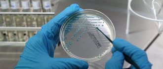
A diagnostic test consisting of taking elements of kidney tissue for morphological examination, kidney biopsy. In other words, this is an intravital examination of a part of an internal organ to identify pathologies. Translated from Greek, the term means “looking at the living.” It was developed in the middle of the 20th century and only in modern conditions has it found widespread use. At the moment, this is the most reliable diagnostic method with no alternative.
There are several types of kidney biopsy:
- Percutaneous
It is controlled using x-rays, ultrasound and magnetic resonance methods.
- Transjugular
It is carried out by catheterization of the renal vein. It is recommended for patients with severe obesity, poor blood clotting, renal abnormalities, and chronic respiratory disorders.
- Biopsy in tandem with urethroscopy
It is done according to indications due to urolithiasis and abnormalities of the urinary tract. Indicated for pregnant women and children, as well as people with an artificially implanted kidney.
- Open
Performed directly during surgery. This type of biopsy is prescribed for patients with operable tumors, frequent bleeding, and one working kidney. The procedure is performed exclusively under general anesthesia. There are practically no complications with this type of biopsy, since it is performed under direct visual control.
What is a kidney biopsy
A kidney biopsy (nephrobiopsy) is the removal of kidney tissue under ultrasound guidance for diagnostic testing. The study is prescribed to clarify the diagnosis if other possibilities of clinical and laboratory examination turned out to be uninformative.
Nephrobiopsy helps:
- identify kidney disease and clarify the diagnosis;
- determine the prognosis of the disease;
- select adequate drug treatment;
- identify kidney dysfunction after transplantation.
There are several types of biopsy:
- Transdermal. The puncture needle is inserted through a skin incision using an ultrasound machine or computed tomograph.
Percutaneous biopsy is performed under mandatory ultrasound or computed tomography control
- Open. It is performed during kidney surgery, when it is necessary to take a large amount of kidney tissue if a malignant tumor is suspected. A laparoscopic biopsy can be performed, which is less traumatic for the patient.
The laparoscopic method of taking a puncture is less traumatic for the patient
- Method of ureteroscopy with biopsy. Performed for stones in the kidneys or ureters. The ureteroscope is inserted into the ureter, then into the lower part of the kidney, followed by sampling.
- Transjugular. It is performed by catheterization of the jugular vein. Indications for such a biopsy are blood diseases with coagulation disorders, patients with large body weight with chronic cardiac and respiratory failure.
Features of biopsy in children
The accumulated experience in performing biopsy in children indicates the need for careful selection for it. In children, the indications do not differ from adults, but the study is always performed under general anesthesia. The preferred method of taking a puncture is percutaneous access.
Renal cell carcinoma (RCC) accounts for about 2% of all tumors in adults (Parkin, 2005). In the USA, RCC was diagnosed in 36,000 patients (22,000 men and 14,000 women) (Jemal, 2005). From 1973 to 1997 the number of newly reported cases of RCC in the United States increased by 43%, and mortality increased by 16%. In Europe, mortality from RCC increased until the 1990s. Since the mid-90s. XX century the mortality rate stabilized. The largest declines were observed in Germany, Norway and the Netherlands (Levi, 2004). In Russia, kidney cancer is diagnosed in approximately 15,000 people per year. In the general structure of oncological diseases, kidney cancer occupies 3% of all malignant tumors (Bray F., 2003, Levi F., 2004).
Biopsy is an important method for verifying the tumor process and strategically determining treatment tactics. However, not all clinics perform a biopsy to determine indications for surgical treatment in patients with kidney tumors.
This is due to several factors:
- Today in urology there are many reliable, highly specific, highly sensitive, effective and accessible diagnostic methods: ultrasound, CT, MRI;
- the specificity of these research methods does not depend on the size of the tumor;
- widespread development and introduction into clinical practice of nephron-sparing methods for the treatment of renal cell cancer, including laparoscopy;
- possible complications of biopsy;
- cost of research.
A dilemma arises: we really actively advocate biopsy when it comes to bladder or prostate tumors, and for some reason we ignore biopsy for kidney tumors.
Today, our surgical activity is very high - we operate on patients with any TNM stage of a kidney tumor, of any degree of complexity (Table 1).
Table 1. Quantitative distribution of kidney tumors by histological structure
| Distribution of kidney tumors by nosology in operated patients in the Stavropol region and the city of Togliatti for the period from 1997 to 2001. | 1997 | 1998 | 1999 | 2000 | 2001 | Total |
| Total operated on | 44 | 42 | 46 | 45 | 72 | 249 |
| Unclassified cancer | 5 | 1 | 3 | 0 | 10 | 19 |
| Papillary cancer types 1 and 2 | 1 | 1 | 3 | 3 | 3 | 11 |
| Oncocytoma | 0 | 1 | 3 | 3 | 4 | 11 |
| Angiomyolipoma | 0 | 6 | 2 | 2 | 8 | 18 |
| Chromophobic cancer | 0 | 1 | 1 | 1 | 1 | 4 |
| Multilocular cystic nephroma | 0 | 0 | 1 | 1 | 1 | 3 |
| Clear cell carcinoma | 38 | 32 | 33 | 35 | 45 | 183 |
Pleshivtsev M.A., Saratov, 2007
It should be noted that, due to the high sensitivity and specificity of radiation diagnostic methods, the percentage of detection of small tumors, which are classified as a separate stage T1a, is increasingly increasing. Treatment of small kidney tumors has become the second most important problem after drug resistance in advanced stages of the disease. Tumors smaller than 3 cm tended to grow slowly and metastasize infrequently. In this regard, there is an opinion about the advisability of performing a biopsy before surgical treatment in order to assess the real need for surgical intervention at this particular moment. If the tumor is poorly differentiated, surgery makes sense at this time. Soon, undoubtedly, new genomic and other methods for analyzing biopsy material will appear, which will make it possible to predict the appropriateness of treatment.
Currently, radiation diagnostic methods are the main ones in detecting kidney tumors. Computed tomography allows us to verify the diagnosis in almost 100% of cases, so we do not think about the need for a biopsy. Unfortunately, the percentage of detection of advanced stages of kidney tumors remains high: in stage T3 - up to 39%, T4 - up to 40% of cases (Kavashima A. et al., 2004).
It is an undeniable fact that all radiation diagnostic methods allow us to verify the diagnosis in almost 100% of cases, so we do not think about the need for a biopsy.
In the mid-50s of the last century, the method of ultrasound examination (ultrasound) first appeared, thanks to which the number of incidentally detected kidney tumors increased from 10% in the 1970s. up to 68% in 1998. If a routine ultrasound reveals a kidney cyst belonging to the third type according to the Bosniak classification, then a biopsy must be performed. However, it should be noted that with the combined use of computed tomography (CT) with ultrasound diagnostics, the diagnosis of kidney cancer can be reliably established without a biopsy.
Angiomyolipomas are also one of the problems most often encountered in outpatient practice as an incidental finding during ultrasound (Figure 1). If you perform a biopsy for angiomyolipoma to exclude the malignant component of the tumor, you may encounter a certain percentage of complications. In this case, the combination of ultrasound and CT diagnostics is also sufficient to establish the diagnosis of angiomyolipoma and not subject the patient to a biopsy (Nelson CP, 2002, Oesterling JE, 1986).
Picture 1. Ultrasound. Angiomyolipoma
Another imaging method used to diagnose a tumor process is magnetic resonance imaging (MRI). The value and availability of MRI in staging the tumor process, assessing the invasion of the fibrous capsule of the kidney, and damage to the lymph nodes is undeniable, but this diagnostic search will still lead us to the realization of the need for surgery.
The information content of MRI and CT also tends to 100%, and this applies to any stage of the disease (Palko A., 1991) (Figure 2).
Figure 2. CT in the diagnosis of kidney tumors
So, the question of a biopsy is especially acute for small kidney tumors, determining further tactics for patients - active surveillance or surgical treatment? As a result of studies, it has been proven that even with small kidney tumors there is a risk of metastasis: there is no safe tumor size. For tumor sizes less than 2 cm, tumor-associated mortality is 4.3%, less than 4 cm – 6.2%; and the frequency of metastasis with such tumor sizes is 5.2% (Tsui KH, 1997). Therefore, even despite the small size of the tumor, it is impossible to guarantee the patient the absence of metastasis. Even tumors that are called benign are conditionally benign.
From a deontological point of view, the situation is also ambiguous. Let's offer a patient with a small kidney tumor a choice of two situations. The first is to suggest a tumor biopsy and, in the absence of a malignant process, recommend dynamic observation. The second is to suggest immediately performing a laparoscopic operation (for example, kidney resection) or an ablative technique (cryoablation) of a tumor in the kidney. If the patient is informed about the possible false-negative result of the biopsy, which, on average, occurs in 5% of cases (Peters C., 2005), its possible complications, and the necessarily benign nature of the tumor, then, most likely, he will immediately choose surgery.
A biopsy, like any invasive procedure, can have complications that either require additional intervention or make the biopsy uninformative (Table 2). These are complications such as inadequate biopsy (up to 10%), micro- and macrohematuria (5-9%), asymptomatic perinephric hematoma (up to 90% of cases), bleeding requiring blood transfusion, injury to neighboring organs (Tang S., 2002, Cluzel P., 2000). In the most serious cases, a kidney tumor biopsy may result in surgery, including nephrectomy.
Table 2. Frequency and structure of complications of kidney biopsy
| Complications | Frequency (%) |
| Inadequate biopsy | 2-3-5-10 |
| Microhematuria | 100 |
| Gross hematuria | 5-9 |
| Asymptomatic perinephric hematoma | 90 |
| Symptomatic perinephric hematoma | < 20 |
| Bleeding requiring blood transfusion | < 3 |
| Bleeding requiring surgery | < 0,2 |
| Trauma to adjacent organs | < 1,0 |
| Infection (in the absence of pyelonephritis) | < 1,0 |
| Intervention-free arteriovenous fistulas | 15-18 |
| Arteriovenous fistulas requiring intervention | < 1,0 |
| Nephrectomy | 0,02-0,06 |
| Fatalities | < 0,1 |
So where is the place for a biopsy? It cannot be categorically stated that a biopsy is not needed for kidney tumors. However, thanks to the diagnostic methods that are available to urologists today, kidney biopsy, as an additional invasion associated with complications, can be avoided in most cases of kidney tumors (Table 3).
Table 3. Complications of biopsy
| Center | Number of patients with serious complications | Number of biopsies | Percentage of serious complications |
| 1 | 4 | 54 | 7,4 |
| 2 | 4 | 28 | 14,3 |
| 3 | 0 | 12 | 0 |
| 4 | 3 | 110 | 2,7 |
| 5 | 5 | 25 | 20 |
| 6 | 8 | 71 | 8,8 |
| 7 | 0 | 36 | 0 |
| 8 | 8 | 26 | 30,8 |
| 9 | 10 | 36 | 27,8 |
| 10 | 6 | 24 | 25 |
| 11 | 7 | 89 | 7,9 |
| Total | 55 | 531 | 10,4 |
F. Hussian, M. Mallik, 2009
Conclusion
• It is possible that a biopsy of a kidney tumor is needed for patients who are planning ablative surgical treatment methods: cryoablation, HIFU therapy, thermal ablation, RFA. But even in this case, a biopsy using the express method can be performed at the time of the operation.
• Patients with radiologically undetectable renal masses are quite rare; however, by conducting a comprehensive examination of such patients, it is possible to make a diagnosis without a biopsy.
• Patients who refuse surgery may be advised to undergo a tumor biopsy. If the histological report reveals malignant cells, then there is a chance that the patient will respond more favorably to the proposed surgical treatment.
• Patients with metastatic disease in extremely serious condition and/or inoperable patients are patients who need to verify the diagnosis with a biopsy and decide on chemotherapy and targeted therapy. I would like to share my own experience gained at the Research Institute of Urology (Table 4).
Table 4. Experience of the Research Institute of Urology
| Operations | year 2009 | 2010 |
| Radical nephrectomy | 48 | 72 |
| Laparoscopic nephrectomy | 6 | 25 |
| Nephroureterectomy | 3 | 11 |
| Kidney resection | 10 | 24 |
| Laparoscopic partial nephrectomy | — | 2 |
| Total | 67 | 134 |
| Histologically – cancer | 66 | 133 |
| Angiomyolipoma | 1- 1,49%- 1,56% | 1- 0,75%- 0,8% |
In 2010, we performed 72 open radical nephrectomies, 25 laparoscopic nephrectomies, 24 open and 2 laparoscopic kidney resections, 11 nephroureterectomies, a total of 134 operations. According to the results of the histological report, out of 134 operations, kidney cancer was detected in 133 cases. One patient was diagnosed with angiomyolipoma, which accounts for 0.8% of cases.
Key words: kidney cancer, kidney biopsy. Keywords: kidney cancer, kidney biopsy
| Attached file | Size |
| 713.23 kb |
‹ Biopsy of kidney tumors: opinion “for” Up Brachytherapy for prostate cancer with Cs-131 sources ›
Indications for kidney biopsy
There are certain indications for performing a kidney biopsy:
- acute renal failure - in this case, a biopsy reveals the cause of the development of the pathology, allows you to determine treatment in the absence of positive dynamics of the disease and select candidates for hemodialysis;
- nephrotic syndrome in glomerulonephritis, rheumatic diseases, tumors - the study is performed if there is no effect from glucocorticoids;
- massive proteinuria - a large amount of protein in the urine, which is usually observed when glomerular filtration is impaired;
- persistent hematuria - the presence of blood in the urine;
- changes in the kidneys with: vasculitis (inflammation of blood vessels of an autoimmune nature);
- periarteritis nodosa (a type of vasculitis that affects small and medium-sized vessels);
- gout (a disease caused by the inability of the kidneys to remove uric acid);
- lupus erythematosus (an autoimmune disease of connective tissue and blood vessels);
A biopsy is performed to determine the structure of the tumor that has appeared in the kidney.
Possible complications
The most common complications of renal biopsy are listed below. Experts include:
- Internal bleeding that goes away on its own over time.
- Serious bleeding requiring a blood transfusion.
- Heavy bleeding. To eliminate it, an operation is performed.
- Breakthrough in the kidney.
- Purulent pyelonephritis, which is accompanied by inflammatory lesions of lipid perinephric tissues.
- Pneumothorax.
- Losing a kidney.
- Muscle bleeding.
- Infections.
Contraindications for the study
Contraindications to biopsy are divided into absolute and relative.
Absolute contraindications include:
- absence of a second kidney;
- chronic renal failure in the terminal phase;
- blood clotting disorder;
- renal artery aneurysm;
- tendency to thrombus formation, presence of renal vein thrombosis;
- hydronephrotic transformation of the kidney;
If the renal collecting system is overstretched due to hydronephrosis, a biopsy is not performed
- infection localized in the pelvis with the formation of pus;
- kidney tuberculosis;
- heart disease accompanied by right ventricular failure.
Relative contraindications include:
- arterial hypertension not controlled by antihypertensive drugs;
- uremia (acute intoxication with your own metabolic products caused by renal failure);
- multiple myeloma;
- atherosclerosis of the renal arteries;
- increased kidney mobility;
- novocaine intolerance.
Advantages and disadvantages
Like any research method, biopsy has its pros and cons. The positive aspects of diagnostic manipulation include the following:
- ease of preparation for the procedure;
- information content;
- is the most reliable and accurate diagnostic method.
The risks of the procedure include the following:
- a large list of contraindications;
- high price;
- possibility of complications after the procedure.
Preparing for the study
The patient must prepare in special ways for the biopsy:
- On the eve of the biopsy, the doctor asks about your health status, allergies and other diseases. Informs about the purpose of the manipulation, the technique of execution, and possible consequences. Consent to perform the procedure is signed.
- In 7–10 days, it is necessary to discontinue medications that affect blood clotting (thinning it):
- Aspirin;
- Analgin;
- Warfarin;
- Cardiomagnyl.
Before the biopsy, you should avoid taking medications containing acetylsalicylic acid and blood thinners.
- A clinical minimum examination is prescribed:
- blood test with platelet count, study of clotting time and bleeding duration;
- general urine analysis;
- blood chemistry;
- Ultrasound of the kidneys and excretory urography (introduction of a contrast agent and subsequent series of x-rays) determines the position and mobility of the kidneys, as well as the structural features of the urinary system.
- On the morning of the procedure, the patient should not take any liquid or food, especially if the examination will be performed under general anesthesia.
Price
Many people wonder how much a kidney puncture might cost. The price depends on the city and on the institution itself. So in small towns, the procedure will cost 1500-2000 rubles. In the capital, in government agencies, prices start from 2,500 rubles. (polyclinic of the Ministry of Foreign Affairs of Russia), 3000 rubles. at the City Clinical Hospital No. 67 named after. L. A. Vorokhobova, 4000 rubles. The study will cost at the Federal Clinical Center for High Medical Technologies of the Federal Medical and Biological Agency, Federal State Budgetary Institution.
In private institutions, prices start from 5,000 rubles. (MedinaMed clinic). In Moscow they can cost up to 60,000 rubles, the average cost is 25-30 thousand rubles. In St. Petersburg, the maximum price for a study is 25,000 rubles; in private clinics you can find options for 10.7 thousand rubles. and below. The lowest price for the procedure is 1,300 rubles at the Leningrad Oncology Center.
Biopsy technique
The procedure is performed in a hospital and takes 30–40 minutes:
- The patient is placed face down on a table with a bolster under the chest and abdomen to ensure maximum access to the kidney area.
- The doctor marks the injection site on the skin, treats the area, and numbs the injection site.
The procedure for collecting kidney tissue takes 30–40 minutes
- An incision is made in the skin and, under the control of an ultrasound machine, a needle is inserted and advanced.
- After inserting the needle into the kidney, the patient must hold his breath. At this point, the kidney tissue is taken. If larger material is needed, the needle may be reinserted.
- An aseptic bandage is applied to the incision site.
After the biopsy, the patient must be monitored for 6 hours. To diagnose complications, the following are measured:
- Body temperature;
- arterial pressure;
- heart rate.
Clinical blood and urine tests are also examined for this purpose. The next day, if the patient feels well, the patient is allowed to go home.
Decoding the results
For diagnosis, a tissue sample measuring 2 mm is sufficient. For quantitative assessment (counting functioning glomeruli), the sample size must be 10 mm. The examination is performed by a specialist using light, immunofluorescence or electron microscopy. After 3–5 days, the biopsy results are ready.
Video: examination of a tissue sample after a biopsy
The result of the procedure and its interpretation
A histological examination of the material obtained from a kidney biopsy is carried out by a pathologist. The result is usually prepared within 7-12 days. The specialist’s conclusion contains a description of the morphological features of the renal tissue: the presence or absence of pathological changes and their nature, as well as other features. Based on these data, a diagnosis is made.
Sources used:
- https://uromir.ru/nefrologija/issledovanie/biopsija-pochki.html
- https://ilive.com.ua/health/biopsiya-pochki_105666i15989.html
- https://www.euroonco.ru/glossary-az/biopsiya-pochki
- https://101analiz.ru/biopsiya/biopsii-pochki.html
- https://testanaliz.ru/biopsiya-pochki
- https://opochke.com/nefrologiya/biopsiya-kak-delaetsya.html
Rehabilitation period
After completing the diagnostic procedure, medical specialists leave recommendations for the patient that allow him to recover after the kidney biopsy.
- Bed rest is recommended for several days.
- Medical experts recommend increasing the amount of fluid consumed per day.
- You should avoid physical overexertion for 2–3 days.
- For preventive purposes, antibacterial and homeostatic drugs were prescribed.
- Heavy physical activity is not recommended for 3 months.
If a patient experiences a jump in blood pressure, sharp pain in the kidney area, an increase in temperature, or purulent or blood impurities in the urethra, it is necessary to seek help from a medical specialist as soon as possible.