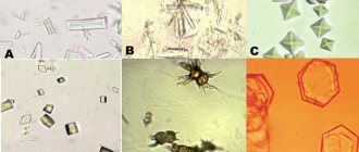
Types of cells
Epithelial cells found in urine can get there from various areas of the genitourinary system. To make an accurate diagnosis, when carrying out work, the laboratory technician must not only determine the type of sediment detected, but also the location of the epithelial cells.
These types of cells are found in urine.
Squamous epithelial cells
- In the female body they line the distal part of the urethra, labia, and vagina.
- In boys, squamous epithelial cells in the urine can only be detected through their migration from the cavernous zone of the urethral canal.
Transitional epithelial cells
Cover the cavity of the renal pelvis, ureter and ureter. In girls they can be found in the proximal zone of the urethral canal, and in boys - in the prostatic zone.
This type of tissue is characterized by the presence of relatively large cells on the surface that have a pear-shaped or flattened appearance. They contain two nuclei, as well as additional fusiform vesicles. This tissue is characterized by invagination of the membrane into the inner part of the cytoplasm.
Renal epithelial cells
They differ in appearance depending on the location in the kidney tubules.
In the proximal zone there is a single-layer type of cuboidal epithelium. Its cells contain one large nucleus, which is located in the central part of the cytoplasm. In addition, they have a brush border in the lumen. In the distal zone, the epithelium is formed by a light type of cubic cells in which there is no brush border.
If squamous epithelium is detected in the urine of a child, the doctor needs to understand the location of the disease that provoked such a symptom.
Disease or not
Let's make a reservation right away. The diagnosis can only be made by a specialist, taking into account all qualitative and quantitative indicators of the analysis of not only urine, but also blood, taking into account the general examination of the patient. The doctor will pay attention to accompanying signs, namely fever, pain in the kidneys or urinary tract and many other diagnostic signs.
Important! Correct collection of material for analysis is the first stage of successful diagnosis.
Increased levels of squamous epithelium may indicate the following:
- the presence of damage to the conduction pathways by the same salt crystals in the urine,
- mechanical damage to the urethra,
- inflammation of the urinary tract and bladder resulting from infection with viral, bacterial or fungal diseases,
- diseases associated with the functioning of nephrons as a structural unit of the excretory system of the kidneys,
- an increase in the amount of epithelium can be caused by taking certain medications (cough medications, for example),
- inflammatory processes in the prostate, which is not so common in boys,
- general intoxication of the body due to various poisonings,
- improper collection of material for analysis.
In any case, in order to correctly diagnose the child’s condition, the doctor, in addition to a thorough examination of the patient, will prescribe an additional clarifying range of tests.
Important! All analysis indicators are considered together and none can be singled out as the main one. A professional will force parents to do repeated or clarifying studies, and only after that can we talk about making a diagnosis and options for tracking the dynamics of changes in indicators.
Norms
Clinical urine analysis takes into account not only the properties of the biological material, but also the microscopic data of the sediment that is contained in it. After the study is completed, a general table of results is created, which allows you to compare the norm with the data obtained in the laboratory for a particular patient.
The number of epithelial cells in a child’s urine directly depends on his age. Normally, their number should be single – no more than 3–10 units.
Microscopic analysis of a child's urine sediment can reveal up to 5 units of squamous epithelium from the ureteral mucosa, as well as up to 3 units. transitional - from other parts of the patient’s genitourinary system.
If a child is over 14 years old, a urine test should not reveal renal cells of the epithelial tissue.
In newborns
In infants, when examining urine, laboratory assistants can detect epithelial cells of three possible types - this is due to the fact that during this period the genitourinary system is adapting to an existence that is not associated with the use of the mother’s body.
In teenagers
During adolescence, the body undergoes a number of hormonal changes. Because of this, in a urine test, the norm in children after 14 years of age suggests the presence of up to 10 epithelial cells of all existing types, except kidney.
For girls
The anatomy of the genitourinary system of the female body ensures that urine comes into contact with the labia during the process of excretion. In addition, hygiene procedures often contribute to significant detachment of epithelial tissue. In this regard, in some cases, it is possible to detect a larger amount of epithelium than the norm in a clinical study of girls’ urine.
Renal epithelium in the child’s urine is normal
Flat epithelium in a child’s urine can be present both in absolute health and in the presence of a fairly wide range of diseases. These cells are components of urinary sediment and are determined by microscopic examination of urine.
The amount of squamous epithelium is often determined by the age of the child, the presence of concomitant pathologies and other factors. Taking into account normal indicators and comparing them with the data of a particular patient, the doctor can diagnose various types of pathologies of both the urinary and other systems of the child’s body.
The surface of the organs of the urinary system is lined with three types of epithelial cells (epithelial cells), which, depending on its condition or the presence of pathologies, may appear in the urinary sediment. These types of epithelium include:
- Flat - is the covering of the inner surface of the urethra (urethra), in girls it lines the outer and inner labia, as well as the vagina.
- Transitional - covers the inner surface of the bladder, the mucous membrane of the ureters, and in boys it lines the ducts of the prostate gland.
- Renal - acts as a covering for the renal tubules, while at the same time being the cells that form the renal parenchyma.
The composition of urinary sediment can change with the growth of the child and the development of his body, as well as with the addition of various pathologies that directly affect the life of epithelial cells.
The amount of squamous epithelium in a child’s urine has important diagnostic significance. There is a generally accepted norm for its maintenance: the number of cells in the field of view should not exceed 5 units. To understand the full picture of the health status, all epithelial cells present are assessed, so upper limits of normal values are also established for them.
In a healthy child, no more than 2–3 units of transitional epithelium are noted, while renal epithelium should not be detected in the child’s urine sediment. Otherwise, we are talking about the presence of pathology in the kidneys. The only exceptions are newborns, because during the postpartum period the activity of the kidneys and the entire urinary system is activated.
When the baby reaches about two weeks of age, the body adapts to life outside the womb, and the norm for the renal epithelium is its absence. All other cases, even single cells, should alert the attending physician.
Reference! As children grow older, squamous and transitional epithelial cells gradually cease to be observed in their urine, provided that they are healthy. Or they are noted in single quantities.
An increase in the number of squamous and transitional epithelial cells, especially in combination with the appearance of renal epithelial cells, indicates the development of pathological processes in the urinary or genital area. If, during the deciphering of the urine test, it turned out that the squamous epithelium is elevated, then, taking into account the specifics of its localization, the doctor will immediately suspect the following diseases:
- Urethritis is inflammation of the urethra (urethra).
- Inflammatory processes associated with hypothermia - in boys, orchitis (inflammation of the testicle), epididymitis (inflammation of the epididymis), vesiculitis (inflammation of the spermatic cord), in girls - vulvitis (inflammation of the vagina).
Urethritis - an inflammatory process in the urethra - one of the reasons for the appearance of squamous epithelium in the urine
And also an increase in these cells can be observed due to improper preparation for the test, namely insufficient quality toileting of the genital organs before urine collection, violation of the basic rules when collecting and transporting biomaterial (urine).
It should be noted that the first two points are most often accompanied by a significant excess of squamous epithelial cells compared to normal values. Whereas in the last two situations, the jump in values is usually not high, and most likely, a repeat urine test will be required with more careful implementation of all necessary recommendations.
- Squamous epithelium in urine. What does this mean?
If in young patients, along with an increase in the number of squamous epithelium, transitional type cells appear, this tells the doctor about the spread of the inflammatory process up the urinary tract. The most common causes of surges in these epithelial cells are:
Uric acid crystals in a child's urine
- Diseases of the lower genitourinary tract of an inflammatory and infectious nature (cystitis (inflammation of the bladder), urethritis).
- Inflammation of the mucous membrane of the ureters, renal pelvis (pyelonephritis, pyelitis), including those developing against the background of urolithiasis as a complication.
- Dysmetabolic nephropathy is the spontaneous discharge of salt accumulations or renal sand, accompanied by damage to the mucous surface of the urinary tract.
There is a high probability that when urine is collected from a child using a catheter, an increased content of transitional epithelium will be detected in the sample. If at least single kidney-type cells are detected in urine, this is regarded as a deviation from the norm and requires further diagnostics to find the cause of the disorder.
An increase in squamous epithelium in the urine of children can be either a single manifestation not related to the disease, or one of a whole complex of symptoms of a certain pathology. If an increase in these epithelial cells is accompanied by one or more signs from the following list, then, in all likelihood, a urinary or reproductive disease is developing in the body.
So, if the child has:
- Pain, burning, cutting during urination.
- Loss of appetite, insomnia or sleep disturbances.
- Drawing, dull or sharp pain in the lower back.
- Weakness, lethargy, urinary incontinence.
- Swelling and increased blood pressure.
- Frequent regurgitation of unknown etiology.
- An increase in temperature that does not decrease under the influence of antipyretic drugs.
You should definitely consult a doctor to examine your child and prescribe additional tests that will help establish a diagnosis. Often with such symptoms in a general urine test, not only an increase in epithelial cells is observed, but also a sharp jump in red blood cells, leukocytes, mucus, bacteria, and protein.
A child's refusal to eat may be caused by a disease of the urinary system.
A clinical blood test reveals an increase in ESR (erythrocyte sedimentation rate), a change in the leukocyte formula and quantity, as well as the quality of blood cells. In order to confirm or refute the alleged diagnosis, a fairly wide range of diagnostic methods is used.
To determine the reason why the child has detected or increased squamous epithelium in the urine, additional research methods are prescribed. It could be:
- Urine analysis according to the method of Nechiporenko, Zimnitsky, as well as a three-glass sample.
- Ultrasound diagnostics of the kidneys, ureters, bladder.
- Computer or magnetic resonance imaging.
- Urography (x-ray examination) of the kidneys.
- Cystoscopy (internal examination of the bladder).
Depending on the child’s health condition and accompanying symptoms, the doctor selects one or more methods that, in his opinion, will help establish a diagnosis and develop the necessary therapeutic strategy. We should not forget that diseases of the urinary system are very dangerous and have a long latent (hidden) course. In addition, their manifestations can be masked by the symptoms of other diseases, for example, such as appendicitis, pneumonia, viral infection, VSD and others. This is especially common in children.
To minimize the risks of urinary system diseases leading to the appearance of epithelial cells in the urine of children, it is necessary to take into account all possible provoking factors. There are many reasons that can increase the content of squamous epithelial cells, as well as transitional or renal cells, and the main ones include:
- Renal epithelium in urine: what tests will tell you
- The hereditary nature of the pathology (the presence of diseases of the urinary system in close relatives).
- Development of toxicosis in early and late periods of pregnancy, insufficient functioning of the placenta.
- Prematurity of the child of varying degrees, and complications that developed against its background.
- Congenital anomalies of the urinary system (structural changes in the kidneys or ureters).
- Asphyxia (oxygen starvation) of the fetus during pregnancy or childbirth.
- Complications of infectious diseases (scarlet fever, as well as diseases caused by streptococcus or staphylococcus).
- Protracted inflammatory diseases accompanied by the accumulation of purulent contents (chronic tonsillitis, erysipelas, furunculosis).
The presence of predisposing factors implies mandatory parental and medical monitoring of the child’s condition, which includes regular urine examination. This simple and painless method allows you to detect the onset of the disease in time, as a result of which the treatment process will be much faster and more effective.
It must be taken into account that pathologies of the urinary system in the early stages are often not accompanied by characteristic signs. Therefore, laboratory tests of the fluid secreted by the kidneys are practically the only way to detect the disease.
One of the fairly common reasons why, during the interpretation of the analysis, it turns out that epithelial cells in a child’s urine are elevated is insufficient quality preparation for collecting biomaterial. Therefore, in order to correctly collect urine for general analysis, parents must do the following. Carry out a thorough toileting of the child’s genitals. Collect a sample from your morning urine sample.
Use a sterile container. Transport the sample no later than 1.5–2 hours to the laboratory. When collecting urine, if possible, take the middle portion, that is, the child flushes the first part into the toilet, then into the container, and the rest into the toilet too. For small children, there are special devices that can be purchased at pharmacies: they look like an oblong plastic bag with a sticky attachment to the skin.
Medical device (urinal) for collecting urine in young children (instructions for use)
This allows you to collect urine without holding the baby for a long time, waiting for his natural desire to urinate. If you use a regular container, you should make sure that it is dry and clean, and ideally also sterile. Following all these simple rules will significantly reduce the risk of receiving false positive results and eliminate the need for a repeat examination.
Memo to parents! Following your doctor's advice on regularly diagnosing children prone to diseases of the urinary system and following the technique of collecting urine for analysis will help keep their health status under control.
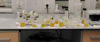
Collection of urine test
A general urine test must be taken during all examinations of the child. It is also indicated for planned hospitalization or suspected diseases of the genitourinary system. In order to obtain objective results of the study, it is necessary to follow the following rules for collecting urine:
- Biological material must be placed in a clean and dry container that does not contain organic residues or detergents. It can be made of plastic or glass. Doctors recommend purchasing sterile containers from pharmacy kiosks to collect urine.
- Before urinating, you must complete all hygiene procedures.
- For the study, you need to use the first portion of urine collected in the morning.
- No more than 90 minutes should pass between the collection of biomaterial and its delivery to the laboratory for analysis.
If there is a suspicion of acute inflammation, the child’s doctor may additionally prescribe a urine test according to Nechiporenko.
The analysis allows you to determine the number of white and red blood cells, as well as cylinders in the patient’s biological material. Detection of the latter is evidence of the presence of renal pathology.
In order to clarify the location, doctors prescribe a sample of 3 glasses. It involves collecting one portion of urine in three containers.
Thus, the first part of the biological material, its main volume and the last part are submitted for analysis.
Diagnostics
If squamous epithelial cells are detected in the baby’s urine and their number is higher than normal, the pediatrician will prescribe an additional examination. Since this is an indirect sign of pathology, especially if additional symptoms are observed.
Symptoms to watch out for:
- Pain and cramps during small hikes.
- Nagging pain in the lumbar region.
- Weakness, apathy, lethargy (the child is sad, unplayful, sleeps a lot).
- Loss of appetite.
- Frequent regurgitation for no reason (in newborns).
- Hyperthermia.
- Swelling of the joints.
If these symptoms are present, the pediatrician prescribes additional diagnostic tests:
- Ultrasound (ultrasound examination of the abdominal organs).
- Additional urine tests (three glasses sample, Nechiporenko analysis, etc.).
- MRI (Magnetic resonance imaging).
- Urinary examination using an endoscope (cystoscopy).
- Examination by specialized specialists (urologist, endocrinologist).
These diagnostic methods help to identify the cause of certain symptoms, make a diagnosis, and prescribe treatment.
Anomalies
- Renal-type epithelial cells in the urine of a child over 14 years old indicate the appearance of nephrosis or nephritis in the acute stage.
- Transitional cells are found when there is inflammation in the ureter, bladder and urethra. The discovery of an increased number of leukocytes confirms this doctor’s assumption.
- If one epithelial cell is detected during the analysis, the laboratory assistant must conduct a second microscopy of this volume of material or take a second portion of urine.
What it is?
Epithelium is the cells that line the surface of organs, providing a protective barrier.
Epithelium is of the following types:
- Flat. Present in the urethra and urethra. Contained in urine in very small quantities.
- Transition. This is the epithelium of the inner surface of the bladder, renal pelvis, and ureters.
- Renal. Covers the kidney tubules.
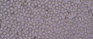
Epithelial cells enter urine as it passes through all urinary organs. Normally there are very few such cells ; a sharp increase in indicators indicates pathologies of the urinary organs. During inflammation, the epithelium actively dies and is excreted in urine.
Reasons for deviations
The most common causes of epithelial cells in urine are:
- incorrect urine collection;
- inflammation in the organs of the urinary system;
- some other diseases;
- appearance and migration of stones;
- taking certain medications;
- failure to comply with hygiene procedures;
- reverse flow of urine;
- intoxication.
An excessive number of transitional cells in a child’s urine occurs in the following cases:
- for inflammation caused by cystitis and urethritis;
- with urolithiasis;
- during catheterization.
Epithelial kidney cells end up in the urine due to kidney pathologies. They can be bacterial or autoimmune in nature.
Cystitis
Inflammation of the bladder mucosa, cystitis is characterized by detachment of transitional epithelial cells in the affected area. Doctors consider infection caused by bacteria or hypothermia to be the main causes of this disease.
In most cases, cystitis is caused by E. coli. The main symptom of this disease is pain when urinating.
Due to the anatomical features of the body, girls develop cystitis much more often than boys.
- Take note: Monurel for children against cystitis
Urethritis
When the urethra becomes inflamed, doctors diagnose patients with urethritis. It can have both infectious and non-infectious etiology. Experts consider the following pathogens to be the most common causative agents of the disease:
- ureaplasma;
- mycoplasma;
- Trichomonas.
Sometimes urethritis occurs due to damage to the mucous membrane by stones passing through the urethra. The disease in this case appears due to autoimmune processes.
The main symptom of urethritis is painful urination accompanied by burning and itching. Some patients experience mucous discharge.
Kidney pathology
Kidney pathologies glomerulonephritis and pyelonephritis are considered one of the most common. Characterized by the occurrence of inflammatory processes in various parts of the organ. Because of this, epithelial cells peel off in areas where the disease develops.
Urolithiasis is a dangerous kidney disease. Stones, while moving through the ureter, can mechanically damage its mucosa, thereby causing exfoliation of the epithelium.
Such injuries ultimately become a gateway to infections and inflammation.
Symptoms
In some cases, an increase in the number of epithelial cells does not manifest itself in any way. This occurs at the beginning of the disease. If the disease is in an acute period or significant damage to the renal tissue has occurred , then the patient experiences the following symptoms:
- Cloudy or discolored urine.
- The appearance of drops of blood in the urine.
- Strong unpleasant odor of urine.
- Swelling of the face and limbs.
- High blood pressure.
- Temperature increase.
- Increased levels of protein in urine.
- Back pain.
- Pain when urinating.
- General deterioration of the patient's condition.
Treatment
Self-treatment is not recommended; there is a risk of adverse consequences.
If an increased amount of epithelium is detected in a child’s urine test, if he does not have symptoms of the disease, the doctor should order additional tests
- additional tests of urine and blood,
- performing ultrasound,
- and other diagnostic techniques.
Depending on the data obtained through a systemic examination, a decision is made to prescribe antibiotics, NSAIDs or other groups of medications.
Rate this article:
Prevention recommendations
Simple preventive measures that should be performed systematically will help to avoid the occurrence of problems or aggravation and complications of the disease (hereditary pathology of the genitourinary system, maternal toxicosis, complicated or premature birth, streptococcal or staphylococcal infection):
- carry out regular diagnostics and submit biological material of the child for laboratory testing;
- carry out hygiene procedures daily, observing the rules and features of care depending on the gender of the child;
- monitor the baby’s diet so as not to create additional stress on the kidneys - limit diuretic products (watermelon, rosehip juice);
- try not to overheat or overcool your legs and genital area;
- strengthen the immune system with hardening, vitamins, and physical exercise.
The reliability of the study depends on how aware parents are of the manipulations during the delivery of biological material. Therefore, do not hesitate to ask your doctor about how to properly prepare for the test.
Also, deciphering the analysis results should be entrusted to a specialist, and not done on your own.
Sometimes self-medication based on research results can cause more harm.
How to get tested correctly?
Often, squamous epithelial cells are detected in urine as a result of improper preparation for the test.
- You need to transfer the container with urine to the laboratory within 1-2 hours after urination.
- During menstruation, it is better to refrain from taking the test.
- Urine collection is carried out only in a clean (preferably sterilized) container; a sufficient amount of urine is 100 ml.
The basic rule that must be followed is to carry out hygiene procedures for the external genitalia before collecting urine.