Cystography is an instrumental method for studying the bladder, which consists of an X-ray image after filling the organ with water-soluble contrast. Depending on the route of administration of the contrast agent, cystography can be ascending or descending. Descending cystography is the last stage of excretory urography, when contrast is injected intravenously, after which it is excreted by the kidneys.
Descending cystography
Ascending cystography is performed after the bladder is filled with contrast through the urethra.
Retrograde (ascending) cystography
Varieties
Depending on the method of administering contrast and the moment of radiography (taking an x-ray), five types of research are distinguished:
- retrograde;
- descending;
- voiding;
- dynamic;
- antegrade.
Retrograde (ascending)
Retrograde cystography, also called ascending cystography, is the fastest X-ray examination of the bladder because it injects contrast material directly into the bladder through a urinary catheter. This X-ray technique allows you to obtain a clear image of the bladder, its contours and volume, as well as identify changes in the tone of the bladder, the presence of tumors, stones, foreign bodies and inflammatory processes in patients.
Descending radiography
The technique of descending (excretory) cystography involves injecting a contrast agent into the patient intravenously, and X-rays are taken after the bladder is filled with the contrast agent. This method takes quite a long time, since its implementation directly depends on the functioning of the renal parenchyma. Most often, this technique is performed at the end of excretory urography (about one hour after urography), which allows to identify abnormalities in the functioning of the kidneys, detect stones and tumors of the kidneys and ureters.
Mixture
Vaccination cystography is most often used for examination in childhood and consists in the fact that after introducing contrast through a catheter into the bladder, the child must empty it, and the radiologist takes X-rays at this time. This technique allows you to identify pathological changes not only in the bladder, but also in the urethra.
Dynamic
The peculiarity of this technique lies not in the technique of contrasting the bladder, but in the time at which the pictures are taken. To perform dynamic cystography, several images are taken while the bladder is filling and emptying, and a fluoroscopic examination is also performed in parallel.
Antegrade
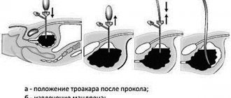
The essence of antegrade cystography is that a radiopaque substance is introduced into the bladder using a special method - through a cystostomy in the lumbar region (a catheter that is inserted directly into the bladder through a small incision in the anterior abdominal wall) or using a puncture of the renal pelvis.
Consequences of bladder examination
Kidney problems are a common occurrence these days, so the question of how cystography is done in children will be relevant. As you know, both adults and children suffer from kidney failure. The cause of the development of such diseases is often poor nutrition, infections and hereditary factors.
At the first suspicion of kidney disease, you should immediately consult a doctor. Only in this case can a correct diagnosis be made in time and appropriate treatment prescribed. The human urinary system can be susceptible to various ailments. To determine what exactly is at stake, the doctor must conduct an examination and prescribe tests.
When it comes to the kidneys, different methods are used to diagnose the disease. The most common of these include urine tests, blood tests and ultrasound.
Its peculiarity is the ability to obtain more accurate data about a disease that has affected such an important organ as the kidneys.
This type of procedure is prescribed to those patients who come to the doctor with symptoms of kidney disease.
However, this type of study allows you to obtain accurate information about the condition of almost all organs of the urinary system. This type of diagnosis is based on the use of X-rays.
As a result, the doctor has the opportunity to obtain the most accurate information regarding the condition of some internal human organs.
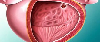
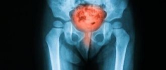
An X-ray of the bladder makes it possible to see possible inflammatory processes or other serious disturbances in the functioning of this organ, for example, ruptures and thickening of the walls.
However, given that during this procedure an X-ray is used, that is, we are talking about a certain amount of radiation, cystography is not often prescribed in childhood, and in one session they try to obtain the maximum amount of information about the condition of the internal organs.
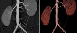
In order to carry out the cystography procedure, it is necessary to introduce a special substance into a specific organ. Often, contrast liquid is injected into the bladder, but in some cases it becomes necessary to inject the solution directly into a specific organ. This usually applies to cystographic examination of the kidneys.
After administration of a special solution, the kidneys acquire clear outlines under the influence of X-rays. This makes it possible to see possible problems hiding inside the body. As a result of the examination, the doctor can discern a violation of the integrity and structure of the walls of the internal organs.
Victory cystography in children deserves special attention. It is carried out at the moment when the patient’s body is cleared of urine. This makes it possible to assess the condition of the urinary tract, as well as see places of fluid leakage, if any.
Just a month ago, the phrases “void cystography” and “vesicoureteral reflux” were empty words for me. I just never heard them, didn’t know what it was and didn’t think at all that it would affect my child.
- In this review I want to talk about this study, since I myself recently looked for information on this topic.
- I tried to describe everything in detail, but if you have any questions, I will be happy to answer.
- By the way, this procedure is done at absolutely any age: both adults and infants.
Background: My son has an enlarged pelvis of his right kidney since birth. More precisely, I learned about this during pregnancy, at about 20 weeks.
When he was 1.5 months old, he fell ill with acute pyelonephritis. We were in the hospital, seemed to have received good treatment and forgot about the kidney problems for a while.
We were registered with a nephrologist from birth, were observed, had periodic ultrasounds, and were constantly tested for OAM and Nechiporenko tests. By the way, the tests were almost always good. The only thing is that my son almost always had a temperature of 37-37.2.
This worried me very much, although outwardly it did not affect his well-being. In the fall of 2020, we had the last ultrasound (as I thought at that time) and according to it we should have been deregistered.
The pelvis did not completely return to normal, but did not enlarge, and this is already good and, in fact, for his age it was an acceptable size (6.5 mm, a little later it became 3.5). The joy knew no bounds! An ultrasound was done on Friday, and on Saturday night my son became ill: he was vomiting and his temperature rose to almost 40.
In the morning I called an ambulance and we were admitted to the hospital. First, by mistake, I was taken to the infectious diseases department (although I said it was KIDNEYS, hear me! KIDNEYS! but no...), two days later I was transferred to the pediatric department.
As I thought, there was an exacerbation of the kidneys, namely acute pyelonephritis again. Relapse His mother...
This time the pyelonephritis was especially severe and the son was on a drip many times, completely refused to eat, and the temperature jumped from 35 to 40.
Since the relapse occurred for the second time in the second year of life (the child was 1 year and 10 months old), we were offered the method of Vaccination Cystography.
Void cystography is a method of x-ray examination of the bladder and adjacent organs of the urinary system during the act of urination.
During urination, a radiopaque substance penetrates the urethra, therefore this diagnostic method allows us to judge the condition of the urethra and bladder.
Also, voiding cystography provides very valuable information in the diagnosis of vesicoureteral reflux.
In order to exclude or confirm a more serious illness - vesicoureteral reflux. That is, urine reflux. If it is not treated, then at least pyelonephritis will become a constant companion. I don’t want to think about a worse development of events, much less talk about it.
Planned hospitalization
https://www.youtube.com/watch?v=oLzddeDXuwg
We were “lucky” and just at the time of treatment the X-ray room was undergoing renovations. They couldn’t do the procedure for us. Naturally, they didn’t keep us, they treated us, discharged us, prescribed treatment and told us to come back in a month. It could have been earlier, but the New Year holidays coincided and there was nothing to catch in the hospital, this procedure was also not done in those days, since all the specialists were absent.
- I don’t know about your hospital, but ours has the following list of documents for admission:
We didn’t need all the documents, only OAM, according to Nechiporenko, a referral from a pediatrician for hospitalization, a certificate of vaccinations, a certificate of epidemic. surrounded, another ultrasound, previous discharge from the hospital, my fluor. All other tests were taken during admission. In any case, it is better to check with your doctor whether you need all the documents on the list.
Under anesthesia or not? In general, it is believed that for boys cystography is much more painful than for girls (the fact is that their genitourinary system is structured completely differently) and it is most often done for them under general anesthesia.
And many small children too, since you need to lie still, and this is difficult to explain to a baby. And the result may not be correct, or the procedure may simply not be possible.
By the way, we were not even offered anesthesia. I didn’t insist. Here, of course, a lot depends on the child, but to be honest, I don’t understand why such a procedure requires general anesthesia. It is not long at all and, although painful, it is tolerable.
It also depends on the doctor. Our doctor inserted the catheter in just a second, without hesitation or delay, thereby not adding any pain to the child.
Yes, the main thing is to insert the catheter, and then it won’t hurt. Well, it’s easy for me to say now, but in general, knowing my son, I had thoughts about anesthesia.
Moreover, several months before this, my son broke his collarbone and they could hardly do an x-ray. He began to panic and become hysterical at the sight of the dark X-ray room. My husband and I could not hold him back, much less distract him.
But this time he took it all calmly. I was pleasantly surprised, I even took a sedative before the cystography, remembering the previous x-ray.
Yes, this is exactly the question that faces everyone who needs cystography. I thought for a long time, assessing the pros and cons of the hospital and the paid clinic.
In a paid clinic in our city, this procedure costs 3,350 rubles (you need to have a doctor’s referral with you for a general urine test). You will be in the office for less than half an hour, and then you will go home. It's tempting, though...
But! Here another question arises: if something goes wrong and the child needs help, then at home you will have to call an ambulance and still go to the hospital.
As for the hospital, cystography is free here.
When we were sent home, the doctor said that we would only need to go to bed for two days in order to be under observation after the study. Just think, two days! Let's lie down.
And then, this doctor is a very well-known nephrologist in our city and I trust her, so I wanted her to carry out this procedure.
In the end, the choice fell on the hospital. We went to bed on Wednesday and, according to my calculations, should have been home on Friday. But a few days before our hospitalization in this department there was no opportunity to go home at night (((And we could not be discharged earlier, as promised in 2 days, because in this case the hospital would not have received compulsory medical insurance payments. A vicious circle .
As a result, we spent 7 days in the hospital. And to be honest, I didn’t regret it, because complications really arose. More on this below.
PreparationPreparation began with me. I’ll say right away that the doctor prepared and set me up, saying that the main thing was my strong attitude and a minimum of emotions. And if we don’t want to, then no one will force us to do it; in general, it’s up to you to decide.
To be honest, with these words she really scared me and even the thought of abandoning this study crept in. I imagined that everything would be much worse and longer.
Indications and contraindications
Cystography, like any instrumental research method, has a number of indications and contraindications, and if the indications are suspicions of the same pathological conditions, then the contraindications differ for different techniques depending on the technique of contrast administration.
| Indications for use | Contraindications | |
| Retrograde cystography | Antegrade and excretory cystography | |
|
|
|
How to prepare?
This procedure does require preparation . This is necessary to obtain reliable results. Preparation includes the following steps:
- A week before the procedure, fatty and fried foods are excluded from the diet. You need to avoid foods that cause gas formation in the intestines. We are talking about legumes, baked goods, milk and carbonated drinks. You should eat more fruits and vegetables.
- If a person suffers from constipation, then a week before the procedure you need to take a mild laxative, since stagnant stool can negatively affect the result of the procedure.
- Two days before the procedure, you should give up tea and coffee and drink clean still water.
- A few minutes before the procedure, you should calm down, sit still, and relax.
- A person must get enough sleep. Lack of sleep and stress can negatively affect the examination result.
When the patient enters the treatment room, the doctor's instructions must be followed.
There is no need to worry or be nervous about the upcoming procedure. When the patient is extremely relaxed, the most accurate results are obtained . These steps are quite enough to properly prepare for cystography.
Preparation and technique
Preparation for various studies, including cystography, is based on eliminating factors that can interfere with the procedure or distort its results. The main obstacle to obtaining clear and reliable images with this research method is gas formation in the intestines. In order to avoid this, patients should follow a special diet two days before the procedure.
It is necessary to exclude from the diet:
- carbonated drinks containing dyes;
- black bread;
- raw vegetables;
- whole milk;
- juices;
- baked goods;
- confectionery;
- cabbage;
- peas;
- beans.
In addition, you must first do a cleansing enema in the evening and in the morning before the study to remove feces and gases. Also, when performing cystography, you should first toilet the external genitalia.
When performing excretory cystography, in order to avoid complications and adverse consequences, the doctor first performs a test to identify allergic reactions to the contrast agent. If an allergy is suspected, the procedure is cancelled.
The main difference in the options for performing cystography is the method of introducing the contrast agent into the bladder. Immediately before the procedure, the patient must empty the bladder.
Technique for retrograde cystography:
- The patient lies down on the couch.
- A catheter is inserted through the urethra into the bladder.
- A contrast agent is injected through the catheter.
- After the bladder is filled with contrast solution, a clamp is applied to the catheter.
- The required number of x-rays are taken.
- The clamp from the catheter is removed and the bladder is emptied.
- After this, a final X-ray is taken.
- After the procedure is completed, the patient is recommended to rest and rest in bed for one day.
Technique for performing descending excretory cystography:
- The patient is seated on a couch or chair.
- A warmed X-ray contrast agent is injected intravenously into the cubital vein for 2 minutes, while the patient is closely monitored so that when the first symptoms of an allergic reaction appear, the procedure is stopped.
- If excretory urography is performed before cystography, then the first images are taken after the administration of contrast approximately ten minutes later.
- After the bladder is filled with contrast, a series of x-rays are taken approximately 60 to 90 minutes later.
- After this, the patient's bladder is emptied and a final image is taken.
Technique for performing voiding cystography:
- The patient is placed on the couch and a urinary catheter is inserted.
- A contrast agent is pumped into the bladder through a catheter.
- The picture is taken after the bladder is full.
- Next, the patient is asked to urinate, while a series of photographs are taken.
- After the end of urination, the last photo is taken.
One day before dynamic cystography is performed, the patient should take a prophylactic dose of a uroseptic, for example Furagin.
Technique for dynamic cystography:
- The patient lies down on the couch.
- Bladder catheterization is performed.
- A radiopaque substance is injected through a urinary catheter until a strong urge to urinate appears.
- At the same time, fluoroscopic observation of the filling of the bladder is performed.
- After it is filled, wait until it completely empties on its own and, while urinating, continue the fluoroscopic examination and take a series of photographs.
The presence of a cystostomy greatly facilitates cystography. To do this, contrast is injected into the bladder through a catheter and the required number of images is taken. However, given the fact that installation of such a catheter requires surgery, it is performed only if there are special indications.
Before performing antegrade cystography, a survey radiography of the abdominal organs is performed through puncture of the renal pelvis. This procedure is minimally invasive and is performed under local anesthesia.
Technique for performing puncture antegrade cystography:
- The patient is placed on his stomach on the couch.
- Based on the plain radiograph, a puncture needle is inserted into the pelvis, sending it a solution of novocaine.
- Some of the urine is removed from the pelvis through a needle using a syringe.
- Next, the contrast agent is injected into the pelvis.
- Pictures are taken.
- After an x-ray, the entire contents of the pelvis are removed and an antibiotic solution is injected into it.
- After this procedure, it is recommended to avoid exercise for one day and maintain bed rest.
Possible complications
Cystography should only be performed by a qualified specialist due to the fact that it can cause complications. One such complication is an allergy to the contrast agent. In this case, the patient experiences shortness of breath, cough and redness of the face. If such symptoms appear, the doctor must provide medical assistance to the patient. The patient is given intravenous hormonal drugs.
In addition to an allergic reaction, the patient may experience a nephrotoxic effect. In this case, the patient’s general condition deteriorates sharply. The patient may complain of severe headache or dizziness. Sometimes you may feel an unpleasant taste of acetone in your mouth. Medical care in this case consists of infusion and detoxification therapy.
When performing retrograde cystography in the case of benign hyperplasia, a metal instrument can accidentally injure the mucous membrane of the urinary canal. One of the complications of retrograde examination may be complete urinary retention. This occurs as a result of irritation of the nerve endings by the contrast agent.
Features of the event
Due to the difference in the anatomical structure of some organs of the urinary system in men and women, there are some differences in the choice of the preferred cystography technique.
Among women
Women tolerate the retrograde cystography method much more easily, which is directly due to the anatomical features in the structure of their urethra - it is much shorter and wider than in men (approximately 30 - 50 mm versus 180 - 200 mm in length and 10 - 15 mm versus 8 mm, respectively ), which allows you to easily insert a catheter into the bladder. Other types of research may have individual characteristics in patients that are not related to gender.
In men
Quite often, men undergo excretory cystography, since the anatomical structure of the urethra and the fact that it passes through the prostate gland (prostate) limit the use of retrograde cystography. In addition, an obstacle to this method is the presence of prostate diseases, in which the urethra narrows, such as prostatitis, tumors, cysts.
In children
The main difference in performing cystography in children is their psychological preparation for the procedure, since many children are afraid of medical workers, and during the X-ray, the small patient must lie still.
Preparation for the procedure in children is carried out in the same way as in adults - foods that increase gas formation are excluded from the diet 2-3 days before the test. In addition, it is recommended to give children porridge and unsweetened tea in the morning; during the day it is recommended to drink tea with fennel or chamomile, dill water; taking sorbents (for example, Activated carbon) and laxatives (Lactulose) also helps to avoid gas formation.
Preparatory measures
You need to carefully prepare for undergoing voiding cystourethrography. The patient will have to perform a number of preparatory procedures:
- Review your diet .
- Stop taking foods that cause increased gas formation. These products include fresh milk, legumes, and sweet soda. To reduce gas formation, it is recommended to drink dill water.
- Immediately before the procedure, you need to cleanse the intestines. To do this, take gentle laxatives or do a cleansing enema.
Preparatory measures depend on the method used for cystography. During a descending study, in addition to bowel cleansing and diet, the patient must prepare a medical syringe and contrast that will be injected into the bloodstream.
With the retrograde method, a person must begin preparation several days before the start of the event. You need to eat right for two weeks before the procedure. Immediately before the examination, take a shower.
The cleaner the external organs, the better the result of the preparation. In addition, the patient must purchase a urethral catheter and contrast agent in advance.
Survey results
The image obtained after cystography is called a cystogram. Using this image, a radiologist is able to determine the size and volume of the bladder, its shape, position in the pelvis and in relation to other organs, the thickness of its walls, the position of the ureters, the passage of the contrast agent solution and its filling of the bladder.
In addition, the method makes it possible to detect the reflux of a contrast agent from the bladder into the ureters, the presence of stones, cysts, diverticula in the bladder, damage to the integrity, fistulas and inflammatory processes of its wall, as well as detect narrowings and strictures of the urethra and ureters.