Preparation for an ultrasound of the bladder and prostate in men is necessary to obtain clear results and data on the condition of the internal organs during the examination. Specialists in the field of urology and andrology often resort to ultrasound examination to determine the diagnosis. This method has a high degree of information content and makes it possible to detect pathological changes in the structure and functioning of the pelvic organs even in the early stages of the development of the disease.
- General rules for preparing for ultrasound
- Preparatory measures for ultrasound of the bladder
- How can a patient prepare for a transrectal ultrasound of the prostate? Types of enemas
- Using glycerin suppositories
- Laxatives
Who is indicated for ultrasound of the bladder and kidneys?
The localization of the organ in the pelvis and its belonging to the urinary system leads to the fact that ultrasound is often performed in conjunction with other organs. In men, the prostate gland is located on the neck, which with age undergoes benign changes and hyperplasia develops. Therefore, bladder ultrasound is often combined with prostate examination.
In women, the organ is located in front of the uterus and covers it; after filling, it becomes possible to examine the genitourinary system. But its targeted study is carried out according to indications. Also, research is required for renal pathologies, regardless of gender, that affect urinary function.
An ultrasound of the urinary system is necessary for the following diseases:
- suspicion of malignant or benign tumors. According to statistics, cancer makes up 3% of all cancer pathologies and is more often observed in men;
- with prostate adenoma or benign hyperplasia;
- prostate cancer;
- acute or chronic prostatitis;
- urolithiasis disease.
Ultrasound is not informative; if the patient has cystitis in acute or chronic form, the examination will show swelling of the organ wall, its thickening.
Research results
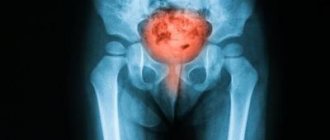
Visualization of internal organs is the main goal of ultrasound. The specialist enters the obtained data into the patient’s chart, then assesses the condition of the genitourinary system. Prostate:
- uniformity or heterogeneity of structure;
- symmetry of outlines (contours);
- dimensional frames: transverse 24-41 mm, longitudinal up to 40 mm, anteroposterior from 25 mm;
- organ weight – up to 18 grams.
- volume 350-750 ml;
- egg shape;
- smooth outlines;
- the walls of the organ are of the same thickness;
- the structure must be echo-negative.
Deviations from standards indicate possible diseases:
- malignant neoplasms;
- benign tumors;
- inflammatory processes, including prostatitis;
- chronic inflammation of the genitourinary system.
If pathology is suspected, additional studies are carried out. Independent interpretation of test results is incorrect; the assessment should be given by a specialist who takes into account the individual characteristics of the patient. For example, these may be genetic or congenital abnormalities of the bladder or prostate.
Preparing for an ultrasound of the bladder
The ability to visualize the urinary organs depends on their fullness. If there is no fluid in a hollow organ, it does not reflect ultrasound, but scatters it. Therefore, preparation for ultrasound of the bladder begins 1-2 hours before the procedure. The patient needs to drink at least 1.5 liters of water without gases. But some doctors recommend following the formula - 10 ml for every kilogram of body weight.
The liquid can be any, but without gases. Warm water or tea is better absorbed. Coffee and other drinks with high caffeine content are not needed. They affect renal blood flow, so they can distort the results of the examination if an ultrasound scan of the kidneys and bladder is performed. During a routine examination, women and men have the same preparation plan.
Ultrasound of the bladder and prostate
For men, examination can be carried out not only through the anterior abdominal wall. This method is used less frequently due to its low information content. When diagnosing pathology of the prostate and bladder, a transrectal ultrasound is performed. It requires special training.
It is recommended to follow a slag-free diet for several days to reduce gas formation. In the evening before the prostate ultrasound, you need to do a cleansing enema or take a laxative according to the regimen recommended by the doctor. It is possible to perform a microenema in the morning shortly before the ultrasound. There are no restrictions on food intake for transrectal diagnosis.
What is the procedure
The principle of the study is based on the fact that the ultrasonic waves emitted by the sensor are reflected from the tissues of the human body and recorded.
After undergoing special processing, they transfer the image to a computer. The specialist sees the full picture and can determine not only the external condition, but also the size, even weight of the organ. For prostate diseases, the following types of diagnostics are performed:
- Transabdominal - in other words, through the stomach. The sensor examines the prostate through the anterior wall of the peritoneum. The method is low informative, the waves pass through the bladder and muscles, the image may be distorted;
- Trasurethral - through the urethra. It is painful, despite the microscopic size of the sensor. It is used most often during surgical interventions.
- Transrectal (TRUS) – through the rectum. A fairly informative method, the study allows you to obtain the maximum amount of data.
If the patency of the rectum is impaired, there are contraindications to TRUS, the sensor can be installed in the perineal area. By examining the area under the scrotum, a specialist can determine whether the size of the prostate is normal and obtain information about its functionality.
Why do you need an ultrasound of the bladder in men?
One of the common pathologies in men is inflammation of the prostate gland, and with age, the likelihood of developing benign prostate hyperplasia increases. It compresses the neck of the bladder, leading to impaired urination, decreased potency and spontaneous leakage of urine.
Ultrasound of the bladder and prostate gland can detect pathological changes in the prostate. It can be:
- signs of sclerosis;
- calcifications;
- increase in volume;
- cysts.
Ultrasound helps to identify tumors in order to subsequently differentiate between a malignant and a benign tumor. Also during the examination, you can notice changes in the seminal vesicle.
The volume of the bubble can vary significantly. To conduct a proper examination, during preparation there should be no feeling of fullness, which turns into pain. The bladder should be moderately full. on average, its volume in a healthy man is about 300 ml. During ultrasound, you can determine the volume of residual urine that remains after emptying. This is one of the signs of pathology.
Norm and deviations
Normally, the prostate gland has a uniform or heterogeneous structure with uneven contours. The weight of the organ does not exceed 18 g. Dimensions: longitudinal corresponds to 4 cm, transverse ranges from 2.7 mm to 4.1 cm, anteroposterior 2.5 cm.
The urinary reservoir is normally ovoid in shape with a volume of 350 to 750 ml, with an echo-negative structure. The contours of the organ are smooth, the walls are of the same thickness.
The presence of pathological processes in the genitourinary system is indicated by:
- changes in organ volumes are a sign of benign neoplasms;
- increased echogenicity is a symptom of oncology, chronic prostatitis or cystitis, as well as urolithiasis;
- decreased echogenicity indicates inflammatory processes and cystic formations;
- enlarged lymph nodes and blurred contours of the organ indicate neoplasms of a benign and malignant nature;
- dark moving areas with low ability to absorb waves - stones, blood clots or pus.
How to do an ultrasound of the bladder
The examination is carried out through the anterior abdominal wall or transrectally. In the first case, a transabdominal sensor is used. The patient is placed on the couch on a clean diaper. A water-based gel is applied to the sensor, which reduces the layer of air between it and the body. The doctor presses the sensor slightly to clearly visualize the structures of the organ. It defines:
- clarity and thickness of walls;
- their smoothness, the presence of outgrowths or uneven contours;
- pathological formations inside the bladder, suspension, calcifications, which may turn out to be stones;
- symmetry of the organ;
- urine volume and residual urine.
An ultrasound of the urinary organs can show the formation of fistulas.
After completing the study, the doctor transfers its results to the conclusion form. The findings are not a diagnosis. Based on these data, as well as other diagnostic methods and the clinical picture, the attending physician will form an idea of the disease and prescribe treatment.
What is ultrasound?
Ultrasound for examining the genital organs and internal organs of a person is safe and absolutely painless. This research method does not use ionizing radiation, but ultrasound – the reflected signals are captured as sound waves with a certain frequency.
This process is also called sonography, where a transmitter (or probe) is used to examine organs through the skin. A gel is applied to the examination area to improve the penetration of sound waves. The transmitter collects incoming sound waves reflected from tissues and transmits them to the monitor. The signal is processed in a computer and an image is obtained, which is observed by a specialist. The image is instantaneous, so ultrasound shows not only the structure of the organ at the time of examination, but also the blood flow through the vessels.
There are three types of ultrasound examination, which is used to identify diseases and pathologies of the genitourinary system of patients. Abdominal (abdominal), rectal for men and vaginal for women.
Price for ultrasound of kidneys and bladder
Ultrasound examination of the urinary system is performed on an outpatient basis. This is a non-invasive procedure that does not disrupt the rhythm of life and does not require a recovery period. Its advantage is that it is safe, has no radiation exposure, and therefore can be performed an unlimited number of times. This feature is useful for dynamic monitoring of patients.
The price of a bladder ultrasound does not exceed 2000 rubles. It consists of the qualifications of the doctor, the equipment used and the pricing policy of the clinic.
In our clinic in Moscow you can undergo an ultrasound of the bladder and kidneys at a low price. We use expert-class devices that have high resolution. Make an appointment by calling the phone number listed on the website or request a call back.
Advantages of ultrasound over other examination methods
During an ultrasound examination of the organs of the urinary system, there is no penetration into the body of the patient being examined, and the specialist receives a three-dimensional image. This procedure is so common that it is done in almost every clinic and hospital. The cost of the manipulation is quite affordable, which makes it accessible to any category of patients. Some examinations prescribed by a therapist are carried out free of charge.
In the case of ultrasound examination, there are some disadvantages, in particular, the image on the monitor is not always clear, and the organ, the prostate gland, is not always visible from all sides. For the most accurate result, before the procedure it is necessary to strictly follow the rules by which the preparation is carried out. It is important to lead a healthy lifestyle and adhere to a diet not only before the examination, but also after it.
Why is preparation so important?
It is important to properly prepare for an ultrasound of the prostate gland, otherwise the diagnostician may incorrectly verify the diagnosis. This cannot be allowed, because inadequate treatment tactics will lead to chronicity of the process, complications and deterioration of men’s health.
You need to carefully prepare for a prostate ultrasound, following your doctor’s recommendations. Preparation usually begins a few days before the study. It includes measures to improve the visualization of the echogenic picture of the prostate gland: dietary recommendations, an enema procedure and some other aspects.
Transrectal method
Preparation for a rectal ultrasound of the prostate will take longer. Since the doctor will conduct an ultrasound examination through the rectum, it is necessary to ensure thorough cleaning of the lower digestive tract. The accumulation of gases and food debris in this area can lead to diagnostic errors in case of prostate cancer or adenoma.
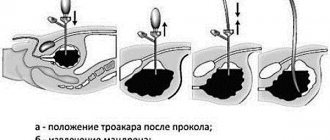
Using a rectal sensor, a clear visual picture of urology is obtained, which allows one to determine the size of the prostate gland, its consistency, possible inclusions and neoplasms. The device is inserted transrectally, moving deep into the intestine by 5–6 cm.
If it is necessary to examine the seminal vesicles, then the advancement of the device is continued another 1–2 cm. In this case, the man will experience discomfort. Unexpressed pain may appear if the pelvic organs are inflamed, or with acute prostatitis. You can view the vessels with a Doppler ultrasound of the prostate. Sometimes an ultrasound examination alone is not enough to make a correct diagnosis. Then they take an analysis of the prostatic juice obtained after a prostate massage. Massage is done through the urethra (on a bougie) or rectally.
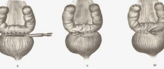
Enema: how to give it
To properly prepare for a prostate examination, it is important to thoroughly cleanse the intestines through an enema. You can do an enema yourself. To do this, you will need an Esmarch mug - a special device for introducing water into the body. It consists of a rubber reservoir and hose and a removable or non-removable tip. As a rule, the rubber hose has a clamp designed to regulate the intensity of the water jet.
Before use and immediately after the procedure, the individual tip is cleaned of fecal residues, washed with soap, and then boiled for 30 minutes. Store dry in the container where the boiling took place.
Sequencing:
- Immediately before the enema, the tip is connected to the hose and thickly lubricated with Vaseline or baby cream.
- The clamp is closed, the tank is suspended at a height of 1.5–1.8 meters and filled with slightly cooled water.
- By opening the clamp and passing water along the entire length of the hose, remove air from the tube. This will help make the procedure easier.
Next, lie on the edge of the bed or couch on your side, bringing your knees as close to your chest as possible. This position is also called the “fetal position”, because this is the position the fetus occupies during intrauterine development.
It is important to try to relax the buttocks as much as possible and carefully insert the tip into the anus, moving it along its entire length. Next, you should open the clamp and wait until Esmarch’s mug is empty.
By removing the tip from the anus, retain as much water as possible inside the body. This is easier to do by taking the knee-elbow position, so the procedure will be more effective. You will have to visit the toilet several times until the feces are evacuated from the man’s body. This enema should be done several hours the day before the proposed prostate ultrasound.
Preparing the patient for the diagnostic procedure can be done without a tedious enema. An enema for a man is an unpleasant manipulation, which is why disposable Microlax microenemas are used.
Microlax is a safe, fast, highly effective laxative, produced in a special tube that allows you to hygienically administer the medicine directly to the desired part of the gastrointestinal tract. Emptying will occur within 15 minutes after the end of the simple procedure.
Taking a laxative
To avoid the unpleasant enema procedure, you can take the laxative Fortrans or its analogues. These drugs are classified as osmotic laxatives, which cleanse all parts of the gastrointestinal tract without disturbing the microflora. Fortrans dose calculation: take 1 sachet of powder per 20 kg of body weight. It is dissolved without residue in 1 liter of water. One package contains 4 sachets of medication.
Preparing for an ultrasound requires special attention. But these are simple steps that can be easily done at home. At your doctor's appointment, you can ask all your questions. A competent urologist will give detailed answers and explain all unclear stages of preparation. This is how high-quality and accurate results of ultrasound examination of the prostate gland in men are obtained.
Price
Prostate ultrasound is performed free of charge upon referral from the attending physician at a local clinic. To avoid waiting in line, you can undergo the examination for a fee. The cost of the procedure is low.
| City | Cost, rubles |
| Moscow | 400-1000 |
| Saint Petersburg | 500-1100 |
| Ekaterinburg | 300-800 |
| Novosibirsk | 400-900 |
| average price | 400-950 |
Prostate ultrasound is used to diagnose acute diseases and monitor chronic pathologies. The procedure is painless, has no contraindications, and even minor deviations are detected.
Useful video
All important issues are covered by a specialist in this video.
The transrectal technique, due to the specifics of its preparation, also has a number of contraindications for its implementation:
- the presence of a malignant process in the large intestine;
- unexplained abdominal pain;
- clinical signs of intestinal obstruction;
- nonspecific ulcerative colitis with damage to the rectum;
- toxic colitis;
- uncompensated heart failure;
- dehydration.
With the transurethral technique, preparation also includes local anesthesia of the urethra to reduce the severity of discomfort. Diagnosis, unlike other methods, is carried out after complete urination.