Humpbacked kidney: causes, symptoms, ultrasound and CT diagnosis, treatment
If, upon detection of convexities of the renal contours, no other changes in the structure or functional activity of the excretory organs are detected, such a morphological anomaly as a humpbacked kidney is not a disease.
In such an organ of normal size and shape, the pyelocaliceal system, normal blood supply and function of parenchymal tissue. The owner of a modified organ may not suspect the presence of such a developmental defect all his life, without feeling any painful symptoms, until a humpbacked kidney is accidentally discovered during preventive examinations or instrumental diagnosis of other diseases.
Additional and more detailed information about the structure and morphological characteristics of the excretory organs will be provided by magnetic resonance and computed tomography studies.
When a modified form of the kidney is detected, first of all, differential diagnosis is carried out with diseases in which a similar change in morphology can be observed. These are tumor-like growths of renal tissue, large cysts that form protrusions of the organ lining.
For diagnostic purposes, predominantly non-invasive instrumental techniques are used. This is an accessible and informative ultrasound study, which in most cases allows one to identify the quality of the structure of the renal tissue.
An invasive research technique (tissue biopsy) is used as a last resort to completely exclude the possibility of the development of malignant neoplasms (tumors), which can form protrusions of the renal contours, similar to the bulges that occur with a humpbacked kidney.
Sometimes, when undergoing an ultrasound or x-ray examination, the patient is found to have protrusions of the renal membrane, which in the conclusion of the doctor who performed the diagnostic procedure will be designated as a “humpbacked kidney.” When making such a diagnosis, it will be important for the doctor to have undamaged parenchyma of the excretory organ, the one-sided nature of the morphological changes, and the absence of signs of tumor development.
Local renal protrusions are found against the background of the normal size of the humpbacked kidney, if other pathological changes do not occur in the organ. Read about what determines the occurrence of such a morphological anomaly and how it can affect the health and functioning of the paired organ.
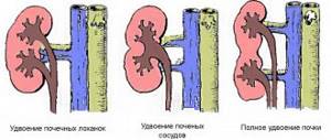
In most cases, with the formation of malformations of the urinary system with fusion of the renal structures, dystopia occurs: a horseshoe-shaped kidney is formed with bilateral prolapse and connection of the organs with the lower poles. This option is the most common - in 90% of cases. Fusion with the upper poles occurs less frequently (about 10%).
Usually the anomaly is typical for a male organism. Often, in addition to the structural features of the genitourinary organs, the doctor identifies concomitant defects of neighboring organs in the child. This combination is explained simply: the formation of the genitourinary and pelvic organs occurs from one embryonic germ; accordingly, a violation of intrauterine development at a certain stage of fetal maturation can cause multiple defects of the genitourinary organs.
A horseshoe kidney may not manifest itself in any way, becoming a diagnostic finding, but this rarely happens. Typically, all symptoms of pathology that occur in a child or adult are caused by the following features of the abnormal relationship:
- dystopic or lowered position of the organs of the urinary system;
- lack of mobility (due to the peculiar shape and many fixing vessels);
- the fused parts of the kidneys are located in front of the large vascular trunks (aorta, vena cava, iliac arteries);
- with severe dystopia, blood flow through the renal vessels is almost always impaired;
- With any injury, due to immobility, the renal parenchyma is much more damaged.
In most cases, symptoms indicating the presence of a horseshoe kidney are caused by compression of neighboring organs, large vessels or nerve trunks. Immobility of the kidney becomes a serious problem for a person with constant pressure on the inferior vena cava.
Manifestation of the clinical picture
Basically, the humpback kidney anomaly occurs without visible symptoms; it is diagnosed during a routine ultrasound examination. In the presence of concomitant diseases, a number of unpleasant sensations appear.
Stages of flow
So, if this pathology is accompanied by urolithiasis, pyelonephritis, cystitis or other diseases, then the symptoms will proceed accordingly. Let's look at the most common signs:
- problems with urination;
- pain in the abdomen, which may radiate to the groin;
- symptoms of the inflammatory process (temperature, intoxication, etc.);
- varicose veins in the lower extremities;
- metabolic disorders;
- problems with bowel movements (constipation, diarrhea).
The risk group includes patients with the following pathologies:
If one of the unpleasant symptoms appears, it is recommended not to hesitate to consult a doctor.
Changes during prolapse and the reasons for their occurrence
The organ in normal condition is located in the retroperitoneal region, the upper part of the lower back. The kidneys are located on the sides of the spine. The right one is 10-15 mm lower than the left one, located at the level of the first lumbar vertebra.
When a person changes body position, organs can move. Their movement in the vertical or horizontal direction is normal if it does not exceed 1-2 cm.
The organ is held in place by a capsule and ligaments that are attached to the spine. They are also held in place by intra-abdominal pressure. But sometimes cases arise when these factors decrease, resulting in organ prolapse. More often it is one-sided. In most cases, the right kidney is prolapsed.
The organ leaves the bed and goes down either into the abdominal cavity or into the pelvic area. The organ can descend as far as the ligaments allow. In this case, not only the kidneys, but also the ureter, nerves and blood vessels descend, their direction changes, which leads to complications. Kidney prolapse occurs for the following reasons:
- Abdominal or back injuries that weaken the abdominal muscles and ligaments. This can cause large hematomas and tendon sprains.
- Sharp weight loss, which reduces the volume of adipose tissue near the kidneys.
- Congenital anomalies of organs or bones that push the organ down (rib injuries, spinal diseases).
- Rapid human growth during adolescence.
- Pregnancy. In pregnant women, nephroptosis develops due to weakness of the muscles of the anterior abdominal wall and a decrease in intra-abdominal pressure.
- Insufficient development of connective tissue, ligaments and fascia can cause nephroptosis.
- The disease can be provoked by visceroptosis (sinking of other organs) and mobile joints.
There are risk groups of people who may develop nephroptosis. This includes people whose work involves lifting heavy objects or requires standing for a long time (loaders, hairdressers, salespeople).
Spongy kidney on ultrasound examination
A spongy kidney is a developmental anomaly - a congenital expansion of the structural components - the collecting ducts. For most patients, this anomaly is harmless and occurs without any clinical manifestations. But in some people, this pathology contributes to the development of an ascending infection that requires appropriate treatment.
With echography, the pyramidal kidneys, which are normally hypoechoic (dark gray), become hyperechoic (white) as a result of an increase in the number of reflective interfaces between the media due to the expansion of the tubes.
With age, secondary calcification of dilated collecting ducts is possible, as well as the formation of cysts in the cortex of the abnormal organ. In this case, the spongy kidney begins to resemble changes in nephrocalcinosis, but without accompanying changes in laboratory tests.
How is it diagnosed?
Diagnosis of the disease presents certain difficulties due to the specific nature of the pathology. To make a diagnosis, a number of studies are required.
Research methods combine laboratory tests and instrumental diagnostics.
Laboratory tests include:
- detailed blood biochemistry;
- bacterial culture of urine;
- general blood and urine analysis;
- urine examination according to Nechiporenko and Zimnitsky.
Instrumental diagnostic methods provide the most reliable picture of pathology. The most commonly used methods are:
- Ultrasound of the kidneys. Using ultrasound, it is possible to detect pathology in the fetus during pregnancy. The doctor evaluates the size, structure of the organ, and the state of blood flow. Unfortunately, this method does not provide a reliable picture, so additional research is required.
- Excretory urography. The patient is injected with a contrast agent into a vein and an x-ray of the kidneys is taken. The images clearly show the patency of the veins and arteries of the pelvic organs.
- Duplex scanning of blood vessels. With its help, it is possible to reliably assess blood flow in organs.
- MRI, CT scan of the kidneys. The layer-by-layer images clearly show the abnormal structure of the organs.
- Retrograde pyelography. It allows you to detect the abnormal location of the organ, the presence of an isthmus, and the condition of the renal pelvis.
- If a tumor is suspected, a fine-needle biopsy is performed.
Read our article to see what an MRI of the kidney shows.
MORE ABOUT: Treatment of stage 2 cervical cancer.
How long do they live after surgery? Correct diagnosis will allow the doctor to decide on the choice of treatment methods.
Manifestations
A humpbacked kidney is not a clinical pathology, since the integrity of the parenchyma and other structures of the organ is not compromised. The organ functions normally, without any symptoms.
A person may not be aware of the existence of an abnormal kidney structure throughout his life if it is not detected by ultrasound and there are no symptoms.
Malfunctions in kidney function will occur if inflammatory processes occur, which are also characteristic of a healthy organ. Symptoms of disorders will depend on the degree of kidney damage and associated pathologies. If a patient develops nephrolithiasis, cystitis, pyelonephritis, then his condition will worsen according to the clinical course of these diseases:
- problems will begin with removing urine from the body: frequent urge to urinate, a feeling of incomplete emptying of the bladder;
- painful sensations will appear in the lower abdomen, radiating to the groin;
- the symptoms of the inflammatory process will increase (high temperature, headache, weakness, loss of strength, anxiety, irritability);
- there will be signs of varicose veins in the legs;
- intestinal function will be impaired, resulting in constipation or diarrhea.
When the first signs of kidney dysfunction appear, you should immediately consult a doctor. The presence of a protrusion in itself does not cause the development of inflammatory diseases of the organ, since it does not affect the functioning of the renal structures.
The cause of pathologies is not an anomaly in the structure of the organ, but other processes associated with the penetration of infection into the body.
Diagnostic methods
If you suspect a horseshoe kidney, you need to undergo a full examination, including the following procedures:
- Ultrasound scanning. Typically, abdominal ultrasound becomes the first screening method in diagnosing any abnormal conditions in the urinary system. A horseshoe kidney may appear as a pseudotumor on ultrasound, making accurate diagnosis difficult. Often the doctor sees changes on the monitor screen and assumes that this is a humpbacked kidney (a small area of bulging along the outer surface) or an abnormal variant of the kidney shape. In any case, all suspicions should be dispelled with the help of more informative examination methods.
- Urography. An X-ray diagnostic method will confirm with a high degree of certainty the presence of one of the variants of the normal renal structure (humpbacked kidney) or help detect a developmental anomaly.
- Scintigraphy. Radioisotope methods are used much less frequently, but they make it possible to detect not only a horseshoe kidney, but also functional disorders in the abnormal organ.
- Arteriography. If a horseshoe kidney is detected, then a prerequisite for treatment will be a study of the vasculature in the renal area. Arteriography will clarify the location of the arterial trunks and detect vascular anomalies.
A horseshoe kidney predisposes to complications:
- inflammatory processes with frequent exacerbations (pyelonephritis);
- kidney stone disease;
- hydronephrotic transformation.
If these problems appear and it is impossible to cope with the disease using conservative methods, the doctor will suggest surgery. The best option is to divide the horseshoe kidney, but you need to very carefully evaluate the vascular connections of the organs.
In a child, the scope of surgical intervention includes dissection of the fusion site and separation (reposition) of the divided kidneys. Surgery will definitely be required if there is pressure on the inferior vena cava, because the resulting venous hypertension can cause severe complications in the vessels of the legs.
A horseshoe kidney is one of the most common types of anomalies in the development of the urinary system and can significantly complicate the life of a sick person. Most often, serious problems and severe complications arise when nearby tissues and organs are compressed.
Complications and symptoms when a horseshoe kidney is detected Link to main publication
In the case where the horseshoe kidney is a single pathology and is not accompanied by concomitant diseases, there are no symptoms. The anomaly is discovered accidentally during a routine ultrasound.
The first symptoms appear in adulthood, but the patient does not associate them with kidney disease. Clinical manifestations depend on the following factors:
- isthmus pressure on the ureter;
- increased innervation of affected organs;
- incorrect location of internal organs due to the isthmus.
All these factors provoke the appearance of the following symptoms:
- pain in the lumbar region or abdomen, aggravated by physical activity;
- indigestion: frequent constipation, flatulence;
- nausea, vomiting;
- loss of appetite;
- weight loss for no apparent reason;
- swelling of the legs and arms;
- disruption of the menstrual cycle;
- phlebeurysm;
- blood in the urine;
- pain when urinating;
- renal colic;
- change in the color and clarity of urine;
- increased emotional background, accompanied by nervousness or depressive symptoms.
The clinical manifestations of this developmental progression depend on individual characteristics, as well as the presence of complications. Some patients learn about the disease by chance: during a medical examination or during a diagnostic examination related to diseases of the urinary system.
Factors determining the presence of clinical manifestations:
- Mechanical compression of the ureter by the isthmus.
- Innervation of an abnormal organ.
- Deviation of the position of the abdominal organs from the anatomically normal one, caused by the isthmus.
Symptoms of pathology can be divided into nonspecific, which are general, and specific, caused by an abnormal structure.
- aching pain in the lumbar area, in the navel area and lower abdomen, which occurs when straightening and bending the torso;
- intrarenal hypertension - appears as a result of compression of blood vessels;
- venous congestion, they lead to swelling of the extremities and varicose veins;
- impaired intestinal motility, which is accompanied by intestinal colic, abdominal pain, constipation and disorders;
- as a result of compression of the lower part of the ureter, stones appear in the kidneys, causing an inflammatory process;
- constant pain, it causes a general emotional disorder of the body, which is expressed in the form of neurasthenia and hysteria;
- menstrual irregularities;
- premature birth;
- presence of blood in the urine;
- formation of cancerous tumors.
Methods of therapy
Depending on the degree and severity of the pathology of the humpback kidney, as well as the addition of concomitant diseases, the doctor decides on the treatment regimen. Very often it does not cause negative symptoms and does not at all interfere with a person’s exercise, going to work, etc.
If unpleasant signs appear, you should immediately contact a specialist. Either drug therapy or surgery is used.
Drug treatment
This therapy is prescribed if the patient is simultaneously diagnosed with other kidney diseases (urolithiasis, pyelonephritis, cystitis, etc.). In this case, it is advisable to use:
- antibiotics, they are selected after precise identification of the causative agent of the infectious process, the course and duration of administration is prescribed individually;
- anti-inflammatory drugs;
- local antispasmodics (to relieve pain attacks);
- uroseptics, which help remove urine accumulated in the body;
- glucocorticosteroids and immunomodulators (used for glomerulonephritis or autoimmune inflammatory processes in the renal parenchyma);
- vitamin complexes to strengthen general immunity.
Surgery
It is used very rarely, only in cases of serious complications that threaten complete disruption of kidney function. For example, in the case of hydronephrosis, a special catheter is installed, with its help excess fluid is removed from the body.
If large mineral deposits are present, the patient undergoes laparoscopic surgery to remove them.
This operation is also prescribed if the inferior vena cava is compressed (in this case, a person’s kidney may fail due to circulatory problems). The doctor decides on the advisability of surgical intervention based on the patient’s clinical picture.
ethnoscience
Since this disease is not a serious pathology, there are no unique folk remedies that are aimed at treating it.
This therapy is used in the presence of concomitant diseases. Most often, patients use infusions or decoctions based on the following medicinal plants:
- rosehip;
- St. John's wort;
- chamomile;
- bear ears, etc.
This anomaly cannot be cured with the help of traditional medicine; in this case, only the removal of acute symptoms is possible. Often these methods provoke the development of serious complications, so you should not get carried away with self-medication.
Reasons for formation
Various diseases are considered to be the main cause of renal lumps. In 60% of cases, pyelonephritis is considered the root cause. During an ultrasound, the doctor pays attention to the uneven edges of the organ and its decrease in size. In addition, an expansion of the heart rate may also be observed.
The main reasons for the appearance of seals:
- Pyelonephritis is a disease in which a person experiences inflammation of the renal pelvis. The cause is various bacteria and pathogens. The disease can develop when the outflow of urine from the body is impaired.
- Hydronephrosis is a disease in which the renal pelvis gradually expands, and the tissue of the organ atrophies, gradually becoming thinner and thicker. Hydronephrosis occurs due to urolithiasis, as well as damage to the urinary system. Kidney stones occur due to poor diet, excess weight, the use of certain medications, and poor drinking habits. With hydronephrosis, the patient may experience strictures. Stricture is a disease in which narrowing of tubular organs occurs. Symptoms in the first stage of hydronephrosis occur unnoticed and are usually accompanied by renal colic.
- Doubling the heart rate does not pose any threat to the patient’s health. Occurs due to the use of certain medications, alcohol and tobacco products. A doubling of the heart rate can also be observed with a lack of certain vitamins in the body. The disease can be congenital; the patient can live his entire life without even knowing about the presence of this anomaly.
MORE ABOUT: Angina
In addition to the kidneys, lumps can form in the lungs and liver. Consolidations in the parenchyma and CL may indicate a tumor. Often the right organ is affected by the tumor. A tumor can be recognized by the following symptoms: blood in the urine, impaired urine flow, prolonged nagging pain in the lumbar region, nausea, vomiting, as well as loss of appetite and sudden weight loss.
To accurately diagnose a growth on the kidney, it is necessary to conduct an ultrasound examination, as well as deep palpation. The tumor is quite often confused with a humpbacked kidney. Humpbacked kidney is a disease in which the contours of the organ protrude. A humpback kidney occurs when the development of the urinary system is impaired or is a congenital defect. At the moment, the exact cause of the anomaly has not yet been clarified.
If there is blood in the urine or if there is prolonged pain in the lower back, you should consult your doctor. Don't delay kidney treatment. The sooner a pathology is detected, the easier it is to treat it.
When examining the kidneys using ultrasound, it is possible to identify almost all pathologies that can occur in the kidneys. In this case, pathologies (changes) are divided into two groups - diffuse and focal. The former affect the entire structure of the organ, while the latter are characterized only by the presence of pathology in a certain area of the kidney, while the rest of its tissue remains healthy and unchanged.
Diffuse lesions include the following pathologies:
- Pyelonephritis. With this disease, no special changes are visible, but a specialist can examine the swelling of the urinary organ, the blurring of the boundaries between its cortex and medulla, and also identify the hypoechogenicity of the kidney. In combination with the patient’s complaints, a diagnosis of “pyelonephritis” is made. If the disease occurs in a chronic form, the affected organs will be reduced in size, the halo of parenchyma will be hyperechoic, and small cystic formations will be visible throughout the structure of the organ. Later, scarring is diagnosed, as evidenced by retraction of the renal cortex.
- Glomerulonephritis. In the acute course of the disease, the kidney parenchyma will be significantly thickened (at least 20 mm). In this case, the organ itself will be enlarged. The parenchyma will have increased echogenicity. If glomerulonephritis has a “long history,” then by the time of examination the affected kidney and its parenchyma will be reduced in size (parenchyma no more than 12 mm).
- Kidney tuberculosis. Characterized by damage to pyramids that contain liquid. In later stages, the parenchyma is destroyed, and the organ itself shrinks.
- Hydronephrosis. Visible in enlarged cups and pelvis, thinned parenchyma or its narrow rim. The kidney itself looks like an overfilled sac with enlarged cups.
Focal pathologies include:
- Cystic formations. They look like anechoic (completely black) formations or spots. The contours of such cysts are smooth and their shape is round. With cysts, the ultrasound signal increases.
- Abscess. Characterized by unclear contours and blurriness. The contents are echogenic. Most often it is pus. In the initial stages of inflammation, the abscess may have hyperechogenicity.
- Angiomyolipoma. It has clear and even rounded contours and increased echogenicity.
- Stones. They look like structures of a certain size that have hyperechogenicity. Concretions (stones) perfectly reflect ultrasound and provide a good shadow.
Important: but on ultrasound only stones larger than 2 mm are clearly visible. Fine sand cannot be diagnosed using ultrasound.
A humpbacked kidney is usually detected in a person during a routine examination, as it does not always give any symptoms. Treatment of the pathology, if it does not cause concern, is also not required, but an accurate diagnosis is of great importance.
In most cases, complications of a humpback kidney include:
- Pyelonephritis;
- Glomerulonephritis;
- Urolithiasis disease;
- Hydronephrosis;
- Cysts.
Uncomplicated cases of pathology have a favorable prognosis. If complications occur, the prognosis depends on their frequency, severity, and timeliness of therapy.
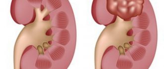
A humpback kidney belongs to the category of kidney deformities and is most often congenital, although acquired changes in the contours of the organ are also not uncommon. Externally, a humpbacked kidney looks like a local protrusion of one of its outer edges, which may initially be mistaken by a specialist for a tumor formation.
Acquired kidney deformities, which are also combined with the term “humpbacked kidney,” may in fact have a different shape. The protrusion on the organ makes it look like a ball, oval, sickle, while the contour can be straight or wavy, the size of the kidney can be normal or enlarged.
Clinical manifestations of the disease are not always present: a person lives without knowing about the existing pathology.
In many children and adults, the symptoms of a humpbacked kidney still occur, and it happens in the presence of the following risk factors:
- Kidney prolapse or other abnormal location.
- Immobility of the organ due to the presence of fixing vessels.
- Fusion of kidney parts with each other.
- Impaired blood circulation in the arteries and veins of the kidney.
- Organ injury during life.
When compression of vessels, nerves or other organs of the retroperitoneal space and abdominal cavity occurs, pain and inflammation may occur, which causes various clinical manifestations. These are aching pain in the sides, in the abdomen, constipation, varicose veins of the legs, pain in the legs. In severe cases, the development of inflammatory and metabolic disorders is possible.
humpback kidney
MRI in St. Petersburg
The reason for examining the kidneys is, as always, the patient’s complaints of lower back pain and hematuria (blood in the urine). In approximately half of the cases, hematuria is a sign of a tumor, not necessarily the kidney, but any part of the urinary system.
In addition to a tumor in the kidney, hematuria from it can be associated with vasculitis, glomerulonephritis, renal vein thrombosis and urolithiasis. Many of the causes of pain and hematuria not related to the tumor can be easily excluded using MRI of the kidneys.
Typically, kidney examination begins with an ultrasound. This is an ideal method for identifying cysts. There are about 80% of these and they do not need further research.
Atypical cysts - with septations, calcifications, thick walls or a solid component require tomographic methods, since the sensitivity and specificity of ultrasound for tumors is insufficient.
In 10% of cases, the cystic form of kidney cancer occurs. In addition, polycystic disease also in some cases degenerates into a tumor.
CT has a sensitivity for tumors of over 90%. However, MRI of the retroperitoneum has obvious advantages: harmlessness, multiplanar examination without reformation, excellent contrast even without the introduction of a contrast agent.
Many MRI centers in St. Petersburg do not have sufficient experience in MRI of the retroperitoneal space. In addition, we usually perform MRI in St. Petersburg abandoned space simultaneously with MRI of the abdominal cavity, which allows us to find metastases in the list and lymph nodes.
MRI characteristics of kidney tumors are based on the principle – cystic-benign-malignant. Renal MRI easily identifies hemorrhagic cysts (cysts containing blood) by the typically high signal on T1-dependent MRI images. Contrast MRI makes it easy to distinguish a cyst from a tumor.
Papillary benign cancers give a mixed signal on T1-dependent MR tomograms due to the admixture of lipids, a cystic component with an admixture of protein and blood. After contrasting, they become more homogeneous. On T2-dependent MRI, these cancers are light and also heterogeneous.
Angiolipoma is a benign kidney tumor. Histologically, they are perivascular epithelial tumors (PECOM). Angiomyelolipomas are sporadic in 80% of cases, 4 times more common in men than in women.
In the remaining 20%, they serve as a manifestation of the disease tuberous sclerosis, neurofibromatosis type 1 or Hippel-Lindau disease, related to phakomatoses. Angiolipomas have a fatty component that is emphasized on out-of-phase MRI.
When fat is suppressed, the signal from it becomes dark.
MRI of the kidney. T1-dependent tomogram, magnification. Angiolipoma.
MRI of the kidney. T1-dependent tomogram with contrast and fat suppression. Angiolipoma.
Another benign tumor, oncocytoma, originates from cells of the collecting system. The formation is solid, homogeneous and weakly and heterogeneously contrasted, often in the shape of a “wheel with spokes”. The tumor often has a ring-shaped pseudocapsule separating the tumor from the surrounding renal tissue. An MRI often reveals a scar in the center of the tumor.
MRI of the kidney. T1-weighted coronal MRI with contrast. Oncocytoma.
Malignant kidney tumors - carcinomas account for approximately 2-3% of all cancers and rank approximately 8th in frequency among tumors. the problem is that they manifest clinically late and up to 50% turn out to be an incidental finding during a diagnostic study. Overall, 85% of kidney tumors are malignant.
Kidney carcinomas belong to various subtypes of adenocarcinomas: clear cell carcinoma - 70-80%, papillary carcinoma 13-20%, other subtypes are observed very rarely, including extremely aggressive sarcomatoid renal cell carcinoma. Typically appear at the age of 50-70 years, the M:F ratio is 2:1.
Cancer staging is carried out using the TNM system or the Robson classification.
Kidney MRI. Coronal T1-weighted MRI before and after contrast. Transitional cell carcinoma.
Kidney MRI. T1-weighted coronal MRI. Renal cell carcinoma in the upper pole of the left kidney with thrombosis of the inferior vena cava.
On MRI, tumors are heterogeneous on T1- and T2-dependent MRI and contain solid, necrotic, and hemorrhagic components. Clear cell cancers are bright on T2-dependent MRI, papillary cancers are dark. However, it must be borne in mind that not all malignant tumors are well contrasted. Enhancement, if present, is rapid in the arterial phase.
The rate of accumulation of contrast agent in MRI does not have such an important diagnostic value as in CT. About 5% of carcinomas contain a cystic component. In very rare cases, carcinomas contain minor fat inclusions and can be confused with angiolipomas. On DWI type MRI, kidney tumors are light, although not as bright as an abscess.
MRI of the kidneys and retroperitoneum can also observe thrombosis of the renal vein and inferior vena cava. Tumor thrombosis is usually contrasted, and it must be distinguished from blood thrombosis in which contrast of the blood clot is not observed.
The prevalence of a thrombus along the inferior vena cava is observed in 4-10% of cases (stage T3), which is very important for the surgeon to know when planning the scope of the operation. The accuracy of MRI in assessing invasion of the inferior vena cava approaches 100%.
Kidney MRI. Coronal T1-weighted MRI with contrast. Tumor of the left kidney with invasion of the renal vein.
Source: https://www.mri-kholin.ru/stati/vyyavlenie-obrazovanij-pochek-s-pomoshhyu-mrt/
Prognosis and possible complications
Despite the absence of pronounced symptoms, the disease can provoke the development of serious complications. Since the isthmus is located in the lumbar region, it puts pressure on the ureter. This provokes the development of pyelonephritis, urolithiasis, hydronephrosis, glomerulonephritis.
MORE ABOUT: Deep vein thrombosis of the lower extremities: causes, symptoms and treatment
Due to the pressure of the isthmus on the intestine, normal peristalsis is disrupted, resulting in chronic inflammation of the intestine.
In addition, patients with an abnormal kidney have an increased risk of developing tumors in the kidneys, including malignant kidney tuberculosis. The patient may develop kidney failure. Compression of blood vessels in the abnormal organ causes varicose veins.
In women, a horseshoe kidney can cause premature birth. In addition, the growing fetus puts pressure on the isthmus, which can result in kidney infarction and hydronephrosis. This condition poses a direct threat to the life of the mother and child.
Concomitant pathological processes develop due to compression of neighboring organs, vessels, and nerve endings.
Possible violations:
- congestion in the pelvis;
- varicose veins;
- accumulation of fluid in the abdominal cavity.
Women of reproductive age have difficulty bearing a child, and premature birth is possible.
Kidney fusion is accompanied by various complications. They develop against the background of impaired urine outflow, which causes changes in the renal pelvis.
Against the background of an anomaly in the structure of the organ, the following complications develop:
- pyelonephritis;
- glomerulonephritis - an inflammatory process in the glomeruli of the kidneys;
- hydronephrosis;
- urolithiasis disease;
- hypertension.
A particular danger is the transformation of kidney tissue cells into cancer cells. Most often, tumors form on the isthmus. Horseshoe kidney is often associated with Wilms tumor, a highly malignant nephroblastoma.
In addition to horseshoe kidney, people often develop other pathologies either together or as a complication. We are talking about the following conditions:
- Stone formation in the kidneys, which can lead to blockage of the ureter.
- Hydronephrosis, as a result of blockage of the urinary tract. Suggests abnormal, progressive changes in the renal pyramid and pelvis.
- Pyelonephritis is an inflammation of the kidney parenchyma, pyramids and renal pelvis.
- Williams tumor is an embryonic neoplasm formed in utero.
- Cancer or polycystic kidney disease.
- Dropsy of the brain (hydrocephalus), developing together or separately with spina bifida.
- Various cardiovascular, gastrointestinal pathologies, malformations of the anorectal system.
- Skeletal abnormalities - severe clubfoot, cleft lip, problems with limbs.
- Arterial hypertension.
Compression of the isthmus of the initial part of the ureter creates an obstacle to the outflow of urine from the pelvis, which leads to the development of pyelonephritis (19.4%), the formation of kidney stones (23.6%), hydronephrosis (41.7%), arterial hypertension (15.2% ). There is evidence that tumor transformation of cells and kidney cancer more often develop in the isthmus of the horseshoe kidney.
Treatment
A correct diagnosis is of decisive importance when choosing methods for treating renal manifestations. If a protrusion on the kidney is detected, it is necessary to clarify the nature of the deformation.
If there are no changes at the cellular level, the tissues are not damaged, the patient does not feel a worsening of the condition, he is observed by a doctor without the use of medication. Uncomplicated cases of anomaly do not require medical intervention.
Therapy with drugs is prescribed when complications and concomitant diseases occur.
The patient is prescribed a complex of drugs, depending on the underlying disease:
- Antibiotics. Prescribed in case of bacterial infection.
- Anti-inflammatory. Their action is aimed at suppressing the inflammatory process in the body.
- Antispasmodic. Prescribed to relieve pain of varying intensity.
- Diuretics. Taking medications helps remove excess fluid from the body.
- Glucocorticosteroids. Prescribed for autoimmune kidney diseases.
- Vitamins. Prescribed to strengthen the immune system.
Modern pharmaceuticals offer a huge number of drugs for the treatment of kidney diseases. Today on the shelves of pharmacies you can find medicines whose manufacturers promise to cure diseases after a few doses.
Do not self-medicate. Therapy for renal pathologies should be carried out under the supervision of a physician. He will prescribe individual treatment, examining the patient to identify contraindications.
Treatment with medications is the main method of treatment. Surgery is rarely used.
It is prescribed if serious complications are detected that are dangerous to the life and health of the patient (hydronephrosis, nephropathy, acute renal failure).
In this case, renal function is impaired, urine is not excreted from the body. The patient is fitted with a catheter, which helps drain the fluid out.
If stones are detected, they are removed during laparoscopic surgery through small holes.
When clamping the inferior vena cava, surgery is also performed laparoscopically to restore blood circulation.
If there are kidney dysfunctions, decoctions and tinctures containing chamomile flowers, St. John's wort, and rose hips will help.
Traditional medicines will not cure kidney diseases, but will temporarily eliminate acute manifestations of functional impairment. Self-prescription and use of decoctions without consulting a doctor can lead to the development of complications.