One of the patients came to me after a control ultrasound examination of the gland. Her face, movements and voice expressed concern about the increasing deterioration.
How is it that I came for an ultrasound, and the doctor began to tell me that I had a very large node and needed to be operated on urgently. That’s what he wrote: a solid knot.
Let me look at the results of the examination,” I suggested.
Please,” the patient replied, handing me the ultrasound report.
I began to carefully study the description of an ultrasound examination of the thyroid gland. In conclusion, in addition to conclusions about the nature of the changes, there was a solid nodular formation... The same node in the ultrasound description was characterized as homogeneous isoechoic.
See how big the knot is!
Yes, indeed, the knot is large. But it is benign...
The ultrasound doctor told me that the node was huge. That's what he wrote - respectable. He said that it was necessary to undergo surgery. I didn’t even sleep well at night after what he told me.
As you said? Solid? — I asked, “This term means fullness, that is, containing not a cavity with something, but biological tissue.” In this case, normal thyroid tissue. And this word is pronounced differently. The emphasis should be placed not on the second, but on the first syllable.
And I was wondering why the doctor wrote this so emotionally...
Using the term, the reputable doctor wanted to show that gland tissue was present inside the focal formation he identified. I think it is not entirely appropriate in the ultrasound protocol for two reasons. Firstly, this can affect the mental state of the patient, who, trying to understand the state of his health himself, carefully reads the description of the ultrasound. Secondly, due to the fact that this term refers to pathological changes and is used by pathologists for histological examination.
Solid kidney formation - what is it?
a) Differential diagnosis of solid kidney formation
:
1. Common diseases
: • Renal cell carcinoma • Angiomyolipoma of the kidney • Urothelial cancer of the upper urinary tract • Pseudotumor of the kidney: o Column of Bertini o Humpback kidney o Focal hypertrophy • Focal pyelonephritis • Horseshoe kidney • Crossed ectopia of the kidney • Kidney lymphoma • Kidney damage in leukemia and multiple myeloma • Metastases • Other kidney tumors: o For example, oncocytoma • Wilms tumor
2. Less common diseases
: • Xanthogranulomatous pyelonephritis • Replacement renal lipomatosis • Renal tuberculosis • Medullary necrosis of the kidney
(Left) Ultrasound, longitudinal scan: solid renal mass, with calcification in the center. The upper pole of the kidney is atrophied and has increased echogenicity due to chronic renal failure. (Right) Ultrasound, longitudinal scan: solid, hypoechoic renal mass, leading to compression of the fatty tissue of the renal sinus. The patient has a disseminated metastatic lesion, the diagnosis was made by performing a percutaneous biopsy of the renal mass.
(Left) Ultrasound, longitudinal scan: a large, homogeneous, hyperechoic renal mass, similar in echogenicity to the fatty tissue of the renal sinus. Presumably angiomyolipoma, CT or MRI is required to confirm the diagnosis. (Right) Axial MPT, T1-weighted image in antiphase in the same patient: multiple low-signal lesions are visualized within the mass, indicating fat adjacent to soft tissue. This confirms angiomyolipoma.
(Left) Ultrasound, longitudinal scan performed for hematuria: solid hypoechoic mass in the upper part of the renal sinus, without blood flow.
The absence of blood flow does not exclude the presence of a tumor; the presence of blood flow confirms the tumor. Unlike Bertini's column, this formation does not develop from the renal cortex. (Right) Ultrasound, longitudinal scan: solid renal pseudoplasm developing from the cortex towards the sinus. The echogenicity is identical to the cortical layer. b) Important information
:
1. Differential diagnosis
: • The main purpose of greyscale ultrasound is to evaluate simple cysts • For non-cystic lesions, assess contour and echogenicity • Lesions with strong distal acoustic shadowing are most likely to contain large amounts of fat: o Calcification or gas should also be considered • Solid lesions should carefully evaluate for the presence of blood flow • It is necessary to use color or power Doppler mode to determine blood flow: o Color loci in solid structures, septa, nodes, foci of necrosis o The presence of internal blood flow allows one to suspect the malignancy of the process • CT or MRI with contrast is usually the following line of examination to characterize solid renal masses and the stage of the process • Contrast-enhanced ultrasound increases the sensitivity of determining tumor perfusion: o Addition to CT or MRI with contrast o Replaces CT and MRI when the patient is contraindicated with iodine or gadolinium contrast • It is necessary to detect the presence of formations located ipsilaterally and contralateral • Take into account the presence of pseudotumors: o Pseudotumors are isoechoic to normal parenchyma and have a normal kidney structure o Pseudotumors are identical to a normal kidney on CT and MRI before and after contrast • It is necessary to visualize signs of malignancy: o Invasion into the renal vein, tumor thrombosis of the inferior vena cava, regional lymphadenopathy and metastatic liver disease • Interpretation of data should be based on clinical information, eg fever, trauma, established malignancy • Risk factors should be considered, eg dialysis, Hippel-Lindau disease
(Left) Ultrasound, longitudinal color Doppler scanning: hyperechoic, wedge-shaped cortical formation with decreased blood flow. (Right) Ultrasound, longitudinal scan: left kidney (between calipers) with moderate dilatation of the pelvis. A solid “formation” develops from the lower pole. The “formation” resembles a normal kidney. No kidney was found in the right bed.
(Left) Ultrasound, longitudinal scan of the kidney: hypoechoic formation in the center with focal expansion of the calyces.
Lymphoma may have a pseudocystic appearance, as in this case. (Right) Ultrasound, longitudinal scan: hypoechoic enlargement of the upper pole of the kidney due to myeloma. The fatty tissue of the sinus is located separately, but is probably involved in the process. 2. Common diseases
:
• Renal cell carcinoma
: o The most common primary malignant neoplasm of the kidney o Variable pattern on gray scale ultrasound: solid, heterogeneous cystic and solid or predominantly cystic structure o Hyperechoic (48%), isoechoic (42%), hypoechoic (10%) o Large tumors are often hypoechoic, exophytic with necrotic anechoic foci o Hypoechoic pseudocapsule o Smaller tumors are often hyperechoic, inferior to angio-myolipoma (gives shading) o May contain dystrophic large areas of calcification o In color Doppler mode: peripheral, intratumoral blood flow o Accompanied by renal vein thrombosis (23%) and compression IVC tumor (7%)
• Angiomyolipoma of the kidney
: o Benign tumor containing fat, smooth muscle and altered vascular tissue o Sporadic cases of detection without concomitant pathology o Multiple in tuberous sclerosis o Echogenic formation, but may have different echogenicity, depending on the ratio of the fatty tissue and soft tissue components of the tumor o Small formations (
(Left) Ultrasound, longitudinal scan: a large solid mass in the lower pole of the kidney, hyperechoic compared with the echogenicity of a normal kidney. Taking into account the size of the mass, nephrectomy was performed. (Right) Axial contrast-enhanced CT scan in the same patient shows a mass-enhancing, solid renal mass with central scar, representing a classic oncocytoma.
(Left) Ultrasound, longitudinal scan of the renal bed did not show the presence of a normal kidney. A large stone with echo-shadow is visualized, surrounded by soft tissues of medium echogenicity. (Right) Axial CT scan with contrast in the same patient confirms the presence of a staghorn calculus surrounded by fat. Almost no residual renal parenchyma is visible.
Subcapsular renal cyst
This formation is localized in the thickness of the renal parenchyma. Typically, its volume corresponds to sizes from two to four centimeters, but in some cases it can be larger.
Inside the cyst there is serous fluid. If there are additional pathologies, blood or pus may be mixed into it.
As a rule, large growth of connective tissue formations or individual bundles does not occur.
Highlight:
- Cystic lesion , which usually does not pose a significant threat to the patient. Sometimes it resolves on its own without treatment.
- Solid formation, as it progresses, entails the development of various complications. Under their capsule there are accumulations of tissue structures that disrupt excretory functions. In the absence of timely medical care, they often lead to serious consequences. Such foci do not stop developing by themselves.
- Cystic solid kidney formation is a combination of the first two types. It includes collections of tissue and fluid enclosed in a capsule.
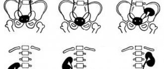
If the presence of a pathological focus located on only one organ is diagnosed, then they speak of a single neoplasm.
When there are several formations, we are talking about a multicystic lesion. If both kidneys are affected, polycystic syndrome is diagnosed.
Accordingly, the lesions are located on one or both sides. When placed in pairs, they can lead to serious consequences associated with severe dysfunction of organs.
Neoplasms in the kidneys
Ultrasound and tomography techniques have become firmly established in clinical practice. It is already difficult to imagine diagnosing diseases of internal organs without the use of these auxiliary examination methods. In the practice of a functional diagnostician (a doctor who performs ultrasound), tumors in the kidneys are often detected. It is very important to understand the nature of the detected changes, because the treatment and prognosis of the disease depend on this.
Studies in metastatic renal cell carcinoma
Chest CT is the most accurate method for detecting lung metastases.
If CT is not available to detect pulmonary metastases, at least a chest x-ray should be performed. Today, it is believed that most bone and brain metastases have some clinical manifestations at the time of diagnosis, so routine skeletal scans and brain CT scans are not usually indicated. If appropriate indications (clinical or laboratory signs) are present, other diagnostic procedures may be performed, such as skeletal bone scans, CT scans or MRIs of the brain. When identifying cystic kidney tumors, it is advisable to classify them. The most effective classification is the Bosniak division.
Classification of cystic neoplasms in the kidneys according to Bosniak
The Bosniak classification, used to evaluate renal cystic neoplasms, divides renal cysts into five categories based on their appearance on CT images to predict the risk of malignancy.
The Bosniak system also provides treatment recommendations for each of these categories (Table 2-1). Table 2-1. Classification of renal cysts according to Bosniak
Cystic cavities
The neoplasm can be benign or malignant. This determines the symptoms of the pathology and treatment tactics. Cysts are isolated separately.
An ultrasound examination reveals a focal formation. It can be not only tumors. Most often a cyst is found. It is a cavity that contains mucinous, serous, and sometimes purulent or hemorrhagic (blood) fluid. The walls are made of epithelial cells. There are simple, complex, single-chamber and multi-chamber cysts.
When there are many cavities, they speak of a cystic kidney. This condition is dangerous because the cysts fill the main space of the kidney, thereby reducing the reserves of nephrons (the main functional units). Acute or chronic renal failure syndrome develops.
If cystically altered tissue is detected in childhood in the plural, then they speak of polycystic disease. The recessively inherited form should be immediately treated with replacement techniques: hemodialysis or peritoneal dialysis. In adult patients, polycystic disease is accompanied by a slow increase in renal failure, as well as increasing persistent pain in the lower back on one or both sides.
According to ultrasound examination, the cyst is a hypodense formation. Functionalists use this term to designate a lesion with a lower density compared to the surrounding tissue. Tomography or a biopsy followed by histological analysis will help confirm that a specialist has a cystic cavity.
Cysts are dangerous because they can reach large sizes. This will inevitably lead to the formation of renal failure. In addition, the cystic cavity may be filled with blood. If it is overfilled, a rupture with clear clinical symptoms is possible.
But the biggest nuisance is malignancy. This is the degeneration of a cyst into a malignant formation. Therefore, cystic cavities should be dynamically monitored using ultrasound or tomography if necessary. If the cyst grows, it is necessary to contact an oncologist and resort to surgical treatment.
Diagnostic measures
The following diagnostic methods are required:
- Ultrasound of the thyroid gland is the main method for diagnosing thyroid diseases. The existing lesion, its size and structure are identified.
- FNA helps determine the benignity and malignancy of neoplasms. The biopsy material is sent for histology. It has been noted that after a puncture and aspiration of the contents from the cyst, in half of the cases its walls collapse and stop accumulating fluid. With very small formations, performing FNA is difficult, so there are also additional research methods.
- Blood test for hormones - T3, T4 and TSH.
Scintigraphy is a scan of the thyroid gland, which is carried out using radioactive isotopes of technetium and iodine. The method determines the level of hormone production in the node and healthy tissue. According to the scintigraphy method, all nodes are divided into 3 groups according to the ability to accumulate isotopes. The fact is that the accumulation of isotopes can be observed in the tissues of the node (TU) and neighboring healthy tissue (HT):
- Warm node – TU=ZT. The node is functioning.
- Hot node - TU is larger than ZT - the node operates autonomously.
- Cold node – TU is distributed only in healthy tissue. The node does not react to isotopes. One in 10 nodes is always cancer. Computed tomography – clarification of the size of the node and its malignancy.
Additional diagnostic methods:
- Laryngoscopy – the larynx and vocal cords are assessed.
- Bronchoscopy or fluoroscopy - examines the trachea.
- Pneumography – determines the presence of nodule sprouting in the lung tissue. For the same purpose, angiography and fluoroscopy of the esophagus are performed.
Tumor formations
On the kidney or in its parenchyma, a hypogene formation may turn out to be a tumor. The decrease in the density of the identified lesion is associated in these cases with the fact that the qualitative composition of the formation differs from the kidney cells that normally line the tubules and glomeruli.
Benign tumor
The most common type of solid renal tumor is an adenoma. It is based on cells with glandular activity. That is why such tumors have a high potential for malignancy—degeneration into malignant tissue.
The growth rate is relatively low. But the peculiarity of kidney adenoma is its large size, which it can acquire over time. Clinically, there may be a urinary disturbance with the development of acute renal failure. But this only applies to large tumors.
Lipoma and angiomyolipoma consist of connective tissue cells of varying degrees of differentiation. Lipomatous tumors contain adipose tissue. Otherwise, they can be called wen. Angiomyolipoma is characterized by a more complex composition and structure. By itself, it is an isoechoic formation. Among fat cells (adipocytes) there are endothelial and muscle cells. They determine the more “variegated” structure of the tumor.
Fibroma is another either iso- or hyper-echoic formation in the kidney. It contains fibrocytes and fibroblasts. The more mature cells, the harder and denser the tumor. With all this, fibroma is considered a benign tumor, which extremely rarely degenerates into cancer.
Clinically, benign formations in kidney tissue manifest themselves extremely rarely. Symptoms are obvious only when the vessels, calyxes and other urinary structures are large and compressed. There is intermittent aching pain in the lower back. The formation of the right kidney gives symptoms of inflammation of the gallbladder or appendix (appendicitis). Only ultrasound together with biochemical tests can make a correct diagnosis. A mass formation in the left kidney imitates manifestations of pancreatitis or splenomegaly (enlarged spleen), intestinal pathology (sigmoid or descending colon).
Malignant types of tumors
Clinically, kidney cancer or sarcoma cannot always be clearly detected. However, there are signs by which one should suspect the appearance of a malignant tumor in the kidney.
- Pronounced weakness, which is difficult to associate with objective reasons.
- Change in body weight (weight loss) without loss of appetite.
- Decreased performance due to unmotivated fatigue.
- Low-grade fever and sweating in the evenings or at night.
- Other manifestations of asthenic syndrome: irritability, emotional lability, tendency to low mood.
Symptoms
This pathology can manifest itself in different ways, depending on its location. Thus, for a cystic-solid formation of the medulla oblongata (remember, this section is located in the occipital part of the head and is a continuation of the spinal cord) the following manifestations are characteristic:
- Dizziness.
- Deafness (usually develops in one ear).
- Difficulty swallowing, breathing.
- Sensory impairment in the trigeminal nerve.
- Impaired motor activity.
Tumors in the medulla oblongata are the most dangerous, as they are practically untreatable. When the medulla oblongata is injured, death occurs.
In general, cystic-solid formations in various parts of the brain are characterized by the following symptoms:
- Headaches, even vomiting.
- Dizziness.
- Insomnia or drowsiness.
- Deterioration of memory, spatial orientation.
- Impaired vision, speech, hearing.
- Loss of coordination.
- Frequent mood changes for no apparent reason.
- Muscle tension.
- Sound hallucinations.
- Feeling like there is some inexplicable pressure in the head.
If a cystic-solid formation of the spinal cord occurs, this is manifested by pain, aggravated in the supine position and at night, descending lumbago, impaired motor function, and paresis.
If at least some of the signs from the above list appear, you should immediately go to the doctor.
Diagnosis and treatment
Confirmation of the diagnosis is based not only on ultrasound techniques. The use of tomography is considered a good help. The biopsy can be performed in a specialized nephrology or urology department of the hospital. It will allow the collection of kidney tissue for examination in the pathology department using cytological and histological analysis. Sometimes doctors resort to laboratory determination of the level of tumor markers. But this analysis cannot be considered informative or specific.
Treatment of kidney cancer, sarcoma or hypernephroma is carried out in a specialized oncology clinic. The radical method is surgical removal in compliance with the rules of antiblastics and ablastics. But radiation and chemotherapy do not lose their position and effectiveness in the treatment of individual histological forms.
Detection of a focal formation requires a wary attitude from the doctor and attention to oneself and one’s own health on the part of the patient. For any changes in the size of the lesion and the appearance of symptoms, you should seek help from a doctor. Timely diagnosis is the key to successful treatment.
Symptoms
A cystic-solid formation of the thyroid gland may not manifest itself at all and may be discovered by chance during a routine examination of the patient. In such cases, the doctor palpates small lumps on the thyroid gland. Many people with this pathology have complaints:
- Difficulty and even pain when swallowing.
- Shortness of breath (which was not there before) when walking.
- Hoarseness of voice.
- Pain (uncharacteristic sign).
The occurrence of a cystic-solid formation in the left or right lobes of the thyroid gland is felt approximately the same. More often they are very small in size (up to 1 cm). However, cases of very voluminous cystic-solid formation (more than 10 cm) have been recorded.
Solid kidney formation - what is it?
Pshikhachev Akhmed Mukhamedovich,
urologist surgeon, oncologist, Ph.D. honey. sciences
If you are reading this page, then you are in search of a doctor who you could trust with your most valuable asset – your Health .
Therefore, you are looking for a specialist who has:
Knowledge - in surgery, urology and oncourology. A broad outlook, combined with narrow specialization and experience, helps to achieve a complete cure for cancer in most patients, remove the tumor, while preserving the kidney, its functions and high quality of life.
Skills - in addition to theory, daily, long-term practice is required. This is the only way to achieve true surgical skill. Minimally invasive interventions, rapid recovery, preservation of all functions are the goals of my work.
Opportunities - the operating rooms of the urology departments of the Lomonosov Moscow State University Medical Center, clinics K+31 and City Clinical Hospital No. 31 are equipped with the latest technology, which allows you to achieve outstanding results and reduce recovery time.
My desire is to improve my professional level. A practicing doctor should never stop developing, and participation in major Russian and international urological conferences, congresses, conventions, symposia and thematic courses helps to be “on the level”. More than 60 scientific papers in the field of oncological urology have already been published, including experience in organ-preserving operations for kidney tumors larger than 7 cm, and this experience helps other doctors.
I devote most of my time to working in the operating urological departments of the Moscow State University Medical Center, clinics K+31 and City Clinical Hospital No. 31 , where the clinical base of the Department of Urology and Andrology of Moscow State University is located. M.V.Lomonosov". Every day I answer calls and emails from patients from regions of Russia. By calling or sending me an email, you can be sure that I will carefully study your situation and will definitely respond to you.
Pshikhachev Akhmed Mukhamedovich, urologist surgeon, oncologist, candidate of medical sciences
Treatment
The discovery of a cystic solid tumor is not a reason to prepare for death. In the vast majority of cases, this pathology is successfully treated. According to indications, the doctor may prescribe drug therapy or surgery. This mainly depends on the location of the tumor. Thus, with a cystic-solid formation on the medulla oblongata, operations are not performed; only treatment with tablets and radiotherapy is practiced. If the tumor is localized in other parts of the brain, surgical intervention using laser and ultrasound is usually prescribed. Chemotherapy and radiation therapy are prescribed only if the tumor is inoperable. For this pathology in the thyroid gland, treatment methods depend on the size of the formation. Small nodules (up to 1 cm) are treated with tablets. If larger formations appear, a puncture may be prescribed followed by removal of the affected part of the thyroid gland.
Where do I operate?
Medical Center of Moscow State University named after M.V. Lomonosov
City Clinical Hospital No. 31. Clinical base of the Department of Urology and Andrology of Moscow State University. M.V.Lomonosov".
Clinic K+31.
Clinic K+31 is a standard medical care, an example of a successful public-private partnership. It combines the experience and traditions of high medicine, high standards of service and equipment with the latest medical technology, including the Da Vinci surgical robot DaVinci. Examination and treatment are carried out according to protocols used all over the world - in diagnostic rooms, hospitals, modern operating rooms, anesthesiology and intensive care departments. Everything to make an accurate diagnosis and carry out successful treatment. Fully equipped 1 and 2-bed wards, 2-room wards for accommodation with accompanying persons. Each room has a comfortable bathroom and shower, a refrigerator, wardrobes, and an alarm button. Cleanliness, disinfection, hygienic patient care, good nutrition. Support after surgery.
Diagnosis of pathology
Today, there are several methods that help diagnose mixed type cysts.
- Ultrasound diagnostics. During the study, it is possible to accurately determine the structure of the formation, its size and location. Ultrasound also allows you to see which structure predominates inside the cyst and make a conclusion about whether it belongs to one of the types. But this type of study does not allow us to determine whether a tumor is benign or malignant. It is this information that allows you to prescribe effective treatment.
- A biopsy is used to determine the malignancy of the tumor. Taking material for analysis from the cyst capsule is quite simple and painless. A thin needle is inserted into the tumor and the contents are drawn into a syringe. Next, it is sent to the laboratory for analysis.
- A blood test can also help diagnose a solid cystic tumor. Based on the results of the analysis and the content of hormones and the ratio of blood components, a specialist can make a conclusion about the presence of pathology and its nature.
- Computed tomography is the main diagnostic method before surgery as a treatment. Using this diagnostic method, you can determine the location of a large tumor in an organ and obtain accurate information about the nature of the pathology.
Depending on the diagnostic results, the doctor prescribes appropriate treatment. It can be either traditional or operational. The method of treatment depends on the size of the tumor and possible complications associated with it.
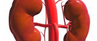
Neoplasms in the kidneys
Kidneys are one of the most important organs in the overall cleansing system of the body. Their main function is to filter the blood and remove harmful substances and waste products. However, they can also be susceptible to various kinds of pathologies, and neoplasms in the kidney are no exception.
The appearance of a space-occupying formation is a consequence of a combination of many factors and causes that can cause such a disease:
- Unfavorable heredity;
- Congenital developmental anomalies;
- Consequences of infectious processes;
- Frequent inflammatory diseases;
- Post-traumatic complications.
Space-occupying formations have a benign and malignant nature of the course of the disease. And, if such a pathology occurs, then, first of all, you need to find out what this neoplasm is.
A mass formation in the left kidney can cause deterioration of venous outflow and dilation of the inguinal veins. Which will lead to swelling of the lower extremities.
Morphological diagnosis
The morphological diagnosis of renal cell carcinoma is established after surgical removal of the kidney tumor or examination of a biopsy sample.
For kidney cancer, the Fuhrman classification according to the degree of differentiation (I, II, III and IV) is generally accepted, representing an important independent prognostic factor for the course and progression of the disease. There are five morphological subtypes of kidney cancer:
• clear cell (80-90%); • papillary (10-15%) subtypes I and II; • chromophobic (4-5%); • sarcoma-like.
These types differ in histological and molecular genetic characteristics, which are presented in table. 2-2. Papillary kidney cancer, in turn, is divided into two different subtypes: I and II, characterized by an unfavorable clinical course (see Table 2-2).
10–15% of renal tumors include a variety of rare sporadic and familial carcinomas, some of which have recently been described, and a group of unclassified carcinomas.
Table 2-2. Main histological subtypes of renal cell carcinoma
Bellini tube carcinoma (collecting system carcinoma)
Collecting system carcinoma is a very rare type of kidney cancer that is often detected late in the course of the disease. Up to 40% of patients have nerve metastases, and most die within 1–3 years of initial diagnosis. Metastases in regional lymph nodes at the time of diagnosis were present in 44% of patients, and distant metastases were found in 32%. The survival rate was 48% at 5 years and 14% at 10 years.
Sarcomatoid renal cell carcinoma
Sarcomatoid kidney cancer is a different type of RCC that has transformed into a cancer of a high degree of gradation (low degree of differentiation), but in itself it is not distinguished as a separate histological variety. In the presence of sarcomatoid changes in kidney cancer, the prognosis worsens.
Unclassified renal cell carcinoma
Unclassified renal cell carcinoma is a diagnostic category used to define a type of renal cell carcinoma that cannot be classified into any of the other categories of renal cell carcinomas.
Multilocular clear cell renal cell carcinoma (multilocular cystic renal cell carcinoma)
There are no clear pathohistological characteristics for the total subtype.
Multilocular clear cell renal cell carcinoma is a well-differentiated clear cell renal cell carcinoma. This subtype accounts for up to 3.5% of removed kidney tumors. To date, no description of metastases has been given for this tumor. According to the Bosniak classification based on imaging criteria, multilocular clear cell RCC is a type II or III cystic neoplasm. However, the same types of neoplasms according to Bosniak are characteristic of a mixed epithelial-stromal tumor of the kidney, cystic nephroma or mulilocular cyst (all of which are benign tumors).
In many cases, preoperative biopsy and intraoperative frozen section examination do not provide a correct diagnosis. Fortunately, the same surgical treatment strategy exists for all of these tumors. For this reason, it is recommended (if technically feasible) to choose organ-preserving treatment if complex multicystic neoplasms with increased density are detected in the kidneys.
Papillary adenoma
Papillary adenomas are tumors of papillary or tubular structure with a low degree of differentiation and diameter. Medullary carcinoma of the kidney Medullary carcinoma of the kidney is a destructive malignant tumor, primarily affecting young people heterozygous for sickle cell anemia. It is also very rare, accounting for approximately 2% of all primary kidney tumors in children and young adults aged 10 to 20 years. In 95% of patients, metastases are detected at the time of treatment.
Translocation carcinoma
Translocation renal carcinomas are rare tumors usually found in children and young adults. The occurrence of the majority of translocation carcinomas (about 90%) is associated with the transcription factor E3 (TFE3), located on chromosome Xp11.2. This type of carcinoma is characterized by relatively slow growth, despite the fact that when patients first present, they often appear at quite late stages of development. There is also information about another rare group of renal cell carcinoma in which the t (6; 11) (p21; q12) translocation is found.
Mucinous tubular and spindle cell carcinomas
These tumors are associated with the loop of Henle. Most mucinous tubular and spindle cell carcinomas behave similarly to low-grade tumors.
Carcinoma associated with end-stage renal disease
Degenerative cystic changes (acquired renal cysts) and a higher incidence of RCC are typical features of end-stage renal disease.
Acquired kidney cysts occur in almost 50% of cases in patients undergoing dialysis, but the frequency also depends on the period of dialysis, gender (3 times more often in men) and diagnostic criteria adopted in the method of assessing the condition. In the case of end-stage disease of one's own kidneys, kidney cancer is detected in 4% of patients. The lifetime risk of developing kidney cancer for such patients is at least 10 times higher than in the general population. Compared with sporadic renal cell carcinoma, cancer associated with end-stage disease and acquired renal cystosis is characterized by multicentricity, bilaterality and less aggressiveness and occurs in younger patients (more often men). However, in patients who have undergone transplantation, this type of cancer is usually characterized by a rather aggressive course, which may be due to immunosuppressive therapy.
Although the histological spectrum of tumors within acquired renal cysts is similar to that of sporadic renal cancer, the majority of these cancers are papillary; in tumors whose development is associated with acquired cysts, it is 41-71%), while in sporadic cancer it is 10%. The remaining tumors are mainly clear cell carcinoma. Patients with end-stage renal disease should have an annual ultrasound to evaluate the health of their kidneys.
Metanephric tumors
Metanephric tumors are divided into metanephric adenoma, adenofibroma, and metanephric stromal tumors. These are rare benign tumors, the main treatment for which is surgical removal.
Epithelial and stromal tumors of the kidney
Epithelial and stromal tumors of the kidney combine two types of benign mixed mesenchymal-epithelial tumors: cystic nephroma and mixed epithelial-stromal tumors. Imaging shows that most of the cystic neoplasms representing this type of tumor can be classified according to Bosniak as belonging to class III, less often - to class II or IV. Although there are few reports of their aggressive nature, both types of neoplasms are generally considered benign and surgical removal is indicated for their treatment.
Oncocytoma
Renal oncocytomas are benign tumors, accounting for 3 to 7% of all renal tumors.
Imaging characteristics alone are not sufficient to distinguish between oncocytoma and RCC. The standard method is histopathological diagnosis. Although preoperative diagnosis can only be made by percutaneous biopsy, this method is not specific for oncocytoma, since its cells are also found in clear cell renal cell carcinoma, granular cell carcinoma, and eosinophilic variants of papillary renal cell carcinoma (type II). In some cases, in the presence of histologically confirmed oncocytoma, one can resort to wait and watch tactics.
Congenital kidney tumors
Congenital kidney tumors can occur in the following cases: von Hippel-Lindau syndrome, congenital papillary RCC, Burt-Hogg Dube syndrome, congenital leiomyomatosis, tuberous sclerosis and constitutional translocation of chromosome 3.
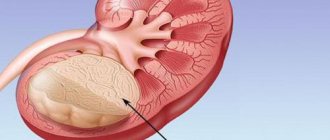
Types of formations in the kidney
A kidney cyst is a space-occupying formation that is a cavity filled with liquid contents with clear, even edges. The capsule of the cyst is formed by tissues of the parenchyma of the organ, which have become denser and formed a cavity. This occurs as a result of the influence of unfavorable factors on the kidney parenchyma. The glomerular excretory ducts become clogged, and primary urine accumulates in one place, forming a capsule. Over time, as fluid contents accumulate, the cyst increases in size.
Laboratory diagnostics
Among the laboratory parameters, the most informative from the point of view of prognosis of the course of the disease is the determination of the amount of serum creatinine, glomerular filtration rate, hemoglobin content, ESR, alkaline phosphatase activity, lactate dehydrogenase and serum calcium concentration.
The functional state of the kidneys especially needs to be assessed in the following situations:
• if there is a risk of a significant decrease in renal function during treatment (tumor of a single kidney, tumor damage to both kidneys); • with a decrease in kidney function, as indicated by an increase in serum creatinine; • in patients at risk of decreased renal function in the future due to the presence of concomitant diseases, such as diabetes mellitus, chronic pyelonephritis, renovasculitis, nephrolithiasis or polycystic kidney disease.
Malignant neoplasms of the kidney
The reasons why tissues degenerate into a malignant process are described in various sources are very relative and include all external unfavorable factors affecting the body. Changes associated with internal problems are most likely genetic in nature and are also associated with changes in the body's immune system.
There are a number of symptoms that suggest a malignant neoplasm:
- sudden unreasonable weight loss;
- intoxication syndrome (unmotivated weakness and loss of strength);
- hematuria of unknown etiology;
- feverish conditions;
- malignant hypertension;
- the appearance of edema of the lower extremities;
- swollen lymph nodes;
- the appearance of pain in later stages.
All clinical manifestations are more typical for the later stages of the disease, when the tumor reaches an impressive size; until this point the disease is asymptomatic. The greatest difficulty of malignant neoplasms is their ability to metastasize. Sometimes metastases that spread to other organs signal the presence of cancer in the body faster than a tumor growing in the kidney.
But if, nevertheless, the patient develops gross hematuria, without attacks of renal colic, without pain, then he should go for a detailed examination of the kidneys. These are laboratory studies with various urine tests, blood biochemistry, cancer markers and instrumental research methods: ultrasound, MRI, CT and radioisotope renography, instrumental methods: cystoscopy, urethroscopy.
Clinical picture
In many clinical observations, the kidney tumor is not palpable until the late stages of the disease.
Currently, about 70% of all cases of renal cell cancer are discovered incidentally during examination for other diseases. The classic triad of clinical symptoms (flank pain, gross hematuria and palpable abdominal mass) is rare today (6-10% of cases).
Extrarsal or iraneoplastic syndromes of kidney cancer are observed in approximately 30% of patients with clinical signs of RCC,
The most common parasoplastic signs are the following:
• arterial hypertension; • fever; • erythrocytosis; • anemia; • hyperkalidemia; • increased erythrocyte sedimentation rate (ESR)
; • cachexia; weight loss; • neuromyopathy; • amyloidosis; • liver dysfunction; • symptoms of metastatic disease, such as bone pain or cough.
Types of malignant kidney tumors
Based on their source of origin, there are 2 types of kidney cancer:
- Primary , growing from epithelial tissue.
- Secondary or metastases of cancer from other organs - adrenal glands, uterus, prostate, lungs, larynx, breast, stomach and intestines, skin.
In turn, primary cancer can be of various types, the main ones are:
- renal cell, growing from the tubular epithelium, accounts for 85% of the total number of cancer tumors;
- transitional cell , originating from the epithelial cells lining the pelvis, makes up 6-7%;
- nephroblastoma or Wilms tumor , originates from the tubular epithelium, is of genetic origin, accounts for 5-8%.
1% of all malignant organ tumors are sarcoma; it is not cancer because it grows not from epithelial tissue, but from connective tissue - muscle, fat cells, blood vessels. This is the most malignant kidney tumor with rapid growth and early metastasis.