Additional vessels in the kidney
blood vessels in the kidney
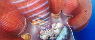
Vascular pathologies include 1–5 additional vessels. Sometimes there are more of them. They are represented by a smaller caliber in relation to the main vessels and can be directed both to the upper and lower poles, and may also have branches from the main rod. The upper polar renal vessels are longer than the lower ones. Those vessels that are directed to the upper part of the organ do not interfere with the overall urodynamics. Mostly they are found randomly during renovasography.
The inferior polar veins are the main causes of failure of urological dynamics and the formation of hydronephrosis. If the vessels begin to move away from the single rod of the renal artery, then disturbances occur in the ureteropelvic region. If they move away from the abdominal aorta, then the passage of urinary fluid fails at the upper part of the ureter. Additional vascular abnormalities that are located behind the ureter have a strong impact mainly on patency and the rapid formation of hydronephrosis.
With pathology, changes in large vessels and arteries may occur. They connect to the ureter, which causes the dynamics to go astray and a reactive expansion of the connective tissue in its walls begins.
If vascular ligation occurs at the time of surgery, this may result in the appearance of ischemic or hemorrhagic growths in the renal system. But if they are not bandaged, then they will subsequently cause active bleeding. The degree of vascular pathology does not affect the strength of morphological changes in the renal pelvis and ureter, but arterial vessels can more quickly form terminal levels of hydronephrosis compared to venous ones.
angiography
Pathological renal vessels in the lower part of the kidney usually cause urodynamic failures. The main symptomatic manifestation is pain, up to the formation of colic in the kidney. Symptoms may be affected by pyelonephritis and urolithiasis. It is possible to diagnose the pathology of the vessels of the kidney of the lower part through the symptoms that are detected during an X-ray examination during excretory urography:
- reduction of space in the area of the ureteropelvic region or ureter in the form of a malfunction;
- similar bending in accordance with the structure of the vessel.
The main way to determine abnormalities of the renal vessels is angiography, which can diagnose the number and location of damaged renal vessels.
Causes
The anomaly occurs during embryonic development; the cause of such deviations is not known for certain. It is assumed that, for unknown reasons, there is a failure of normal development, as a result of which the renal artery may experience duplication.
There are several types of pathologies of the renal vessels - arteries, depending on their number:
- Double and multiple. Double accessory artery is rare. The second artery, as a rule, is reduced and is located in the pelvis in the form of branches on the left or right.
- Multiple arteries are found in normal and pathological conditions. They depart in the form of small vessels from the kidney.
Types of accessory renal artery
Intrarenal type constricting vessel. Anomalies of the shape and structure of the renal arteries
In cases of development of the intrarenal type of vessels, one of them presses on the isthmus of the upper calyx. Before complications occur, the only symptom is aching pain on the side of the kidneys, which begins to increase when the person is moving or just standing still. The vascular type of obstruction may not have any symptoms indicating its presence. In some patients, arterial compressor disorder is discovered incidentally. Mostly people begin to complain of pain on the side of the kidney and costovertebral part. With physical activity, the pain increases rapidly.
In situations where the anomalies do not have any particular complications, doctors do not prescribe treatment. Indications for surgery exist when such serious consequences as urolithiasis, pyelonephritis, and hypertension are possible. These complications require resection of the upper region of the kidney.
renal vein aneurysm
The knee-shaped kidney vessel is a congenital elongated and knee-shaped change in the vessels of this organ. It contributes to malfunctions in the urinary organ, which leads to the development of renovascular hypertension. Renal arteriography is used to diagnose pathology. An aneurysm is a sac-like dilatation of renal vessels that can be located outside the kidney. In most cases, this anomaly is unilateral, and bilateral ones are rare. They are usually based in the branches of the bud. Sometimes an aneurysm is diagnosed in additional arteries. There are almost no symptoms, but quite high blood pressure is observed. It should be noted that aneurysm rupture leads to death in 80% of cases.
What is kidney dystopia and what is the danger of the anomaly?
There are many anomalies in the development of the genitourinary system. All of them are formed during embryonic development.
One of them is dystopia of one or both kidneys.
This pathology is quite common, it occurs in 3% of the world's population.
Most often, with this defect, the kidney is located in the pelvic cavity. Also, it can be located in the sacral region and move towards the anterior abdominal cavity.
In rare cases, organs of the urinary system are found above their normal location, namely in the mesogastric, epigastric region, and even in the chest cavity.
Kidney dystopia, in contrast to acquired pathologies, is characterized by the fact that the organ is firmly fixed and cannot independently move to a typical place.
Anomaly classification
Depending on the location of the vessels, kidney dystopia occurs:
- Lumbar is the most common. It is characterized by the fact that the renal artery arises at the level from the 2nd lumbar vertebra to the division of the abdominal aorta into the iliac vessels. With this arrangement, the renal organs are located slightly below their normal location.
- Pelvic is diagnosed in 20% of cases of the total number of this anomaly. In this case, the renal artery arises from the internal iliac artery, resulting in a shorter ureter than usual. Such dystopia can be confused with pelvic diseases, since in this case the kidney is located in the pouch of Douglas in women, and in men - between the rectum and bladder.
- Ileum is less common. Unlike the pelvic one, with this option there are several renal arteries, each of which branches off from the common iliac. As a result of this anomaly, the renal organs are located below their normal location and are palpated on the anterior surface of the abdominal wall, in the same place where the ovaries or appendix are located.
- Thoracic is characterized by the fact that the kidney is located in the chest cavity. In this case, its artery departs at the level of the 12th thoracic vertebra and above. The consequence of this is elongation of the ureter.
In addition to classification by location, homo- and heterolateral dystopia are distinguished.
In the first case, the displaced kidney is localized on the typical side. In the heterolateral version, it is on the opposite side.
Also, dystopia of the right kidney (more often) and the left one stands out. Bilateral crossed dystopia is less common.
Why does the disorder develop?
Currently, there is no reliable data on the causes of renal dystopia.
The abnormal location of the urinary organs is associated with a genetic predisposition, as there are cases of inheriting the defect from parents.
However, dystopia can occur without a family history.
During the formation of embryonic primordia, the renal organs are located in the pelvis. Due to various factors, they do not move to their typical location, as they should under normal conditions.
Provoking influences include bad habits of the mother during pregnancy (alcohol abuse, smoking), exposure to radiation and chemical compounds, stressful situations and past pathologies.
Features of the clinical picture
Most often, a sign of dystopia is intermittent, aching pain in the lumbar region.
Symptoms of kidney dystopia can be very diverse, they depend on the type of anomaly:
- With a bilateral crossover anomaly, the signs of the disease are more pronounced. Chronic renal failure, arterial hypertension and urinary syndrome develop more often.
- Pelvic dystopia is characterized by pain in the rectal area and lower abdomen. Unpleasant sensations intensify with palpation of the bladder and digital examination. They may be accompanied by defecation disorders - frequent urge or, on the contrary, constipation. In addition, there are dysuric disorders, which are manifested by frequent or rare urination, pain, or false urges.
- Ileal anomaly is characterized by pain localized in the lower abdomen. More often, unpleasant sensations cover one side. For this reason, this variant of the anomaly can be confused with an ovarian cyst, appendicitis or tumor.
- Thoracic disorder, in most cases, is associated with a hernia of the diaphragm. The following symptoms are identified: pain in the chest, dysphagia, belching, difficulty breathing.
Diagnostic methods
Diagnosis of the anomaly is based on physical and instrumental examination data.
Patients with this pathology do not always complain, since in the lumbar variant the anomaly can be asymptomatic.
If a person is bothered by pain in the kidneys or pelvis, then an experienced specialist may suspect this disease.
Depending on the anatomical region in which the dystopic kidney is located, the doctor makes a differential diagnosis:
- With lumbar dystopia, the pathology is compared with inflammatory diseases of the tubules or glomeruli of the kidneys. A distinctive feature is noted during palpation, since the kidneys can be felt above their typical location.
- Ileal anomaly is differentiated from gynecological and oncological pathologies.
- With the pelvic variant of the anomaly, palpation of the kidneys is not available, but unpleasant sensations during rectovaginal examination allow one to think about this disease.
- Thoracic dystopia is the most difficult to diagnose, since it is extremely rare and is often confused with a mediastinal tumor.
An accurate diagnosis can only be made using instrumental methods. An ultrasound of the kidneys will allow you to determine the location of the organs, as well as their structural changes.
Modern methods of therapy
Patients with renal dystopia are subject to dispensary observation by a urologist. With an asymptomatic course of the disease without impaired renal function, treatment of the anomaly is not required.
The main goal of the doctor is timely diagnosis and treatment of possible complications.
Conservative treatment
Conservative therapy allows you to get rid of pyelonephritis and small kidney stones.
Inflammatory diseases of the renal tubules require antibacterial therapy.
For this purpose use:
- cephalosporin antibiotics: Ceftriaxone, Cefuroxime, CEF IV;
- fluoroquinolones: Ciprofloxacin, Ofloxacin, Pefloxacin;
- macrolides: Azithromycin.
If the glomeruli of the renal vessels are involved in the infectious process, then they resort to therapy with Prednisolone and cytostatics.
Diagnosis and treatment of accessory renal artery at Best Clinic
To make an accurate diagnosis and subsequently prescribe effective treatment, our specialists use the most modern types of diagnostics:
- digital subtraction angiography;
- color echo Dopplerography;
- magnetic resonance imaging;
- ultrasonography;
- excretory urography.
After conducting a differentiated diagnosis, the doctor prescribes adequate treatment in strict accordance with the physiological, genetic, psychological and other characteristics of each patient. The goal of therapy is to restore normal urine flow from the kidney.
Modern equipment
Diagnostics
To detect an additional renal artery, different diagnostic methods are used:
The last method is considered the most accurate. Dopplerography gives a complete picture of the condition of the left and right kidneys, and also monitors blood flow (pressure, direction). But, if the fluid flow is slow, the device will not be able to record its movement. Aortography reveals abnormalities of the solitary artery.
Symptoms of the disease
Additional vessels that are located in the upper part of the organs do not cause any clinical manifestations. If they are located in the lower region of the kidneys or near the ureter, their pressure causes a disruption in the flow of urine from the kidney. This is dangerous, since the patient subsequently develops hydronephrosis (enlargement of the renal pelvis). In addition, the removal of fluid from the body is disrupted, which leads to increased blood pressure. With prolonged hydronephrosis, parenchymal atrophy and renal infarction occur. Constant injury to the vascular walls causes increased thrombus formation.
Symptoms of kidney malformation
- If one kidney is defective, there may be no symptoms.
- Pain in the lumbar region (pulling, constant).
- Increased blood pressure.
- Attacks of “colic” - sharp, periodic pain in the lumbar region.
- Hematuria (presence of blood in the urine).
- Changes in urine (it becomes cloudy, the color of “meat slop”, foamy).
- Increased body temperature, chills.
- Tendency to edema.
- Varicocele (dilation of testicular veins).
- Thirst.
- Headache.
Kinds
Accessory arteries are classified by number into:
- Double . They are rare. The second vessel is usually reduced and is located in the pelvis in the form of a branch on the right or left. In some cases this is the normal limit.
- Multiple . Many small vessels branch off from the kidney.
These varieties do not always lead to pathology. But they are often combined with other renal anomalies. For example, with polycystic, double, horseshoe-shaped, dysplastic or dystrophic kidney.
To achieve this goal, Best Clinic specialists can resort to:
- resection of the accessory vessel itself, the affected kidney, as well as the sclerotically changed area of the urinary tract;
- resection of the area of urinary tract stenosis followed by plastic surgery. This operation is performed in cases where partial removal of the accessory artery is impossible due to the fact that it supplies a significant part of the kidney.
Best Clinic specialists have been successfully diagnosing and treating developmental anomalies of the genitourinary organs for many years. You can find out all the necessary information and sign up for a consultation by calling or sending us a request.
To make an appointment, call +7 (495) 530-1-530 or click on the “Make an appointment” button and leave your phone number. We will call you back at a convenient time.
Diseases of the urinary system affect approximately 35% of the entire world population. Approximately 25-30% are associated with kidney abnormalities. These include: renal artery aneurysms, multiple or double renal arteries, solitary artery, accessory renal artery, fibromuscular stenosis, etc.
Developmental disorders
Anomalies of the renal arteries in 80% of pathologies cause congenital diseases.
An additional trunk in the kidney provides nutrition to the paired organ. The additional vessel has a smaller diameter than the main one and is directed to the upper or lower pole of the organ. Often the pathology is localized on the right side. An accessory artery can originate in various parts of the bed, but the most common site of development is the abdominal aorta.
Doppler of renal vessels is normal
The normal diameter of the renal artery in adults is 5 to 10 mm. If the diameter is 65 mm, an accessory renal artery is likely present. With a diameter of the main renal artery of 15 mm, an additional renal artery is almost always present.
The renal artery should be assessed at seven points: at its exit from the aorta, in the proximal, middle and distal segments, as well as the apical, middle and lower segmental arteries. We evaluate peak systolic (PSV) and end-diastolic (EDV) blood flow velocities, resistivity index (RI), acceleration time (AT), acceleration index (PSV/AT). See Vascular Doppler for more details.
The normal spectrum of the renal arteries has a pronounced systolic peak with antegrade diastolic flow throughout the cardiac cycle. In adults, the normal PSV on the main renal artery is 100±20 cm/sec, EDV is 25-50 cm/sec, in young children PSV is 40-90 cm/sec. In segmental arteries, PSV drops to 30 cm/sec, in interlobar arteries to 25 cm/sec, in arcuate arteries to 15 cm/sec and interlobular arteries to 10 cm/sec. RI at the renal hilum Developmental disorders
Anomalies of the renal arteries in 80% of pathologies cause congenital diseases.
The reason for the appearance of incorrect localization of blood vessels is considered to be the preservation of the embryonic vascularization of the kidney. The problem is quite often combined with organ pathologies. The following anomalies of the renal artery occur:
- accessory - the departure from the aorta of a vessel smaller than the main artery to the kidney;
- lumbar - low branch from the aorta;
- aneurysm - expansion;
fibrous stenosis - narrowing of the vascular lumen;
Clinical picture
The disease is usually asymptomatic. It appears only when the urinary tract is crossed by an accessory artery.
Due to this crossing, the outflow of urine from the kidneys becomes difficult, resulting in the following clinical manifestations:
Hydronephrosis is a persistent and rapid expansion of the renal pelvis, resulting from a violation of the outflow of urine. Arterial hypertension is high blood pressure (BP). A jump in blood pressure occurs due to a decrease in fluid content in the body, the vessels narrow, blood flow becomes more difficult, and as a result, pressure increases. Kidney infarction. With prolonged hydronephrosis, gradual atrophy of the renal parenchyma occurs, which subsequently leads to infarction of the entire kidney. Formation of blood clots and bleeding at the intersection of the accessory artery and the urinary tract.
The kidney increases in size. There may be blood in the urine, and going to the toilet becomes painful. Patients complain of aching lower back pain and high blood pressure.
On palpation, a pain syndrome develops in the form of attacks of renal colic; pain can also radiate to the ribs, both during physical activity and at rest.
Clinical significance
Renal artery stenosis, or narrowing of one or both renal arteries leads to hypertension as the affected kidneys release renin to increase blood pressure to maintain perfusion for the kidneys. RAS are usually diagnosed with duplex ultrasonography of the renal arteries. Treated with balloon angioplasty and stents if necessary.
Atherosclerosis can also affect the renal arteries and can lead to insufficient renal perfusion leading to decreased renal function and possibly renal failure.
- exogenous intoxication (ecology, taking medications);
- endogenous damaging factors (severe toxicosis of pregnant women, transient functional kidney failure during pregnancy);
- genetically predetermined (hereditary) influences on the formation and development of individual anatomical formations.
Let's work together to make the unique material even better, and after reading it, we ask you to repost it on a social network convenient for you. net.
Types of vascular abnormalities in the kidneys
Abnormally formed renal vessels are most often arteries, although some pathologies of the development of the venous bed are also distinguished.
Abnormally formed renal vessels are often arteries, although some pathologies of the development of the venous bed are also distinguished. Among all arterial anomalies, the following malformations of the vessels supplying the kidney are distinguished:
By accessory renal artery we mean an accessory arterial vessel that is much smaller in diameter than the main artery. The accessory artery can arise from the aorta, main renal artery, iliac, phrenic, adrenal arterial vessels and flow into the lower or upper renal segments. Often the accessory artery is reduced and does not serve as a blood supply, although it may be functionally viable. With the superior location of the accessory artery, any pathological changes in the functioning of the organ usually do not occur. The lower localization of the abnormal vessel can be dangerous when it compresses the ureter, which leads to atrophy and sclerosis of the urethral canal, difficulty in urine drainage and accumulation of fluid in the cavity of the pelvis.
- Multiple (double) arteries are the main vessels supplying the kidneys, and regardless of their number, they are approximately equal in cross-section and flow into the kidney in one place. Often, the presence of an abnormally developed multiple renal artery does not affect the function of the organ. However, a combination of such an anomaly with some renal pathologies, such as polycystic, dystopic, double or horseshoe-shaped kidney, is not excluded.
- The solitary artery is a rare anomaly when both organs (left and right) are supplied with blood by one common vessel. It rarely affects kidney function, except in cases where the solitary artery is abnormally located and can interfere with the drainage of urine by compressing the ureter.
- An artery aneurysm is its abnormal expansion, which occurs due to the absence of muscle tissue in the choroid. The vessel wall, made only of connective tissue fibers, is not able to contract and regulate the lumen. Pathological expansion disrupts hemodynamics in this area, leading to a slowdown in blood flow and the formation of areas with turbulent fluid movement. An aneurysm, which can be located inside the organ or extrarenally, leads to disruption of the normal blood supply to the renal tissue. With a large area of insufficient supply, the aneurysm often becomes the cause of a serious illness - renal infarction.
Areas of fibromuscular stenosis are most often located in the distal third of the renal artery and represent alternating narrowing and dilatation of the vessel. The cause of this pathology is the excessive development of fibrous or muscle fibers in the choroid. This anomaly affects the blood supply to the renal tissue to a lesser extent, but causes arterial hypertension, which is difficult to correct with antihypertensive drugs.
Renal artery stenosis
Renal artery stenosis is a dangerous pathology. Stenosis is essentially a narrowing of the diameter of blood vessels. During normal functioning, blood filtration leads to the formation of primary urine. When the walls narrow, the blood volume decreases; the stronger the narrowing, the less blood the kidneys feed. Lack of blood leads to increased blood pressure, and the organ cleans the blood much worse.
Renal artery stenosis completely disrupts the functioning of the organ. With a critical decrease in blood volume, as well as with poor nutrition for a long time, the kidneys stop functioning normally and urine is not formed or excreted. Stenosis occurs due to certain diseases. Stenosis can be provoked by atherosclerosis, diabetes mellitus, aneurysm, some inflammatory processes, as well as neoplasms in the renal arteries.
In order not to provoke the appearance of stenosis, this disease has an extremely negative effect on the condition of the kidneys, as well as on the general health of a person, and there is a risk of a very serious illness. If therapeutic measures are not applied in a timely manner, stenosis can lead to hormonal imbalance, decreased protein levels, swelling and a decrease in fluid secretion, and a decrease in the amount of plasma.
Reasons for appearance
Deviations from the normal development of an organ are established during embryogenesis. Exactly what factors lead to the appearance of this pathology have not yet been precisely established. Only a hereditary predisposition to the abnormal structure of the bloodstream has been identified. A connection between the disease and the influence of various teratogens on the embryo has also been established.
There is an assumption that bad habits of a pregnant woman and poor ecology can lead to the formation of an additional renal vein in the child.
Renal arteries of the elderly
Arterial walls throughout the body tend to thicken with age. The renal arteries thicken more slowly than others. In old age, the thickness of the renal arteries is finally formed. This happens from the moment of birth. If the right renal vein has thickened significantly, then the same process is observed in the left one and vice versa.
In newborns, the inner shell of the hyperplastic thickening bifurcates into two membranes. As the body matures, the elastic lamina is divided into membranes many times. There is an increase in the number of membranes at the beginning of the arteries, as well as at the site of the first division into two separate branches, then this spreads along the entire perimeter of the bifurcated arteries.
In older age, changes lead to the appearance of an elastic layer with connective tissue and elastic fibers.
Age-related changes do not always lead to the development of pathological processes in the human body. Thickening occurs in any person and leads to the formation of sufficiently thick walls that can withstand damage. The simple structure of the blood supply in newborns copes well with small loads and small volumes of blood, but as the body grows, all processes become much more complex, and accordingly, the thickening of the walls, inherent in nature, is advisable.
Treatment of the disease
After identifying this developmental anomaly, experienced surgeons at the El.En. clinic choose the necessary tactics. The goal of treatment is to normalize the outflow of urine, prevent and treat complications.
Effective restoration of urine outflow in case of anomalies in the development of renal vessels is only possible through surgery.
At the clinic "El.En." The following methods are used.
1. Resection (removal) of the accessory vessel, the affected area of renal tissue, and, if necessary, the kidney.
2. Resection and plastic surgery of the narrowed area of the urinary tract. It is performed when removal of an additional vessel is impossible.
If you notice signs of a disease of the urinary system, consult a doctor immediately. Only timely diagnosis and treatment will achieve the desired effect!
Make an appointment with a urologist using the form.
Or call: +7.
Treatment
What to do and how to treat is determined only after a complete diagnosis of the disease. Treatment is based on restoring the physiologically normal flow of urine from the body. This effect can only be achieved through surgery.
Resection of the accessory artery. Removal can be complete or partial. Partial - the accessory artery and the damaged area are almost removed. Complete removal—removal of both the accessory artery and the entire kidney.
Resection of the urinary tract. This operation is performed when resection of the accessory artery is impossible. The narrowed section of the urinary tract is removed and stitched back together.
The method of surgical intervention is determined by a urologist-surgeon individually for each patient.
Treatment of the anomaly
After a full examination, the doctor prescribes specific treatment for each case, based on the data obtained. The main goal of therapy is to restore healthy urine flow from the kidneys. This is achieved by resection of the kidneys or resection of sclerotically changed areas of the genitourinary tract, using ureterouretero- or ureteropyelostomy.
Do not forget that the diagnosis of “accessory renal artery” poses a danger to the body as a whole and its individual systems. You need to monitor changes in your body, consult a doctor for preventive purposes, and especially with symptoms such as: pain in the head; a sharp increase in blood pressure; pain in the lumbar regions; changed color, volume and other visible properties of urine; swelling of the face in the morning. Ignoring them is dangerous to health.
Parapelvic kidney cyst: causes, symptoms and treatment
Have you been trying to cure your KIDNEYS for many years?
Head of the Institute of Nephrology: “You will be amazed at how easy it is to heal your kidneys just by taking it every day.
Our readers successfully use Renon Duo to treat kidneys. Seeing how popular this product is, we decided to bring it to your attention. Read more here...
Parapelvic kidney cysts are one of the types of simple formations that are benign in nature. Such cysts are localized at the gate of the kidney or, as doctors call it, the renal sinus. In appearance, they resemble an oval-shaped sac filled with a clear liquid (in some cases, the liquid may be yellowish in color).
This type of cyst is very rare and is diagnosed in older people. Most often, education does not bother a person and does not show any signs of its development. Also, this kidney pathology (Kidney (anatomy) is an organ of the excretory (urinary) system of animals and humans) (Kidney (anatomy) is an organ of the excretory (urinary) system of animals and humans) is single in nature and in most cases a parapelvic formation is found on the left kidney. Multiple spread or damage to the kidney on the right side is diagnosed extremely rarely. With increasing education (a single purposeful process of upbringing and training, as well as the totality of acquired knowledge, skills, values, experience and competence in the broad sense of the word), professional treatment is needed (a process whose goal is to alleviate, relieve or eliminate symptoms and manifestations a particular disease or injury, pathological condition or other disability) to reduce the risk of complications.
Reasons for the development of the disease
Today, the causes of parapelvic cysts (a pathological cavity in tissues or organs, having a wall and contents (liquid and/or non-viable cells)) of the kidneys (Kidney (anatomy) is an organ of the excretory (urinary) system of animals and humans) are being carefully studied, and so far most scientists tends to believe that this pathology is a congenital disease. Most likely, during the development (this is a type of movement and change in nature and society associated with the transition from one quality, state to another, from old to new) of the embryo, anomalies occurred in the formation of the tissues of the left or right kidney.
An infectious disease of the genitourinary system can also provoke an increase in the resulting cyst.
Symptoms of the disease
In the early stages of formation, parapelvic formations are asymptomatic, so it is very difficult to identify them. Even if a small cyst is detected, treatment is not prescribed. Most often, they begin to cause discomfort when they increase significantly in size. The growth of the formation creates pressure on the vessels of the organ and pelvis, which can provoke sharp pain in the side, which is paroxysmal in nature.
Symptoms
Usually the pathology does not have pronounced manifestations. Unpleasant symptoms develop only if the accessory artery crosses the urinary tract. This leads to difficulty in the flow of urine from the kidneys. The following symptoms appear: