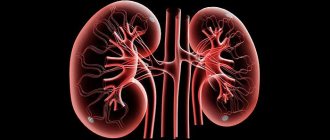
What is kidney prolapse?
In medical practice, the phenomenon of kidney prolapse is called nephroptosis. With it, kidney mobility exceeds the physiological norm. In the absence of pathologies, this organ, during breathing and changing body position, can move vertically from the area of the renal bed by a maximum of 2-3 cm. If a child has prolapse, the kidney moves by 6-8, and sometimes by 10 cm. There are situations when it falls into the pelvic cavity. Popularly, such a mobile organ is often called vagus.
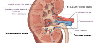
The organ is held in its correct position by structures such as fascia and ligaments. They form the renal bed and fascial fat capsule, fatty tissue between the kidneys and adrenal glands, regulate intra-abdominal pressure (it is created by the diaphragm and the anterior abdominal wall),
Doctors distinguish several stages of kidney prolapse. There are three of them:
- The first stage - the prolapsed organ can be felt through the abdominal wall during inhalation. When you exhale, the kidney returns back under the costal edge and it is no longer possible to palpate it.
- The second stage - the kidney can be completely palpated slightly below the hypochondrium line if the patient is in an upright position. When the patient lies down, the kidney is located in the hypochondrium.
- The third stage - regardless of the position of the body, the kidney completely emerges from the hypochondrium area. In advanced cases, the organ sinks into the pelvis.
Mixing of the kidneys can occur vertically and rotationally (moves in a circle), pendulum-like or around the renal pedicle. When moving to more severe stages, the main vessels of the organ (arteries and veins) can twist and stretch. When this happens, the diameter of these vessels decreases.
Due to such processes, the blood supply to the renal structures is disrupted, hypoxia occurs, lymphatic drainage is disrupted and venous pressure increases. Pathologies in lympho- and hemodynamics are fraught with the development of chronic pyelonephritis.
When stage 3 nephroptosis occurs, the ureter becomes bent. Because of this, the pelvis in the kidneys begins to expand and problems arise with urine excretion. First, inflammatory processes occur in the kidneys, which then develop into adhesive processes (perinephritis occurs). Adhesive formations fix the organ in an incorrect position.
Most often, kidney prolapse is diagnosed in girls. The right kidney is mainly affected. This is due to the fact that the left kidney has a strong ligamentous apparatus. In addition, the right kidney is normally located slightly lower. Some babies have bilateral ptosis, called ptosis.
Symptoms
The clinical picture can be of a different nature: complicated nephroptosis, clinically manifest and asymptomatic. There are common symptoms that are observed in almost all patients:
- Pain. It is paroxysmal and dull in nature, localized mainly in the hypochondrium, abdomen (radiating to the back) and lower back. The intensity of this symptom increases when the child moves, coughs, and plays sports.
- Urinary incontinence at night and during the day.
- Problems with appetite and other bowel dysfunction. This occurs due to the course of general pathological processes in the body.
- Increased pressure. Occurs due to the entry into the blood of angiotensin, which is synthesized during vascular spasms.
- Severe swelling. Mostly the area under the eyes swells. Swelling becomes especially severe if the baby drinks a lot of liquid.
- Renal colic. The processes inside the kidney are disrupted and severe spasms occur.
- Nausea and vomiting. Appear at the extreme stages of the disease.
- Blood and traces of protein in the urine.
If the child’s condition is very advanced, he develops psycho-emotional disorders - neurasthenia, depressive states, increased excitability. This situation is characterized by respiratory failure.
Please note: in the absence of proper treatment for nephroptosis, toxic substances will begin to accumulate in the child’s blood plasma fluid, which can lead to brain damage.
Degrees of the disease
Experts divide nephroptosis into several stages, in particular on which the clinical picture of the disease will depend. Thus, there are 3 degrees of nephroptosis:
- The disease, which occurs in the 1st degree, is accompanied by a shift of the organ by 1 vertebra. This pathology does not require emergency medical care, as it only indicates renal mobility.
- With the 2nd degree of nephroptosis, increased signs of deviation from the norm appear and problems with the blood circulation of the kidney are observed. In this case, the mobile kidney descends by 1.5–2 vertebrae and requires close monitoring and corrective measures.
- The last, 3rd degree of nephroptosis is characterized by a shift of the kidney by 3 vertebrae and organ dysfunction. Often diagnosed in teenagers.
Depending on the degree of the disease, complaints in young patients become more frequent and pain intensifies. That is why it is important to consult a doctor in time and undergo a special examination. In most cases, nephroptosis is right-sided and is extremely rarely found on the left side. In isolated cases, it occurs bilaterally.
Symptoms and first manifestations
The problem of nephroptosis is the absence of clinical manifestations at the initial stage of the disease and therefore the disease can only be detected by X-ray examination and palpation. However, in most cases, mild pain symptoms are still observed at the site of the prolapsed kidney. The child complains of paroxysmal pain in the hypochondrium, lower back or abdomen, which appears with the slightest physical activity.
The child has a deterioration in appetite and lower back pain.
With the development of nephroptosis, the patient experiences a deterioration in appetite and lower back pain. Subsequently, blood pressure increases, as angiotensin, which is formed during vascular spasms, enters the blood. In children and teenagers, you may notice severe swelling under the eyes in the morning, especially after drinking a large amount of liquid before bed. In addition, renal colic may be present, and the 3rd stage of the disease is characterized by nausea, vomiting, increased blood pressure and blood in the urine.
In advanced forms of nephroptosis, depression, neurasthenia, respiratory and circulatory disorders, difficulty urinating and increased excitability are observed. If nephroptosis is not treated, there is a risk of brain damage due to the accumulation of toxins in the blood plasma.
Kidney prolapse in a child - causes and consequences
There are many reasons for this phenomenon, but most often nephroptosis occurs due to:
- Kidney diseases caused by infection - rickets, bronchitis, tonsillitis. With them, deformation of the spine often occurs.
- Excessively intense physical activity or heavy lifting.
- Congenital anomalies in skeletal development. This means inadequate development or absence of the lower ribs.
- Anatomical features of the child. Due to age-related body imbalances, the baby’s muscle tissue develops unevenly.
- Rapid and significant loss of body weight, due to which the fat capsule around the kidneys becomes thinner.
- Predisposition to stretching of connective tissues (for example, Ehlers-Danlos syndrome).
- Injury to the lumbar region with subsequent damage to the ligamentous apparatus and the appearance of hematomas in the perinephric tissue.
Children with poor tone of the muscles located in the anterior abdominal wall, an asthenic physique and poorly developed subcutaneous fat are susceptible to kidney prolapse.
The process of kidney prolapse itself does not have a pronounced clinical picture until changes in hemodynamics and urine output begin. At the same time, already at the initial stages of the disease, pathological processes of structural damage to the renal tissue begin. This phenomenon can cause the patient:
- pyelonephritis – inflammation of the kidneys;
- myocardial infarction and stroke - due to changes in blood circulation and increased arterial and venous pressure;
- urolithiasis - difficulty draining urine stimulates the development of infections in the kidneys, as a result of which stones can form in the renal pelvis.
Root causes of nephroptosis in children
Kidney mobility is a pathology that is characterized by their displacement from their original location. The following factors influence the development of nephroptosis:
- a sharp jump in growth;
- weak back muscles and abs;
- congenital anomalies in which ribs are missing or are not fully developed;
- heredity;
- injury;
- thin constitution;
- fast weight loss;
- past illnesses: bronchitis, rickets, tonsillitis.
Return to contents
Diagnosis of the disease
Since the initial stages of nephroptosis have virtually no pronounced symptoms, it is almost always discovered by chance. To fully diagnose the disease, the following methods are used:
- taking an anamnesis - the patient is questioned about complaints, previous injuries and diseases;
- palpation (palpation) of the abdomen, sides and back;
- various x-ray examinations (radiography of the kidneys and excretory urography);
- ultrasound examination of the kidneys (lying and standing);
- laboratory tests of urine and blood (they also help to identify possible complications);
- renography (to study the completeness of kidney functionality);
- radioisotope scanning or scintigraphy to further clarify the position of the kidney.
Who to contact and diagnostic methods
If symptoms of nephroptosis occur, you should contact a nephrologist. Based on the clinical manifestations, the doctor will conduct an examination.
First of all, the specialist conducts an external examination and palpation examination. Also assigned:
- blood and urine tests;
- X-ray examination;
- Ultrasound;
- angiography of renal vessels;
- scintigraphy.
Based on these research results, a diagnosis is established, the degree of development is established, and a course of therapy is prescribed.
How is the pathology treated?
Kidney prolapse is treated comprehensively. The following activities are used:
- diet
- orthopedic treatment
- belly massage
- physiotherapy
Feeding a child with kidney prolapse
It should not put extra strain on the kidneys or irritate them. The baby should eat up to 6 times a day, portions should be small. The lion's share of the diet consists of fruits and vegetables, consumed boiled, baked or raw. Seafood dishes will be very healthy. It is important that the child drinks a sufficient amount of ordinary water - at least 1.5 liters.
The red list includes strong broths, pickles, all canned foods, legumes, sweets, soda, and sweets. If a patient has developed chronic renal failure, it is necessary to constantly monitor the amount of protein consumed per day (up to 25 g). There are a lot of them in cereals, beans and flour. Many metabolic products are released from them, which are excreted by the kidneys and thereby load them.
You should also not consume a lot of phosphorus and salt. They are found in nuts and cocoa. Sodium-containing foods should also be limited, because they stimulate increased blood pressure and retain fluid in the body.
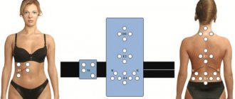
Orthopedics
To support the muscles, you can wear special bandages. They are put on in the morning and worn throughout the day. It is important that a specialist selects a belt for the child and that he explains the technology for putting it on.
Exercises
They are effective only in the initial stages of dysfunction. Performed daily, the lesson lasts at least half an hour. All exercises are performed lying on your back. To make it easier to do them, place a soft towel cushion under the child’s lower back.
- Bend your knees. As you inhale, stick your stomach out and exhale, draw it in. 10-15 repetitions are done.
- Raising straight legs 5 times each.
- Pulling the bent leg towards the stomach, 5 times each leg.
- Exercise “bicycle”, done for about 3 minutes.
- Bringing your knees together with a ball between them. Up to 5 reps.
- As you inhale, raise your outstretched legs, and as you exhale, lower them. Up to 10 times.
- Heels and knees together, lifting straight legs up. On inhalation, separate them; on exhalation, cross them. Up to 10 repetitions.
You cannot run, stretch, jump or hang on horizontal bars and parallel bars.
Other treatments for nephroptosis
Treatment with medications is used only when the child already has some complications. These include kidney stones, pyelonephritis and arterial hypertension. In cases where conservative treatment methods do not help a small patient, surgical intervention is used. Main indications for surgery:
- persistent pain;
- recurrent or chronic pyelonephritis;
- orthostatic hypertension;
- loss of any physical activity;
- hydronephrosis.
The intervention is aimed at artificial fixation of the kidney. Most likely, the child will undergo surgery via laparoscopy. This is the best option, because the method is almost non-traumatic, the risk of complications is minimal, blood loss is also minimal, and the recovery period is easy.