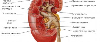
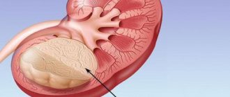
One of the organs of the urinary system is the kidneys. This paired organ is located on the sides of the spine at the level of the last two thoracic and first two lumbar vertebrae.
They are bean-shaped, slightly rounded at the upper and lower poles, with an average weight of 120-200 grams.
The main function is to purify the blood and produce urine, so the organ suffers from all the negative processes occurring in the body, in addition, all the medications and foods that a person consumes throughout his life affect its functioning.
Therefore, it is not surprising that there are many diseases that affect the kidneys, and the most dangerous of them is cancer.
General characteristics of the disease
Kidney cancer refers to a malignant neoplasm consisting of the cells of the organ itself. Cancer is a mass of cells that begins to divide uncontrollably, and the main function of the cells is lost.
The faster cell division occurs, the faster they spread through the lymphatic and circulatory system and the more dangerous the disease becomes.
According to the International Classification of Diseases, 10th revision, the disease was coded as C64 and C65.
However, clarification is possible for each of the codes, for example, C64.0 is cancer of the right kidney, and C64.1.0 is cancer of the left upper segment of the left kidney, and the coding C65 means malignant neoplasms of the renal pelvis.
Thus, the code makes it possible to determine the location and nature of the tumor.
Kidney cancer: ICD 10 code - what the coding says
Kidney cancer is one of the most common oncological diseases (occupies 10th position in the list of existing oncologies), which is marked by an annual increase in the number of cases, the complexity of therapy and rehabilitation, and most importantly, the danger of death.
For all patients, this term is quite clear if it is written down in words, but doctors also put a certain code next to it, which immediately raises a lot of questions - how does kidney cancer differ according to the ICD, what clarifications are hidden behind such a coding and why is it needed?
What does "ICB" stand for?
General information
The International Classification of Diseases (abbreviated as ICD) is a standard, an approved document that represents a statistical basis in the healthcare system, the purpose of which is to group, systematize, register and compare data on morbidity and mortality in all regions of different countries.
Coding is used to translate verbal formulations of diagnoses made in different countries into uniform designations - codes (alphanumeric, in alphabetical order from A to Z), which makes it convenient to work with data and carry out their analysis. For example, in ICD 10 kidney cancer is under the letter “C” - “cancer” (translated as “cancer”).
This classification is subject to regular review, clarification and adjustment once every ten years. Today, the Tenth Revision is used in international medical practice.
For your information! WHO specialists are currently working to compile the Eleventh Revision, which should be introduced in 2020.
Kidney oncology coding
When considering the disease “kidney cancer,” the ICD includes several codes at once, but all of them are needed solely for statistical purposes and in order to facilitate the understanding of diagnostic data between doctors of different clinics. At the same time, the designations according to ICD 10 will not in any way affect the diagnostic methods performed or the chosen type of treatment for kidney cancer.
Etiology and pathogenesis
The causes of kidney cancer are not fully known, so everyone may be at risk, regardless of age or gender.
At the same time, oncologists and urologists have identified several factors that increase the risk of developing cancer pathology:
- smoking, it is believed that cigarettes increase the risk of tumors by 2.5 times;
- men, their risk is 2 times higher than women;
- excess weight;
- uncontrolled long-term use of drugs;
- severe pathologies of the genitourinary system, especially in patients on dialysis;
- injuries and falls, mechanical damage to the kidney;
- some genetic pathologies;
- working with chemicals: herbicides, asbestos, cadmium, gasoline, organic solvents;
- high blood pressure;
- representatives of the Negroid race have a higher risk;
- diabetes;
- estrogens.
In addition, the disease is characterized by pronounced changes in blood vessels and rapid metastasis to other organs, usually the lungs, liver and brain.
Causes of kidney stones in children
Have you been trying to cure your KIDNEYS for many years?
Head of the Institute of Nephrology: “You will be amazed at how easy it is to heal your kidneys just by taking it every day...
Read more "
Kidney stone disease, or nephrolithiasis, is the stagnation of undissolved salts and various compounds in the human kidneys.
Today, kidney stones begin to appear in children no less frequently than in adults. In addition, the incidence rate among newborns is increasing.
Impaired excretion of salts and an increase in their concentration in the kidneys occurs due to problems with metabolism.
As a result, the salts “stick” to each other and turn into compactions – stones. Initially they are small in size and similar to sand, but over time they can grow to several centimeters.
Stones come in different shapes, but the most dangerous are crystalline ones, which cause severe pain.
In order to promptly identify kidney stones in a child, it is necessary to listen carefully to his complaints.
If a child over 4 years old can independently describe his symptoms, then with kids the situation is much more complicated. In this case, you should pay attention to crying when urinating.
But, unfortunately, the disease often does not make itself felt, and this can lead to complications.
Symptoms of the disease
Symptoms of stones in children differ little from those in adults, but renal colic is less common at an early age.
Kidney stones can only be accurately diagnosed through a medical examination - blood and urine tests, ultrasound, x-rays or MRI.
But various symptoms will presumably help identify kidney stones:
- intermittent urination;
- burning and pain when urinating;
- renal colic - pain in the lumbar region;
- increased body temperature and nausea (rather rare symptoms);
- bright yellow sediment or blood in the urine;
- high blood pressure;
- pain in the genitals and lower abdomen;
- excretion of stones or sand with urine.
Stones appear mainly in one kidney, less often both are affected. The localization of renal colic also depends on this: the pain is concentrated either on one side or along the entire lower back.
Sometimes pain is felt in the hips, tailbone and genitals. This symptom rarely occurs in children. They often suspect the disease based on pronounced sediment in the urine.
Children who cannot speak become whiny and touch their genitals and stomach. In this case, it is necessary to consult a doctor as soon as possible and undergo all the necessary tests.
Blood comes out in the urine mainly in the presence of sharp crystalline stones. They cut the kidney walls with their edges, so the blood comes out.
In advanced cases, the wounded surfaces become infected and begin to rot. Then you can see pus in the urine.
It is worth knowing that if kidney stones are not treated in a timely manner, the affected organ can suffer severe damage and even stop functioning altogether.
Causes of the disease
The main cause of kidney stones is considered to be metabolic disorders.
But there are many factors that can lead to these disorders, which subsequently cause an increase in the concentration of mineral salts in the kidneys:
- poor nutrition;
- increased calcium content in food;
- bone diseases;
- insufficient water intake;
- congenital pathologies of the genitourinary system;
- bacterial infections;
- drinking too hard water with a high content of mineral salts;
- hot climate;
- narrowing of the ureter;
- sedentary lifestyle;
- heredity;
- excessive alcohol consumption;
- frequent use of diuretics.
Frequent consumption of too salty, fried and fatty foods can lead to nephrolithiasis.
The children's body is very sensitive to harmful substances in food, so doctors and nutritionists recommend maintaining a separate diet for babies.
This applies not only to the prevention of urolithiasis, but also to the health of the entire body.
Lack of fluid in the body leads to an increase in the concentration of salts in the kidneys, which then turn into stones. A hot climate causes the disease according to the same principle: water leaves the body through sweat.
Physical inactivity, or lack of activity, is also a cause of pathology. Sedentary children have problems with joints and bones.
As a result, calcium is removed from the bones into the blood and urine, and its increased content in the kidneys can lead to the formation of stones.
The folk way to cleanse the kidneys! Our grandmothers were treated using this recipe...
Cleaning your kidneys is easy! You need to add it during meals...
This is why excess calcium is harmful for children, especially in combination with low mobility.
Some diseases of the genitourinary system prevent the timely outflow of urine, which causes harmful substances and salts to accumulate in the kidneys. These later form into stones.
Diuretics, on the contrary, remove too much fluid. After them, the body becomes dehydrated, and the kidneys cannot fully dilute and remove salts. Diarrhea causes the same symptoms.
It should be remembered that if parents or grandparents have kidney stones, the child also has a chance of getting this disease.
Treatment of the disease
If kidney stones are detected in a child, under no circumstances should you treat yourself. This can only be done by a qualified doctor.
Stones up to 10 mm can be removed from the body with the help of medications. The child is prescribed a special diet, sufficient fluid intake and, if necessary, pain medication.
The time of such treatment depends on the stage of the disease.
Surgery is required if the stones are large enough, cause persistent pain, or interfere with the flow of urine.
Today, there are several gentle surgical techniques. This is especially important for children, as they are very sensitive to anesthesia and severe pain.
One such technique is the destruction of stones by laser or ultrasound shock waves. The stone, turned into sand, leaves the body in urine within a few days.
Another method is to insert a special endoscope into the kidney through a small incision in the skin. The rarest and most traumatic method is classic open surgery.
Clinical picture
Kidney cancer is one of the most insidious oncological diseases, since its characteristic symptoms often go unnoticed and occur in late stages, which is why the diagnosis is made by chance during routine examinations.
The main symptom that forces people to see a doctor is hematuria, that is, blood appears in the urine.
This usually happens unexpectedly, without any apparent reason, and can be either isolated or short-lived - appearing and disappearing suddenly.
The further hematuria progresses, the greater the chance that the cancer becomes inoperable, and the patient begins to develop complex anemia.
In addition to hematuria, the following symptoms appear:
- pain in the lumbar region;
- secondary arterial hypertension;
- chronic fatigue;
- sudden loss of body weight;
- pain during urination;
- lack of appetite;
- swelling of the legs;
- deep vein thrombosis;
- disturbances in liver function;
- increase in body temperature.
Subsequently, the patient palpates a tumor in the enlarged kidney. For children, this symptomatology is not typical; the tumor is usually detected during routine examinations.
Signs characteristic of men
In the early stages, cancer in men does not show any symptoms and is usually detected during an ultrasound examination of the abdomen.
A characteristic symptom specifically for men is the appearance of varicocele - that is, dilation of the veins of the testicle and spermatic cord; a concomitant symptom is dilation of the veins of the legs and the appearance of hemorrhoids.
In advanced cases, testicular atrophy, suppression of spermatogenesis and, as a result, infertility can occur.
For women
In women, the symptoms of cancer in the early stages are also not very pronounced, so the diagnosis is also made by chance when going to the hospital about a general malaise or pressure surges.
For women, the characteristic symptoms are sudden weight loss, fatigue, and with further development of the disease, severe lower back pain appears.
The most common symptom for women is varicose veins or venous thrombosis of the legs, and this may be the main symptom of kidney cancer.
Further development of the disease is manifested by a specific symptom - “jellyfish head” - a vascular pattern appears on the surface of the abdomen.
Stages of flow
The disease does not begin spontaneously; symptoms simply do not appear for a long time. So at the first stage of development of the disease there are no symptoms; rare pain in the lumbar region and pain when urinating may be isolated manifestations.
At the next stage, the patient experiences constant severe pain in the lower back, drops of blood may appear in the urine, and the patient may experience chronic fatigue and lack of appetite.
At the next stage, the patient experiences a sharp decrease in body weight, lower back pain worsens, swelling of the legs, increased body temperature, and hematuria may be observed.
In the final stages, in addition to the symptoms described above, the patient may experience a cough due to the appearance of metastases in the lungs, headaches and dizziness, and constant pain in the abdominal area. Usually these symptoms appear in advanced stages of the disease.
Kidney cancer code according to ICD 10 and its features
According to the latest statistics, kidney cancer code ICD 10 has become diagnosed 3 times more often than several years ago. The disease can affect anyone, regardless of their age, but it affects the elderly the most.
What is kidney cancer and what causes it?
Under normal conditions, the body controls cell division. Nature has established that when a cell ages, it dies. But there are cases when the natural process is disrupted, which causes uncontrolled division of structural elements. As a result, a tumor forms, which is not normal. Such diseases are usually called oncological. For example, kidney cancer according to ICD 10 is listed under codes C64, C65. It can also be found under other names, such as: adenocarcinoma, hypernephroma, Gravitz tumor.
There are two types of tumor.
- If we are talking about renal cell carcinoma, then a painful formation is formed from the initial part of the proximal tubules. They are responsible for removing urine from the kidneys.
- Transitional cell carcinoma means that the collecting system is affected. The structure of this tumor resembles the one that forms in the bladder.
Important! Given the different structure of adenocarcinomas, kidney cancer is listed under different codes in the ICD. C64 - a malignant neoplasm can be located everywhere except the renal pelvis, C65 - nowhere except the collecting system.
Scientists have not been able to identify the exact causes of the disease, but they still have an idea of the risk factors. They are:
- smoking tobacco products, alcoholism;
- unhealthy diet that causes obesity;
- ionizing radiation;
- harmful conditions for professional activity;
- uncontrolled use of certain groups of medications.
The functioning of the urinary system is negatively affected by: genetic disorders in the functioning of the body, immunodeficiency states, diseases of local organs. It has long been noticed that kidney cancer according to the ICD, which has several codes, affects older men more.
A few words about the goals and objectives of the ICD
The International Classification of Diseases, which is a document used as the basis for the operation of the health care system, is revised periodically. Its main goal is to create optimal conditions for such actions in relation to mortality and morbidity data as:
- systematic registration;
- analysis;
- interpretation;
- comparison.
Often this data is used to formulate a diagnosis, for example, cancer of the right kidney in ICD 10 is listed under code C64, and it is indicated in the medical history. The alphanumeric value makes the process of storing, retrieving, and analyzing data more convenient. Due to the existence of an international classification, it is possible to ensure the unity of methodological approaches, as well as the comparability of materials at the international level.
Clinical picture
Common manifestations of the disease are considered to be:
- the presence of streaks of blood in the urine;
- pain in the lumbar region;
- rapid weight loss;
- increased body temperature;
- Constantly bad mood, general weakness.
Attention! Cancer of the left kidney in ICD 10 is listed under code C64.1, but its symptoms exactly coincide with those that appear when the right side of the urinary organ is affected. And one of the main things is to palpate the painful tumor in the lumbar area.
Most patients complain of pain not only in the lumbar region, but also in the abdomen. Moreover, renal colic may be accompanied by hematuria. But if this phenomenon is one-time or short-term, then it is simply not given importance, which is very dangerous. As the disease progresses, metastases appear. If the left kidney is diseased, secondary lesions are located in the para-aortic lymph nodes; if the right one is affected, then the aortocaval, precaval, and laterocaval lymph nodes are primarily affected.
Diagnosis and treatment
Regardless of which ICD 10 code kidney cancer is listed under, a number of studies are carried out in order to accurately diagnose it. The first step is an ultrasound, followed by urography and computed tomography (magnetic resonance imaging). Contrast X-ray examination of blood vessels (angiography) and biopsy of the diseased organ also take place and give good results.
Attention! Doctors emphasize that kidney cancer is one of those diseases that are prone to spontaneous remission. This is the reason for the practice of watchful waiting.
If there is an increase in the size of the malignant tumor, then its surgical removal is urgently performed. If you seek help early, it is possible to perform organ-preserving surgery. In order to exclude cases of metastasis, lymph nodes must also be removed. The most important “law” in the treatment of kidney cancer is that treatment should begin no later than 2 weeks after an accurate diagnosis is made.
One of two types of surgery is performed:
- nephrectomy - complete removal of an organ. Allows the patient to completely get rid of the disease and reduce the likelihood of its reoccurrence. If the tumor is small, the operation is performed using a laparoscope.
- resection - the kidney is excised at the site of the painful tumor. This operation is performed if there is only one functioning tumor.
Modern technologies allow a person to live for many years even without kidneys. For this there are: hemodialysis, transplantation, peritoneal dialysis. But if complete removal of the organ is impossible, it makes sense to carry out embolization (the vessel feeding the tumor is blocked) or ablation (the pathologically formed body is destroyed under the influence of low temperatures and ultrasound). Such procedures do not eliminate cancer, but prolong the patient's life. In the presence of inoperable tumors, radiation and chemotherapy are effective.
The prognosis of kidney cancer with ICD 10 code C64-65 depends on the stage at which treatment was started. The survival rate ranges from 50-90%, with stage 4 it does not exceed 10%.
worldmed.info
Classification of the disease
To choose the right treatment in modern medicine, there is a classification of kidney cancer.
Depending on the course of the disease, the structure of the neoplasm and the degree of its development, doctors have developed the international TNM classification, where:
- T – the ability of specialists to give a competent assessment of the primary focus of a malignant neoplasm;
- N - assessment of the condition of the patient’s lymph nodes;
- M – Possibility to detect the presence of metastases in a patient.
In addition to the international classification, there is another systematization of the disease “Robson”, which distinguishes 4 stages of the disease:
- The size of the tumor does not exceed 70 mm in diameter, the neoplasm is located within its capsule and does not extend beyond the edges of the kidney, which does not allow the tumor to be detected by palpation. The stage passes completely unnoticed by patients. There are no metastases.
- The tumor increases significantly in size , but is limited to the organ, so the tumor is still difficult to diagnose.
- The tumor continues to grow, but does not extend beyond the renal capsule . The tumor can spread to the adrenal glands. At this stage, cancer cells have invaded the lymph nodes, renal veins, or inferior vena cava, but there are no distant metastases.
- At this stage the tumor grows rapidly , the tumor grows into the renal capsule, and patients develop metastases in various organs - lungs, liver and others. Requires urgent surgical intervention.
Thus, the earlier the diagnosis is made, the less the degree of organ damage and, as a result, the survival prognosis is greater.
N25—N29 Other diseases of the kidney and ureter
N25 Disorders resulting from renal tubular dysfunction
- N25.0
Renal osteodystrophy - N25.1
Nephrogenic diabetes insipidus - N25.8
Other disorders due to renal tubular dysfunction - N25.9
Renal tubular dysfunction, specified
N26 Shriveled kidney, unspecified
N27 Small kidney for unknown reason
- N27.0
Small kidney, unilateral - N27.1
Small kidney bilateral - N27.9
Small kidney, unspecified
N28 Other diseases of the kidney and ureter, not elsewhere classified
- N28.0
Renal ischemia and infarction - N28.1
Acquired kidney cyst - N28.8
Other specified diseases of the kidneys and ureter - N28.9
Diseases of the kidney and ureter, unspecified
N29* Other lesions of the kidney and ureter in diseases classified elsewhere
- N29.0*
Late syphilis of the kidney A52.7 - N29.1*
Other lesions of the kidney and ureter in infectious and parasitic diseases classified elsewhere - N29.8*
Other lesions of the kidneys and ureters in other diseases classified elsewhere
Add a comment Cancel reply
List of classes
- Class I. A00-B99. Some infectious and parasitic diseases
Excluded: autoimmune disease (systemic) NOS (M35.9)
disease caused by the human immunodeficiency virus HIV (B20 - B24) congenital anomalies (malformations), deformations and chromosomal disorders (Q00 - Q99) neoplasms (C00 - D48) complications of pregnancy, childbirth and the postpartum period (O00 - O99) certain conditions arising in the perinatal period (P00 - P96) symptoms, signs and abnormalities identified during clinical and laboratory tests, not classified elsewhere (R00 - R99) trauma, poisoning and some other consequences of external causes (S00 - T98) endocrine diseases, nutritional disorders and metabolic disorders (E00 - E90).
Note. All neoplasms (both functionally active and inactive) are included in class II. The corresponding codes in this class (for example, E05.8, E07.0, E16-E31, E34.-) can, if necessary, be used as additional codes to identify functionally active neoplasms and ectopic endocrine tissue, as well as hyperfunction and hypofunction of the endocrine glands, associated with neoplasms and other disorders classified elsewhere. Excluded: certain conditions arising in the perinatal period (P00 - P96), some infectious and parasitic diseases (A00 - B99), complications of pregnancy, childbirth and the puerperium (O00 - O99), congenital anomalies, deformities and chromosomal disorders (Q00 - Q99 ), diseases of the endocrine system, nutritional disorders and metabolic disorders (E00 - E90), injuries, poisoning and some other consequences of external causes (S00 - T98), neoplasms (C00 - D48), symptoms, signs and deviations from the norm identified in clinical and laboratory studies, not classified elsewhere (R00 - R99).
Methods of therapy
Conventionally, methods of treating kidney cancer can be divided into surgical and therapeutic.
The use of each of them is discussed at an oncology conference, in which doctors from various fields take part, with the obligatory participation of an oncologist-urologist.
In each individual case, treatment is selected individually.
Traditional methods
In most cases, patients require surgical treatment; the type of surgery largely depends on the stage of the disease and the extent of the process. The most radical treatment method is nephrectomy - removal of the kidney, which is often accompanied by removal of the lymph nodes.
A partial nephrectomy can also be performed, in which only the cancerous formation in the kidney is removed, along with the required amount of surrounding tissue.
For less trauma, laparoscopic operations can be performed, the main advantage of which is less trauma, reduced hospitalization and rehabilitation time.
In addition to surgical treatment, oncologists also use other constructive techniques:
- hormonal therapy;
- chemotherapy;
- radiation therapy;
- immunological therapy.
In recent years, doctors have been trying to preserve the organ for the patient, for which they use:
- cyberknife;
- cryoablation;
- radiofrequency ablation.
Chemotherapy is prescribed in advanced cases - at stages 3-4. Drugs are prescribed according to a specially defined scheme; usually this method brings positive results in combination with other methods.
The main function of this procedure is to affect not only cancer, but also metastases. The following drugs are usually used: Nexavar, Sutent, Inhibitor.
Another method of treating this pathology is targeted therapy, in which specific drugs provoke the death of cancer cells, while having virtually no harmful effect on the affected organ and nearby organs.
Targeted drugs reduce the growth of a malignant tumor at the molecular level, preventing cancer cells from growing into a healthy part of the organ.
ethnoscience
Quite often, when a malignant neoplasm is detected in the kidneys, patients turn to traditional medicine; such therapy can give a positive effect only after consultation with a doctor and in combination with traditional treatment.
For tumors, medications such as:
- infusions of medicinal herbs;
- balms;
- herbal decoctions;
- compresses;
- ointments.
The most effective medicinal herbs in the fight against cancer are:
- cinquefoil;
- yarrow;
- tansy;
- plantain;
- celandine;
- mistletoe;
- sagebrush.
If the medicinal components are selected correctly, it is possible to normalize the functioning of organs and systems in the body, and also remove toxins and products of cellular breakdown of a cancerous tumor. Each of the folk recipes must be approved by the attending physician.
In addition to medicinal herbs, beekeeping products can also be used - honey and propolis, rich in vitamins and microelements.
Kidney cancer - international classification - ICD-10
ICD 10 is a general regulatory standard approved by the international health organization, which provides a detailed classification of diseases into groups and types. It is designed for the convenience of statistical recording, comparison of treatment results, systematization and simplification of the exchange of information between clinics around the world.
For the convenience of specialists from different countries, the coding of the disease is translated from the field of ordinary linguistic description into a combination of numbers and letters, each of which has its own meaning, which prevents misunderstandings and confusion when deciphering the diagnosis.
This system is not static - it develops, is adjusted, supplemented and refined as medical science develops. At the same time, the database is reviewed annually and at this stage we use ICD 10, which means the 10th revision.
Classification of kidney cancer according to ICD
According to this classification, there are two main groups:
- ICD 10 - C64.0 or C64.1, describes the state of oncopathology in the kidney tissues;
- ICD 10 - C65.0 or C64.1 speaks of the growth of cancer cells in areas of the renal pelvis.
The numbers 0 or 1 characterize the side of the organ affected. If the ICD is 0, it means the right part of the kidney is affected, and 1 indicates pathology on the left side.
Each of these two groups is divided into subgroups that describe the patient’s condition in more detail - the type of tumor, its differentiation and the location of the growth focus. In the case of C 64.0, the detailing provides information about the area of the affected segment of the right kidney:
- ICD 10 - C64.0.0 - upper;
- ICD 10 - C64.0.1 - medium;
- ICD 10 - C64.0.2 - lower;
- ICD 10 - C64.0.8 - several segments or the entire organ are affected.
Information about the localization of the tumor focus in the left kidney is completely similar, only coded with the number one, for example, a tumor in the upper part of the organ has the designation - ICD 10 - C64.1.0
Symptoms
The disease in its initial stage is secretive, so it can be detected mostly by chance, for example, during a routine medical examination, usually during an ultrasound examination of the kidneys or peritoneum.
The first signs of tumor growth are fatigue, loss of appetite, weight and drowsiness. As the tumor develops, it manifests itself with very specific signs:
- Hematuria – the appearance of blood in the urine. This is a sign of vascular damage;
- Aching lumbar pain, intensifying at night and difficult to relieve with analgesics;
- Swelling of the legs;
- Increased blood pressure and temperature.
At the advanced stage, uncontrolled varicose veins appear and develop, most often on the legs.
Treatment of kidney cancer with different ICD codes
The type of therapeutic therapy primarily depends on the degree of development of the pathology, the histological structure of cancer cells, their differentiation and the general health of the patient. These factors are purely individual and are not subject to any classification, therefore the ICD 10 code has minimal relevance to the development of treatment measures.
The best results are achieved by complex treatment, which is based on surgical removal of the tumor and its metastases. The extent of surgical intervention depends on the size of the tumor, its location and the degree of damage to neighboring tissues and organs.
The initial stages of the disease are characterized by small tumors that must be removed using the laparoscopic method - a special probe inserted through a small incision into the affected area of the kidney. Along with the tumor, it is customary to remove part of the tissue surrounding it, which minimizes the risk of relapse. After the operation, to consolidate the result, a course of radiation or chemical therapy is carried out.
In advanced, late stages, it is necessary to resort to radical removal of the tumor along with the kidney. Most often, regional lymph nodes and adrenal glands are also removed. After the operation, a course of chemotherapy is required, and after its completion the patient is under constant supervision of an oncologist.
Separately, it is worth dwelling on a relatively new method – immunotherapy. Its effectiveness is considered questionable by many experts, however, as an additional treatment, immunotherapy has the right to life. Its essence lies in stimulating the natural protective functions of the body with the help of a group of immunological drugs.
Embolization
This is the name of the newest method of treating kidney cancer. Since the kidney is an organ very densely supplied with blood vessels, it is possible to block those that supply the tumor without causing harm to healthy tissues. To do this, under hardware control, substances that block them are injected into certain vessels. At the same time, cancer cells lose their nutrition and die.
rak03.ru
Dietary requirements
If a patient has been diagnosed with an oncological tumor, then he needs to eat properly and exclude from his diet foods that can harm the body.
It is strictly forbidden to consume such products as:
- smoked, cured and pickled products;
- carbonated drinks;
- strong coffee and tea;
- canned foods;
- legumes;
- fish and meat broths;
- lard and fatty meats;
- alcohol.
The following products must be present in the patient’s diet:
- cereals;
- dairy products;
- eggs;
- fruits, vegetables and herbs;
- lean meat and boiled fish.
Cancer patients should follow a drinking regime, drinking no more than 1 liter of water per day to get rid of excess stress on the kidneys.
Renal cell carcinoma
List of basic and additional diagnostic measures: Basic (mandatory) diagnostic examinations carried out on an outpatient basis: · General blood test; · General urine test; · Biochemical blood test (total protein, urea, creatinine, bilirubin, glucose); · Ultrasound of the kidneys and retroperitoneal space; Ultrasound of the abdominal organs;
Additional diagnostic measures carried out on an outpatient basis:
· Coagulogram; · Blood test for acid and alkaline phosphatases, blood ions (K, Na, Ca, Cl); · cytological examination of urine sediment; · ECG; · ECHO KG - Ultrasound Dopplerography of the vessels of the kidneys and the inferior vena cava (if the presence of thrombus), and/or MRI; · Ultrasound of the pelvic organs; · X-ray of the chest organs in two projections; · CT of the chest; · CT of the pelvic organs; · CT of the brain; · CT and/or MRI of the musculoskeletal system ;· CT (or MSCT) and/or MRI of the abdominal cavity and retroperitoneal space;· MRI of the abdominal cavity and retroperitoneal space;· X-ray of skeletal bones (depending on the affected area);· Osteoscintigraphy;· spirography;· Fibroesophagogastroduodenoscopy.
The minimum list of examinations that must be carried out when referred for planned hospitalization: in accordance with the internal regulations of the hospital, taking into account the current order of the authorized body in the field of healthcare.
Basic (mandatory) diagnostic examinations carried out at the inpatient level (in case of emergency hospitalization, diagnostic examinations are carried out that were not carried out at the outpatient level):
· UAC;
Additional diagnostic examinations carried out at the inpatient level (in case of emergency hospitalization, diagnostic measures are carried out that were not carried out at the outpatient level):
· MSCT of the abdominal cavity and retroperitoneal organs with/without contrast; · MRI of the abdominal cavity and retroperitoneal organs with/without contrast; · CT of the chest; · CT/MRI of the skeletal bones, spine; · CT/MRI of the brain; · MRI of the small pelvis; Ultrasound of the abdominal organs, retroperitoneum, pelvis, pleural cavity; Ultrasound dopplerography of vessels, heart; EchoCG; Excretory urography; Angiography of the vessels of the kidneys, inferior vena cava; Fibrocolonoscopy; Irrigography; Renal scintigraphy or radioisotope Renography of the kidneys; Fibroesophagogastroduodenoscopy.
Diagnostic measures carried out at the emergency stage: no.
Diagnostic criteria for diagnosis:
Complaints and anamnesis. As a rule, in the initial stages there is no clinical picture. As the tumor process grows, the following may be observed: · pain; · hematuria; · increased blood pressure; · palpable formation in the projection of the kidney. Extrarenal symptoms: · IVC compression syndrome: swelling of the legs, varicocele, dilation of the subcutaneous abdominal veins, deep vein thrombosis of the lower extremities, proteinuria - develops in 50% of patients with tumor thrombosis of the IVC or with compression of the IVC by a tumor and enlarged lymph nodes. Paraneoplastic syndrome - cachexia, weight loss, fever, neuromyopathy, amyloidosis, increased ESR, anemia.
Physical data:
with small formations, an objective examination does not reveal any pathology characteristic of RCC.
As the kidney mass grows, the following may be detected: · palpable mass in the projection of the kidneys; · palpable enlarged cervical and supraclavicular lymph nodes; · non-disappearing varicocele or bilateral edema of the lower extremities, which indicates tumor invasion of the IVC.
Laboratory research
· Complete blood count – the most typical is the presence of anemia, of varying severity; increased ESR; General urine analysis - macro- or micro-hematuria, or changes in the analysis may be absent; Biochemical blood test (total protein, urea, creatinine, bilirubin, glucose) - possible increase in urea creatinine with signs of renal failure; Coagulogram – There may be signs of a bleeding disorder.
Instrumental studies:
· Ultrasound of the kidneys and retroperitoneal space, ultrasound of the abdominal organs - identification of formations at the initial stage, on the basis of which further in-depth examination is decided; · CT of the abdominal organs and retroperitoneal space - clarification of the nature of the process, the presence of mt formations and the condition of the abdominal organs. When conducting a study with contrast enhancement, it is possible to determine the excretory function of the kidneys and perform a differential. diagnostics of education. It is a mandatory examination method for making a diagnosis; · CT scan of the chest - necessary to clarify the extent of the process in the lungs, pleura, bones of the chest; · CT scan of the brain - if there is a suspicion of mts in the brain, in the presence of any brain symptoms, since one of Frequent localizations of mts in RCC are the brain; · In the presence of pain in the bones of the skeleton (most often the bones of the upper and lower extremities, spine), in order to exclude mts in these organs, radiography and/or CT/MRI of these localizations is indicated; · Excretory intravenous urography (X-ray signs of formation - afunction or decreased function of the kidney on the affected side, deformation of the maxillary joint - shifting, pushing aside of the calyces, pelvis, amputation of the calyces, enlarged contours of the kidney, etc.). In the case of bolus-enhanced CT, excretory urography may not be performed; MRI of the abdominal organs (with contrast) is an additional examination method to determine the nature of the tumor, study the extent of the tumor thrombus in the inferior vena cava, if it was not possible to obtain clear information from a CT study . also indicated for patients with an allergy to intravenous contrast, and pregnant women without impaired renal function; Ultrasound of the kidneys and IVC - to assess the spread of a tumor thrombus and the state of blood flow in the studied area; EchoCG - used for pathology of the heart or suspected presence of a blood clot in the atrium ;· Angiography of the renal vessels and IVC - have limited indications, are used as additional diagnostic tools in individual patients;· Isotope renal renography - indicated for patients with decreased renal function for a complete assessment of renal function in order to optimize the planned treatment, for example, if preservation is necessary renal function; · With a locally widespread process, or concomitant pathology of the gastrointestinal tract, there is a need to conduct an examination of these organs, where endoscopy, irrigoscopy, and fibrocolonoscopy are used.
Indications for consultation with specialists:
· consultation with a cardiologist - for all patients over 50 years of age and with concomitant pathology of the cardiovascular system; · consultation with a gastroenterologist - with concomitant gastritis, gastric ulcer; · consultation with a vascular surgeon - for patients with varicose veins of the lower extremities, as well as with tumor thrombi of the venous system of the kidneys; · consultation with a cardiac surgeon - in the presence of a tumor thrombus extending into the atrium, or if the patient has a history of operations on the heart vessels, pacemakers; · consultation with a neurologist - in the presence of mts in the brain, spinal cord, and also if the patient has a history of was registered with a neurologist regarding a stroke or other neurological diseases; · consultation with a pulmonologist/thoracic surgeon - if the patient has concomitant pathology of the lungs, or MTS in the lungs;
· consultation with an endocrinologist – in the presence of diabetes mellitus or other endocrine diseases.
Source: https://diseases.medelement.com/disease/%D0%BF%D0%BE%D1%87%D0%B5%D1%87%D0%BD%D0%BE-%D0%BA%D0% BB%D0%B5%D1%82%D0%BE%D1%87%D0%BD%D1%8B%D0%B9-%D1%80%D0%B0%D0%BA/14366
Prognosis for recovery
The prognosis for cancer patients largely depends on a timely diagnosis and the speed of treatment. The outcome of the disease depends on the stage of the disease and the presence of metastases.
A five-year survival rate after surgery is considered favorable:
- Thus, when diagnosed at stage 1 of cancer, the prognosis is most favorable - 90% of patients are completely cured.
- stage 2 cancer are similarly high, with 73% of patients achieving a five-year survival rate.
- Stage 3 cancer significantly reduces the likelihood of a successful outcome, so if the tumor has metastasized to the lymph nodes, then the survival rate is 53%.
- Stage 4 cancer , unfortunately, leaves almost no chance of recovery; only 8% of patients survive to the five-year rate.
Thus, thanks to modern techniques, the survival rate has increased significantly; after cancer is detected, about 43% of patients live another 10 years after surgery, but this figure consists of patients with stage 1 and 2 cancer.
Causes
Until now, it has not been possible to determine exactly what causes the tumor to appear. However, experts recommend paying attention to those factors that can lead to the appearance of angiomyolipoma in the right or left kidney. These include:
- genetic predisposition: the disease can be inherited;
- period of bearing a baby. This tumor is a hormone-dependent neoplasm. During pregnancy, there is a sharp increase in estrogen and progesterone, and this is the trigger for its development;
- presence of other tumors;
- the presence of concomitant diseases (diabetes mellitus, renal failure, genitourinary tract infections).
The combination of such factors often ends in cell degeneration, resulting in the formation of myoangiolipomas.
Preventive measures
The main prevention of the disease is giving up bad habits, proper nutrition and undergoing a mandatory annual preventive examination, which includes an ultrasound of the abdominal organs.
Kidney cancer is not a death sentence; the sooner the disease is detected, the greater the chance of returning to normal life after treatment.
If at least one of the symptoms occurs, you should immediately consult a doctor, as sometimes a simple visit to a therapist can save a life
Diagnosis of pathology
Diagnosis of cancer of the localization in question always includes:
- medical examination (visual examination, medical history, prescription of diagnostic procedures);
- laboratory tests (blood sampling for tumor markers, creatine, uric acid);
- Ultrasound, MRI, CT;
- retroperitoneoscopy (it is performed to remove a piece of affected tissue for biopsy);
- pyelography (x-ray examination with a contrast agent) may be prescribed.
Order a cost estimate for treatment in Israel


