Normal ultrasound of the heart in a newborn
Echocardiography of a newborn is included in the list of required examinations that need to be done for a newborn baby before he turns one year old. Despite this, there is a list of indications for such an ultrasound:
- Murmurs in the baby’s heart detected by auscultation;
- Immaturity of the newborn;
- Heart problems in the mother or any family member;
- Problems with feeding in the baby, such as redness around the mouth and blue discoloration of the nasolabial triangle.
Ultrasound examination standards for a month-old child will be as follows:
- LV size (diastole): for male infants - from 1.9 to 2.5 cm, for female infants - 1.8 - 2.4 cm;
- LV size (systole): approximately the same in both sexes - from 1.2 to 1.7 cm;
- The thickness of the pancreas wall is from 2 to 3 mm;
- The septum between the ventricles has a thickness of 3 to 6 mm;
- LA in diameter: in boys-infants from 1.3 to 1.8 cm, in girls-infants - from 1.2 to 1.7 cm;
- LV diameter: in male infants - from 0.6 to 1.4 cm, in female infants - from 0.5 to 1.3 cm;
- Posterior wall of the LV: in both sexes its thickness is 3–5 mm;
- The speed of blood passing near the pulmonary valve is 1.3 meters per second.
LV – left ventricle, RV – right ventricle, LA – left atrium.
What does the doctor look for during an ultrasound examination?
The doctor takes measurements, checks the condition, quantity and structure of the excretory organ:
- Estimates quantity. The norm is that a person has a pair of kidneys, on the left and on the right.
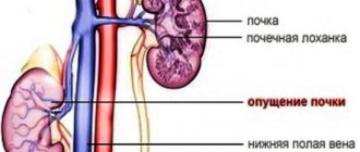
- Visualizes positioning and mobility. In normal condition, the kidneys are located at the level of 1-2 lumbar vertebrae and 12 thoracic vertebrae. The left one may be slightly higher than the right one, but its mobility should not be excessive. Otherwise, nephropathosis can be assumed, which will require treatment.
- Study the size and shape of a paired organ. The bud is bean-shaped, concave on the inside and convex on the outside. The surface is permeated with arteries and vessels. If it is increased, there is a possibility of stagnation or inflammatory disease. A small kidney is characteristic of chronic diseases and dystrophy.
Standard indicators for an adult:
Thickness – From 4 to 5 cm
Length – from 10 to 12 cm
Width – from 5 to 6 cm
- The structure of organ tissue should be homogeneous, the thickness of the parenchyma is normally from 1.5 to 2.5 cm. It becomes thinner with age and reaches 1.1 cm at 60-65 years.
- There should be no stones or sand visible in the renal pelvis (cavities). If during ultrasound diagnostics the doctor discovers stones, then his task is to measure them, because they can cause blockage of the ureter when moving.
Ultrasound of a newborn at 1 month: normal brain parameters
An ultrasound of the newborn’s brain, the decoding (normal) of which is carried out only by a doctor, is also done based on the following indications:
- Labor that was too fast or too slow;
- The weight of the born child is less than 2 kg 800 g;
- Penetration of infections into the mother's womb during pregnancy;
- The birth of a baby before the thirty-sixth week of pregnancy;
- Lack of crying at the moment the baby was born;
- Birth trauma and stay in intensive care after it;
- Brain hernia in a newborn;
- The presence of brain pathology during a mandatory ultrasound scan during pregnancy;
- Caesarean section operation;
- Paralysis, strabismus and paresis.
Ultrasound of the head in newborns: the norm and deviations from the results are as follows:
The ventricles should be cavities containing cerebrospinal fluid. Goals and indications necessary for the study. If the ventricle is dilated, it may indicate hydrocephalus, which means a buildup of cerebrospinal fluid in the skull.
Get a free doctor's consultation
Ultrasound for children: norms
Ultrasound for children: norms. What are they? What can an ultrasound detect? First of all, it should be noted that these figures may differ depending on the age of the child and some related factors. However, it is worth familiarizing yourself with the norms of ultrasound diagnostics of children for a deeper understanding and interpretation of the results of the ultrasound diagnostics obtained.
Norms of cardiac ultrasound in children
Heart ultrasound in children is designed to determine the size of the heart chambers, their condition and integrity. The thickness of the atria, ventricles and the condition of their walls are also studied. The study can also detect a large number of heart diseases.
Ultrasound (sizes) in children are normally as follows (for newborns):
| Left ventricle (side) thickness | 4.5 mm; |
| Pancreas side thickness | 3.3 mm |
| The septum (length) between the ventricles | from 3 to 9 mm |
| LV ejection fraction | >from 66 to 76 percent |
| Heart rate | 120-140 beats per minute |
For a one-year-old child, the norms will be as follows: diameter (at relaxation) of the left ventricle – 20-32 mm; diameter (with contraction) of the LV – 12-22 mm; the posterior side of the LV in thickness is 3-6 mm; aorta diameter – 10-17 mm; left atrium diameter – 14-24 mm; the middle wall of the pancreas in thickness is 1-4 mm; The diameter of the pancreas is 3-14 mm.
The same data for a child from six to ten years old will be as follows: the diameter of the left ventricle at relaxation is 29-44 mm; LV diameter during contraction – 15-29 mm; The posterior side of the LV is 4-8 mm thick; aorta diameter – 13-26 mm; left atrium diameter – 16-31 mm; the thickness of the median wall of the pancreas is the same as in the previous case; The diameter of the pancreas is 5-16 mm.
RV – right ventricle of the heart, LV – left ventricle.
Norms of kidney size according to ultrasound in children
In what cases can a doctor prescribe an ultrasound scan of the kidneys for a child? These are swelling, pain in the abdomen and lower back, problems with urination, colic, test results that deviate from the norm. Diagnostics is also indicated for injuries. Preventive examination should be carried out in children aged one and a half months.
If the contours of the kidneys are smooth and the fibrous capsule is clearly visible, we can speak of normality.
The size of the kidneys is directly dependent on the age and height of the child being diagnosed. If the height is no more than fifty to eighty centimeters, the length and width must be measured. No preparation is required for this.
An ultrasound of the kidneys in children (normal) shows that the kidneys on the left and right differ in size. The length of the kidney on the left should be between 4.8 and 6.2 cm, on the right - from 4.5 to 5.9 cm. The left kidney (wide part) should be between 2.2 and 2.5 cm, the right - from 2.2 to 2.4 cm.
The full parenchymal renal thickness on the left is 0.9 - 1.8 cm, and for the kidney on the right - 1 - 1.7 cm.
If the diagnosis mentions intestinal pneumatosis, this indicates that the child has increased gas formation and special preparation for the study was not carried out.
Norms of liver size according to ultrasound in children
Diagnosis of the liver by ultrasound in childhood is indicated in the following cases: hepatitis, yellowing of the skin and whites of the eyes, pain on palpation, as well as pain in the right hypochondrium.
“Ultrasound of the liver (normal in children): table” - this method helps to quickly find the necessary parameters and compare them with the existing results. Below are normal indicators for the main age groups of children. What can an ultrasound detect?
| For a one year old child | norms of the right hepatic lobe – 60 mm, on the left – 33 mm; normal portal vein – from 2.91 to 5.7 mm |
| For a child of five years old | liver lobe on the right – 84 mm, left – 41 mm; portal vein – from 4.3 to 7.6 mm |
| For a nine year old child | the norms of the right lobe will be 100 mm, the size on the left will be 47 mm; Norms for a fifteen year old teenager: right hepatic lobe – 100 mm, left – 50 mm. |
Thyroid gland (children): normal ultrasound
The thyroid gland can influence many processes in the body, and for a child its normal functioning is especially important, since thyroid hormones are responsible for normal physical development, growth, metabolism and the functioning of the nervous system.
From birth until the age of two years, the volume of the organ should not be more than 0.84 ml. Before reaching six years of age, growth up to 2.9 ml is considered normal. During puberty, the thyroid gland begins to grow very actively. In boys, by the age of fifteen, the volume of the organ reaches 8.1 - 11.1 ml. As for girls at this age, the volume will be 9 - 12.4 ml.
Ultrasound of the thyroid gland is extremely necessary, since if pathology is detected in childhood, there is a favorable prognosis for treatment. How is it done?
Norms of the spleen according to ultrasound in children
Ultrasound diagnosis of the spleen in childhood is most often prescribed if there is a suspicion of enlargement of this organ.
As for the standards for ultrasound diagnostics, it should be noted that a lot depends on how the ultrasound is performed and you should not forget that in a newborn the size of the spleen is extremely small: 4.5 cm in length and 2 cm in thickness.
If the spleen of a one-year-old child is examined, its length is slightly more than 5 cm and its thickness is 2.5 cm. As for a five-year-old child. The length of his spleen within the normal range should be 7.5 cm, and its thickness should be 3.5 cm.
For a ten-year-old child, the length will increase to 10.5 cm and the thickness to 5 cm.
Ultrasound of the abdominal cavity in children (normal)
The main indications for performing an ultrasound of the peritoneum in children are:
- Painful sensations in any part of the peritoneum;
- Constipation and gas;
- Repulsive odor from the mouth and belching;
- Jaundice;
- Internal organ injuries.
This ultrasound includes diagnostics of the liver, kidneys, children's spleen, pancreas and gall bladder. The parameters of the first three organs were discussed above.
As for the norms of the pancreas, they will be as follows for a newborn: 10 by 14 mm (head of the gland), 1 by 1.4 cm (tail of the gland). 0.6 by 0.8 cm (body).
For a child from one to five years: 1.7 by 2 cm (head part), 1.8-2.2 cm (tail part), 1 by 1.3 cm (body). Interpretation of an adult prostate ultrasound will be completely different.
The normal dimensions of the gallbladder in children are as follows: from two to five years: the long part is about 5.1 cm, the wide part is 1.7 cm; for a child from 9 to 11 years old: long part 6.4 cm, wide part 2.3 cm. It is also convenient to use tables for independent interpretation of ultrasound results.
Source: https://ultra-sonographi.ru/normyi-uzi-detyam.html
Ultrasound of the hip joints in a newborn
Ultrasound of the hip joints of newborns: normal angles and other indicators can only be fully deciphered by a doctor, but it is also useful for parents to know this information in order to understand what is happening with the child’s body.
The main objective of this examination is to detect dysplasia. This pathology is a situation where the development of joints does not proceed correctly. Ultrasound is indicated in such cases as:
- Breech presentation of the baby;
- Infections and poor nutrition of the mother during pregnancy;
- Oligohydramnios and toxicosis during pregnancy;
- Constant exposure to bad environmental conditions on the mother.
Based on these and other data, which are verified in accordance with the tables, the existing type of dysplasia in the newborn is identified.
Symptoms of hyperechogenicity
Syndrome of hyperechoic renal pyramids causes pain in the lower back of a cutting, stabbing nature
Hyperechoic renal pyramid syndrome has a number of symptoms:
- Body temperature changes;
- Pain in the lower back of a cutting, stabbing nature;
- Changes in the color and smell of urine, sometimes blood droplets are observed;
- Abnormal stool;
- Nausea, vomiting.
The syndrome and symptoms indicate a clear kidney disease that needs to be treated. The release of pyramids can be caused by various organ diseases: nephritis, nephrosis, neoplasms and tumors. Additional diagnostics, examination by a doctor and laboratory tests are required to establish the underlying disease. After which the specialist prescribes therapeutic treatment measures.
Ultrasound of the newborn's kidneys: normal
Ultrasound examination of the kidneys in a newborn is extremely necessary, since today about five percent of children are born with pathologies of the kidneys and urinary system. In addition, such a study is included in the list of required examinations of the newborn and the timing of this procedure.
As for the size of the kidneys in a newborn, they will be as follows:
- The width of the right kidney is from 14 to 29 mm, its length is from 37 to 59 mm, and its thickness is from 16 to 27 mm;
- The width of the kidney on the left will be from 14 to 27 mm, the length of the kidney on the left will be from 36 to 60 mm, and the thickness from 14 to 27 mm.
The right kidney is located lower than the left in the newborn due to its location under the liver. The outline of the kidneys in newborns may be uneven and slightly lumpy because the structure of the kidneys is not yet complete.
The renal parenchyma must differentiate into cortical and medulla layers. It is not possible to examine the pelvis using ultrasound diagnostics. The diameter of the cups and the thickness of the pelvis should not exceed the norm.
In any case, the ultrasound examination should be interpreted by a qualified specialist, who can be asked any disturbing questions about abnormalities.
What are the performance standards for children?
The procedure for diagnosing kidney ultrasound in children is technically carried out in the same way as in adults. Children are placed on their backs on the couch, while the abdominal area to the pubis and the lateral surfaces of the body are opened for the sensor. The gel is a little cool and if the child understands the words, then you can explain to him that this will not last long and then the gel will be wiped off with a napkin. Also, a child from 1.5 years old can already turn around, stand up and breathe.
If ultrasound diagnostics is performed on a very young child who does not yet understand words, then he should be distracted.
The paired organ is examined for a full bladder. Making a full bladder for children is not easy, but doctors recommend an algorithm: give a newborn and a child under 1 year 100 ml of liquid (water, juice) in 20 minutes, and over two years the volume of liquid should be based on his weight, 10 ml per 1 kg of weight .
Before giving your baby something to drink, let him urinate!
To prevent gases from interfering with a normal ultrasound examination, you should not feed your child fruits, raw vegetables, bread and dairy products a day before the procedure.
It is normal for children to do kidney ultrasounds separately, left and right, since the organs are still developing. They also pay attention to the baby’s gender, age, height and weight. Results in girls may differ from kidney sizes in boys.
By the time the baby is born, his kidneys are not yet fully formed! Only closer to six months, the surface of the baby’s kidneys acquires normal size and shape.
When parents receive a kidney ultrasound report, the interpretation should be carried out by a specialist in the field of pediatric urology or a nephropathologist with experience working with children.
| age | index | Left kidney size, mm | Right kidney size, mm |
| First 2 months of life | thickness length width | 13,6-30,2 40-71 15,9-31 | 18-29,5 39-68,9 15,9-31,5 |
| 3-6 months | thickness length width | 19-30,6 47-72 17,2-31 | 19,1-30,3 45,6-70 18,2-31,8 |
| 1-3 years | thickness length width | 21,2-34 55,6-84,8 19,2-36,4 | 20,4-31,6 54,7-82,3 20,9-35,3 |
| Up to 7 years | thickness length width | 21,4-42,6 67-99,4 23,5-40,7 | 23,7-38,5 66,3-95,5 26,2-41 |
Encyclopedia of Ultrasound and MRI
Currently, ultrasound of the kidneys in children is the most frequently prescribed instrumental method for diagnosing the pathology of this area. It has many advantages, including non-invasiveness, painlessness, speed, fairly high accuracy, and relatively low cost. This method has wide application not only in the presence of symptoms of kidney diseases, but also as the first method for detecting congenital anomalies and kidney diseases in a child.
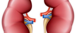
Different sizes of kidneys in a child
The position of the kidneys in children is slightly asymmetrical. The right one is slightly lower than the left one by standard. Over time, their position changes. The “renal pedicle” is initially long in the baby. All vessels are located obliquely, and this is the norm.
In modern medical institutions there are special types of diagnostics, which are based on determining the renal dimensions. They reveal changes in size, as well as symmetry of children's organs. At the same time, the doctor examines the medical history, palpates the organs, and clarifies the symptoms. In some cases, changes can be detected by touch.
Indications
Kidney ultrasound in children can be performed on children of any age, starting from the first days of life. The most common indications for performing an ultrasound examination of the kidneys are:
In addition, ultrasound is performed as a screening examination in newborns at 1-2 months, if the parents have kidney anomalies or the child himself has congenital defects of other systems. If during pregnancy any abnormalities in the structure of the kidneys or hydronephrosis were discovered in the fetus, then an ultrasound scan is performed for such a child immediately after discharge from the maternity hospital or inside it.
Parameters and indicators studied
- Quantity. A healthy person has two kidneys. There are cases when one is removed surgically due to certain reasons. Anomalies in the number of these organs are possible: complete absence or doubling.
- Dimensional data. Using ultrasound, the length, width and thickness of the organ are measured. Kidney size varies depending on a person's age, weight, and height.
- Localization. The retroperitoneal location of the organs is normal. The right kidney (D) is located just below the left (L). The location of the right kidney is considered normal at the level of the 12th thoracic vertebrae and the 2nd lumbar vertebrae, and the left kidney - at the level of the 11th thoracic and 1st lumbar vertebrae.
- Shape and contours. The bean-shaped shape is considered normal. The structure of the tissue is normal - homogeneous with smooth contours.
- The structure of the renal parenchyma, that is, the tissue that fills the organ. In a healthy person, its thickness ranges from 14 to 26 mm. With age, the parenchyma becomes thinner, and for older people the norm for this indicator is 10-11 mm. An increase in this parameter indicates inflammation or swelling of the organ, a decrease indicates dystrophic changes.
- State of blood flow. When analyzing renal blood flow, a color image is used on the monitor of an ultrasound machine. Dark tones indicate that the patient's blood flow is normal (50-150 cm/sec). Bright spots indicate increased renal blood flow.
The adrenal gland of an adult weighs at least 12 g, length – 40–60 mm, width – up to 30 mm, thickness – 4–7 mm. Some people are born with only one adrenal gland. The adrenal gland of a newborn weighs a maximum of 7 g, and this is almost twice as much as the mass of the organ of a one-year-old child.
This is explained by the fact that the mass of the organ has decreased due to the thinning of the cortex, which is in the process of restructuring. By the age of five, the mass of the adrenal glands returns to the initial value, after which it gradually increases. The cortex of the organ is formed by the age of 12.
By the age of 20, the weight of the adrenal gland becomes greater, the maximum size indicators are reached - up to 13 g. In the future, neither the size nor the mass of the adrenal tissue undergoes changes. Women's adrenal glands are slightly larger in size compared to men's. During pregnancy, the adrenal gland enlarges by 2 g.
In the eighth decade, there is a decrease in the mass and size of the organ.
The adrenal glands are located asymmetrically: the left one is slightly behind the right in size and weight
What pathologies can be detected during an ultrasound examination of the kidneys?
An ultrasound of the kidneys can give the doctor a lot of information about the organ, its structure and, indirectly, about its functioning.
Using echography, you can identify congenital kidney defects:
- absence,
- doubling,
- dystopia (abnormal location),
- shape anomalies,
- size,
- abnormalities in the structure and location of the renal vessels,
- congenital disorders of the organ structure (polycystic disease, tubulopathies, hypoplasia, embryonal tumors).
Ultrasound helps in diagnosis:
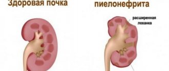
- inflammatory kidney diseases (pyelonephritis, glomerulonephritis),
- abscess,
- injuries,
- volumetric formations,
- urolithiasis,
- kidney damage due to diabetes mellitus,
- nephrosclerosis,
- obstructive lesions of the urinary system (hydronephrosis).
As a rule, nephroptosis (prolapse) or increased mobility of the kidneys in children is also first detected during an ultrasound examination.
What diseases does ultrasound detect?
Ultrasound examination allows you to accurately determine the following kidney diseases:
- Kidney size in an adult: table, norm in children, asymmetry
- nephroptosis;
- reduction of ureters;
- neoplasms;
- pyelonephritis;
- glomerulonephritis;
- hydronephrosis;
- kidney dystrophy;
- inflammatory processes in blood vessels;
- abscess;
- diverticula;
- the presence of air bubbles in the collecting system;
- amyloidosis;
- vascular dysfunction;
To accurately determine the diagnosis, it is necessary to properly prepare for an ultrasound. To do this, you need to follow a special diet for several days and not eat anything for about 10-14 hours. Increased gas formation (flatulence) may distort the results.
The doctor who conducts the ultrasound examination does not make a diagnosis, he records all the indicators. A urologist or nephrologist uses them to determine the presence or absence of diseases. During diagnostics, ultrasound detects 96% of tumors in the kidneys.
How can I prepare my child for a kidney test?
An ultrasound of the kidneys of a small child can be performed without any special preparation. If the examination is scheduled for a teenager, then 2-3 days before the visit to the doctor, foods that cause bloating in the intestines should be excluded from his diet. On the morning of the test or the day before, you should have a bowel movement.
Carrying out an ultrasound scan of a child's kidneys
In some cases, as prescribed by a doctor, a kidney examination is performed against the background of a full bladder, and then after urination. For this purpose, the child should not be forced to drink huge amounts of liquid. Usually the bladder fills well on its own 1.5 - 2.0 hours after breakfast with tea or compote.
Infants are examined 15-20 minutes after feeding.
In emergency situations where immediate visualization of a full urinary tract is needed, the child's bladder is filled in a medical facility using a catheter with sterile saline.
Preparing a child for ultrasound diagnostics
For the most reliable result of this diagnosis, it is necessary to prepare the child for the study:
- children under 2 years of age need to drink no more than 100 ml of clean water;
- up to 7 years of age - the amount of liquid is not more than 300 ml;
- Preparation for a kidney ultrasound involves drinking up to 500 ml of a non-carbonated drink from the age of 10 to the age of 15;
- If there is gas formation in the child’s stomach, be sure to use a carminative.
Water in the child’s body will straighten the walls of the bladder and allow ultrasonic waves to penetrate as much as possible into the organs of the urinary system and kidneys.
Gas formation in the body distorts the reliable result of the study, so preparation for an ultrasound of the kidneys in this condition involves taking medications with a carminative effect.
It is advisable to perform an ultrasound in the first half of the day and not on a stomach full of food.
Normal values
In normal condition, a child should have two kidneys, located on both sides of the spinal column at the level of the XI-XII rib - I-III lumbar vertebrae, depending on age. In newborns and infants, they are located lower, since the spine is shorter than in older children.
In newborns, the kidney continues to rise from the small pelvis and by the age of 2 its upper pole reaches the level of the first lumbar vertebra. The right one is normally located slightly lower than the left one, since it is located under the liver.
The left kidney is usually larger than the right. The permissible difference between them is within 1 cm. In newborn full-term children, the length of the kidney is on average 4.5 cm. By 1 year it reaches 6.2 cm. Then the kidney grows evenly and normally adds about 3 mm every year. Normal kidney sizes are determined using special tables in accordance with the age or height of the child.
The contour depends on the age of the child. In newborns and infants, it is clear, but may be uneven (lumpy), which is associated with the lobulation of the kidney due to its incomplete structure. In younger and older children it becomes even.
When scanning longitudinally, the kidney has an oval shape. There may be a local bulge in the area of the lateral contour – the so-called “humpbacked kidney” or in the area of the medial contour – the so-called “pseudotumor” (with a normal echostructure of the kidney). On a cross section, the shape of the bud is round.
Normally, a child should have a clear differentiation of the renal parenchyma into the cortical and medulla layers. The echogenicity of the kidney parenchyma in children after 6 months is slightly lower or comparable to the parenchyma of a healthy liver - these are indicators of healthy kidneys.
Usually the pelvis is not visualized. If they are visible and located intrarenally, then their thickness should not exceed 3 mm in children under 5 years of age, 5 mm in children under 10 years of age, and up to 7 mm in adolescents. If the pelvis is located extrarenal, then its thickness should not exceed 6 mm in children under 5 years of age, 10 mm in a child from 5 to 10 years of age, and 14 mm in older children.
The diameter of the cups, if visible, should not be greater than the thickness of the pelvis at the appropriate age.
Kidney sizes according to ultrasound in children: table with standards
The kidneys are an organ of the urinary system, which is a kind of filter that rids the body of harmful substances. They are responsible for regulating fluid balance and also influence homeostasis.
And if a child begins to complain of pain in the lumbar region or problems with urination, this may indicate the presence of a pathology of the urinary system.
To confirm or refute the presence of a problem, an ultrasound examination of the kidneys is prescribed. The procedure is absolutely safe and allows you to see a complete picture of the condition of the organ.
When the kidney ultrasound has been done, the doctor interprets the results of the study. But it is possible to do this yourself, relying on a table with standards, which indicates the normal size of the kidneys according to ultrasound in children.
When and to whom is it prescribed?
Kidney ultrasound is prescribed at any age if indicators are available. Possible indications for testing in newborns are:
- difficult pregnancy, childbirth;
- the presence of congenital diseases of the genitourinary system in parents;
- detection of stones in urine;
- excess bilirubin in the blood;
- the presence of deviations in the development of external organs;
- dysbacteriosis;
- increased blood pressure;
- swelling of the limbs.
Kidney ultrasound is prescribed at any age if indicators are available
Indications for ultrasound diagnostics in children over 1 year of age are the presence of the following symptoms:
- pain in the lumbar region;
- painful urination;
- change in urine color, hematuria;
- headaches, migraines;
- renal colic;
- discomfort after injuries to the lower back, abdomen;
- causeless high body temperature;
- increased concentration of salt in urine;
- neoplasms diagnosed by touch.
It is necessary to conduct a study if there is a suspicion of kidney inflammation, disorders of the endocrinological system, or diseases of the cardiovascular system have been detected.
An ultrasound should be done when preparing the baby for surgery.
How to prepare a child?
For the results of the study to be accurate, the baby must be prepared for the procedure. First of all, he needs to explain that an ultrasound is a painless procedure that does not cause any discomfort, and that there is no need to be afraid.
The child must understand that during the examination you need to listen to the doctor and not be mischievous. 8 hours before the procedure, the child must refuse to eat, and 3 days before the session it is necessary to follow a special diet aimed at reducing the formation of gases in the intestines, improving the visibility of the cavity.
Ultrasound is a painless procedure that does not cause any discomfort to the child.
You need to adhere to the principles of the diet:
- stop consuming legumes, cabbage, dairy products and carbonated drinks;
- limit the consumption of sweet, fried, fatty foods;
- after meals, drink activated carbon at the rate of 1 tablet per 10 kg of weight;
- If you have flatulence, you need to take an Espumisan tablet 2-3 hours before the session.
To ensure that organs are clearly visible during an ultrasound, you must come to the procedure with a full bladder. To do this, half an hour before the start of the study, you should drink a certain amount of water equal to the volume of your bladder.
For children under 2 years old, the amount of liquid drunk should be no more than 100 ml, for children 2-7 years old - 200 ml, at the age of 7-11 years old you need to drink 400 ml, and at an older age - at least 0.5 liters of water. Before going to the hospital, it is recommended to purchase a diaper and wipes.
Rules for conducting a pediatric ultrasound
An ultrasound of the kidneys is performed by a sonologist in a specially equipped room, in the presence of parents. The child lies down on the couch, freeing the lower back and stomach from clothing. If the baby is a newborn, then during the study he is in the arms of his mother. Sometimes the procedure is performed in a standing position.
After the patient is positioned on the couch, the doctor applies a special gel to the area being examined, which is absolutely harmless and can be easily removed from the skin with a napkin.
The specialist then moves a sensor over the body, observing a three-dimensional image on the screen. For a better overview of the organ, the organ is examined while moving in different planes, for which the doctor may ask the child to turn around, stand up, or sit down. The baby must follow all the sonologist’s instructions. The duration of the procedure is 5-30 minutes.
At the end of the session, parents are given ultrasound results, which are interpreted by a pediatric urologist or nephrologist. If it is not possible to see a doctor as soon as possible, you can decipher the ultrasound results using the table of standards.
What does a kidney ultrasound show?
Ultrasound diagnostics of the kidneys in a child allows you to assess the number and size of organs, their location and shape, and shows the parameters, structure, and condition of the tissues. This study allows you to detect pathology of the kidneys and organs of the urinary system.
A doctor can decipher an ultrasound of the kidneys
During an ultrasound, the doctor pays attention to the following points:
- Number of kidneys. There will be 2 of them: on the right and on the left. Sometimes the position of the kidneys changes, as a result of which they may be located one under the other or descend into the pelvic area.
- Organ performance. Both kidneys must function equally.
- Form. A healthy organ has the shape of a bean grain. Due to abnormal development, the kidney can take the shape of the letter S or L, the appearance of a horseshoe. Sometimes the buds grow together.
- Mobility. In healthy kidneys, it is no more than 1.5 cm. If the organ descends or begins to wander, then mobility increases.
- Presence of sand, stones, stagnation in the cavity. In a normal state, none of this should happen.
In addition, during an ultrasound, attention is paid to the size of the organ, the thickness of the walls, the condition of the structural tissues, as well as some other important parameters that make it possible to diagnose the presence of abnormalities.
Decoding and standards
A pediatric urologist or nephrologist interprets the results of kidney ultrasound in children, in strict accordance with the normal table. This table with standards includes features and possible deviations.
For each age, the norm varies. Ideally, the contours of the organ should be smooth, with a clearly visible fibrous capsule. The dimensions of the pelvis should not exceed 6 mm. The normal rate of renal blood flow is 50-150 cm/sec.
When identifying pathological conditions, the size of the kidneys is of great importance, which varies depending on the age and height of the child.
In a newborn baby, until he is 2 months old, the kidney size is 49 mm, and the renal pelvis is 6 mm. From the age of three months until the baby reaches 1 year of age, the organ increases by 13 mm and is 62 mm.
Until a child reaches 12 years of age, the size of the kidney increases by approximately 13 mm every 5 years. The weight of the organ in a newborn baby is 12 grams, by the age of 5 it is 54-56 grams, and by the age of 18 the weight of the organ is 120 grams.
The exact indicators of the length, width and thickness of the left and right kidneys can be determined using a special table, taking into account the height and age of the baby.
What diseases can be detected?
Based on the results of ultrasound diagnostics of the child’s kidneys, the presence of pathologies can be determined:
- pyelonephritis;
- urolithiasis disease;
- nephrosclerosis;
- hydronephrosis;
- nephroptosis;
- congenital anomalies;
- narrowing of the ureters;
- cystosis;
- glomerulonephritis;
- tumor neoplasms;
- acute cystitis;
- circulatory disorders;
- abscess.
Kidney ultrasound allows you to determine the presence of any structural changes in organs, inflammation or any other deviation from the norm.
You can make an approximate interpretation of the diagnostic results yourself, but an experienced specialist with knowledge of terminology can make an accurate diagnosis and prescribe treatment.
Source: https://urologia.expert/pochki-i-mochetochniki/razmery-pochek-v-norme-po-uzi-u-detej
Features of the results in a newborn
The perception of kidney ultrasound results in newborns has its own characteristics, because they retain the features of an incomplete kidney structure.
In newborn children, the kidneys are located lower than in older children, and almost parallel to the spine, and then gradually rise higher to the diaphragm and come together at the upper poles.
The outline of the kidneys in newborns is usually lumpy because they have a lobular structure. It can last up to 2 years, and according to some sources, up to 5 years.
In children under 6 months of age, the echogenicity of the cortical layer of the parenchyma is higher than in older children, and exceeds the echogenicity of the liver and spleen.
In a newborn, the echographic sign of “white pyramids” may be detected, manifested by the hyperechogenicity of several pyramids in the renal parenchyma. It is defined as the norm for up to 1-2 months. The collecting system on echograms, if it is not expanded, is not visible in newborns and children under 6 months of life
Sizes of the pelvis on ultrasound
The ureter, minor and major calyces are normally not visible on ultrasound. There are three types of location of the pelvis: intra-, extrarenal and mixed type. With an intrarenal structure, the lumen of the pelvis at an early age is up to 3 mm, at 4-5 years - up to 5 mm, in puberty and in adults - up to 7 mm. For extrarenal and mixed types of structure - 6, 10 and 14 mm, respectively. When the bladder is full, the pelvis can increase to 18 mm, but 30 minutes after urination it contracts.
Drawing. Regardless of the filling of the bladder, ultrasound shows a pelvis of mixed (1) and extrarenal (2) location, as well as under the fibrous bridge (3).
Take care of yourself, Your Diagnosticer!
Pathologies detected by ultrasound examination
Ultrasound can reveal signs of many kidney abnormalities and diseases in children. We will briefly consider the typical echographic signs of the most common detected pathologies and the terms used in this case, which the ultrasound doctor can add to the conclusion. In children, various developmental options may occur: an additional kidney, an intrarenal septum, a hypertrophied renal column or Bertin's column. All of them are anatomical features of a particular child and do not pose any danger to his health.
Extra kidney
An additional kidney is defined as a formation similar in shape and structure to a normal kidney, but significantly smaller in size. In this case, the doctor carries out a differential diagnosis with a tumor.
Ultrasound signs of diseases
Ultrasound scanning makes a certain contribution to complex diagnostics:
- acute and chronic pyelonephritis and glomerulonephritis;
- abscess and carbuncle of the kidney;
- tuberculosis;
- anomalies in the development and location of the kidneys;
- urolithiasis;
- chronic renal failure;
- micro- and macrohematuria;
- traumatic kidney damage;
- resistant to all treatment methods for arterial hypertension, etc.
Changes in the size of the kidney, the thickness and structure of the parenchyma, as well as the expansion of the pelvicaliceal region are of great importance for the diagnosis of inflammatory processes occurring in the organ. Thus, with pronounced acute pyelonephritis, ultrasound scans show:
- kidney enlargement;
- sharp limitation of physiological displacement or complete immobility of the organ;
- thickening of the parenchyma (up to 3 cm) and renal pyramids, while the size of the cups remains normal (this is especially noticeable with unilateral lesions);
- diffuse or focal heterogeneity of the parenchyma;
- thickening and layering of the wall of the renal pelvis;
- a halo of irritation around the inflamed kidney caused by swelling of the fatty tissue surrounding the organ.
In acute pyelonephritis, hypoechogenicity, focal and diffuse heterogeneity of the parenchyma, as well as a decrease in the tone of the pyelocaliceal system are noted.
Large foci of heterogeneity of the parenchymal structure are typical for renal carbuncles. In the photographs they appear darker compared to healthy areas of parenchyma.
In this part, the outer contour of the kidney is unnaturally deformed and bulges outward. The carbuncle on sonograms is represented by a hypoechoic (dark) focus with blurred boundaries and contours.
On Doppler scanning, there is no vascular pattern.
In the photo, number 1 indicates a carbuncle, and number 2 indicates unchanged parenchyma.
A rounded dark inclusion with homogeneous contents and uneven thick walls usually represents an abscess. Bubbles of gas or suspension may be visible in its cavity.
Both the kidney carbuncle and its abscess can easily be confused with a cyst, which has similar signs on echograms. In cases of difficulties with making a diagnosis, a puncture of the formation is performed under ultrasound control with further examination of the obtained material.
With apostematous nephritis, ultrasound sonograms show numerous echo-negative areas measuring 2–3 mm, clearly visible on the highly echogenic adjacent parenchyma. They are usually located just below the kidney capsule. The organ is enlarged in volume, has a pattern in the form of nostrils, the renal pyramids are poorly visualized.
The boundaries of the kidney are indistinct and uneven in areas. During respiratory movements, the physiological displacement of the organ is reduced. From abscesses located near the surface of the kidney, infection can spread to its capsule and surrounding tissues. As a result, there may be an echo-negative border around the organ.
On the ultrasound image of apostematous nephritis, arrows indicate anechoic dark zones - apostematous
Unfortunately, there are no specific echographic symptoms of chronic pyelonephritis. Only during an exacerbation of the inflammatory process can a slight increase in the size of the organ and a decrease in the echogenicity of its parenchyma be recorded.
However, with a long-term, protracted course of chronic pyelonephritis, the following features can be seen on ultrasound:
- reduction in the longitudinal size and volume of the kidney;
- thinning of the parenchyma;
- uneven contours of the kidney, formed as a result of scar-sclerotic retractions in places of former foci of inflammation of the parenchyma during exacerbations of the disease;
- increased echo density of the parenchyma in retraction zones;
- blurred edges between the medulla and cortex, poor visibility of the pyramids.
Long-term chronic pyelonephritis is characterized by thinning and diffuse compaction of the parenchyma, uneven contours of the kidney and its reduction up to wrinkling.
In a patient with acute glomerulonephritis, on an echographic image the size of the kidneys is significantly increased, their contours are blurred. The echogenicity of the parenchyma is noticeably increased, as a result of which it appears light. The pyramids against its background clearly appear in the form of low-echoic (dark) spots.
The ultrasound picture of chronic glomerulonephritis in the stage of subsiding inflammation in many patients does not differ from the norm. If the ultrasound was done in the phase of exacerbation of the inflammatory process, then the echographic image of the affected kidney will be similar to that in the acute form of the disease.
The ultrasound picture of chronic glomerulonephritis in the active stage does not differ from that in the acute form of the disease
Often, against the background of long-term, sluggish glomerulonephritis, chronic renal failure (CRF) develops, the ultrasound signs of which are listed in the table below.
Anatomical and functional features of the kidneys in children
Location: lumbar region, on both sides of the spinal column. The functional unit of an organ that ensures its functioning is the nephron. The kidneys are the main organs of the urinary system. Their main functions:
- excretion of water and water-soluble products of nitrogen metabolism (urea);
- maintaining a constant internal environment of the body - acid-base balance, content of potassium and sodium ions;
- regulation of blood pressure using the renin-angiotensin system;
- participation in the processes of metabolism of glucose and amino acids;
- stimulation of the formation of red blood cells in the red bone marrow.
Differences in the structure of the urinary system organs in children
In newborns, the weight of the kidney is approximately 10% of the weight of an adult - 10–12 g. During the infant period (up to 12 months), their weight reaches 37 g, and their size increases by 1.5–2 times.
The main difference in the structure of organs in newborns and children no older than 1 year is the immaturity of the cortical layer, in which the filtering part of the nephron is located - Bowman's capsule, as well as the ascending and descending loops. The thickness of the cortex does not exceed 2 mm, while the medulla is 8 mm. As one matures and grows, the cortical layer increases by approximately 4–5 times.
Externally, the kidneys of infants have a pronounced lobular structure, characteristic of animals. As they grow older, the tuberosity and lobules disappear and in adolescents the organs have a smooth outer surface, just like in adults. The renal pelvis of a newborn is wide and ampulloform.
Growth in weight and size occurs mainly in the first 12 months after birth. From 6 to 9 years, and then in the period of 16–17 years, the cortical layer develops and forms as much as possible, while the medulla stops growing by 13 years.
Features of the location of the kidneys in children
The organs of the urinary system are located significantly lower in children than in adults:
- Left kidney : upper edge – 12th thoracic vertebra, lower edge – 4th lumbar vertebra.
- Right kidney : upper edge – 1st lumbar vertebra, lower edge – 5th lumbar vertebra.
Features of operation
Underdevelopment of the cortical layer is a small area for the primary filtration of blood in the glomeruli of nephrons. Indeed, renal filtration in children is significantly lower than in adults. She reaches the norms characteristic of an adult only by the age of 16–17. Therefore, to remove toxins and waste products, children need much more water than adults.
The immaturity of the nephron loops, in which water is absorbed back into the bloodstream (reabsorption), ions and mineral salts, leads to the fact that the urine of children is low concentrated. With insufficient fluid intake or excessive fluid loss, dehydration occurs very quickly, especially in infants under 12 months of age.
Despite the immaturity of the excretory system, newborns and infants maintain a constant internal environment and blood composition due to breast milk, the composition of which minimally loads the excretory organs. Early introduction of age-inappropriate complementary foods creates excess stress on the urinary system.
Ultrasound of the bladder in children.
It is incredibly informative for identifying pathology or an inflammatory process. How much fluid a child needs to drink before the procedure depends on age:
- Up to 2 years – 100.0.
- Up to 7 years -200.0 – 300.0.
- Up to 11 years – 500 ml.
- Up to 15 years – 700.0 – 800.0.
This examination is carried out in two stages. First they look at him with a full bladder, then they ask him to go to the toilet and look at him again. Ultrasound of the bladder is prescribed in the following cases: suspicion of urolithiasis, assessment of the shape of the bladder, inflammatory diseases, assessment of the amount of residual urine.
How does ultrasound of the bladder occur in children?
The child lies on his back, the abdominal area is freed from clothing, the doctor applies the gel to the lower abdomen, the bladder area, and begins the examination by moving the sensor over the abdomen. What can a doctor find during an examination? Attention is drawn to the shape of the bladder; its asymmetrical shape may indicate neoplasms. A small size indicates chronic cystitis in the acute stages, fibrosis (overgrowth of connective tissue due to inflammation). The bladder may contain pus, blood clots, sand, or suspension. The child is then asked to urinate and the examination continues to determine the volume of residual urine.
Normal bladder volume in children:
- Up to a year 35 – 50.0
- Up to 3 years 50 – 70.0
- Up to 8 years 100 – 200.0
- 8 – 10 years 200 – 300.0
- 10 – 14 years 300 – 450.0
Residual urine after urination is unacceptable and indicates a dysfunction of the bladder. This may be due to a small force of contraction of the bladder walls or, most often, due to the sphincter - the muscle that closes the urinary canal. The presence of residual urine indicates a violation of the emptying of the organ, this is manifested in a frequent urge to urinate, poor urine stream, urinary retention, urinary incontinence and recurrent urinary tract infection. The presence of residual urine is the only sign of vesicoureteral reflux (reverse reflux). In general, for an ultrasound, the bladder must be completely empty, but up to 10% urinary retention is acceptable:
- Newborn 2 -3.0
- Up to year 3 – 5.0
- 1 – 4 years 5 – 7.0
- 4 – 10 years 7 – 10.0
- 10 – 14 years 20.0
- Over 14 years old up to 40.0
If an ultrasound of the bladder in children reveals an excess of residual urine, it means that the child has a problem with urology.
Reasons for increased residual urine volume during ultrasound of the bladder in children.
Problems with nervous regulation, spinal injuries, hypothermia in the pelvic area, weakened tone of the bladder muscles, impaired conductivity of the urinary canal, urolithiasis of the bladder, cystitis, tumors.
To decipher an ultrasound of the kidneys and bladder in children, you need to contact a pediatrician; if there is any pathology in the examination, in this case you will need to consult a specialist doctor - a nephrologist, urologist.
The parameters of the child’s urinary system have significant age-related characteristics. This must be taken into account when interpreting diagnostic results. The article describes the normal size of kidneys in children - table.
Content
Ultrasound examination of the kidneys in children
Normally, ultrasound of the kidneys in children is performed during preventive examinations at the age of 3 months, when performing a comprehensive ultrasound examination of the abdominal organs, as well as during a school examination at 6–7 years. An unscheduled ultrasound is prescribed in the presence of the following complaints:
- prolonged increase in body temperature to 37.3–37.5 0C for no apparent reason;
- pain in the lumbar region;
- febrile fever 38–39 0C, not associated with ARVI or respiratory diseases;
- swelling of the face, which occurs mainly in the morning;
- pain when urinating;
- high blood pressure;
- abnormalities in urine tests (detection of protein, red blood cells, white blood cells, bacteria, mineral salts).
Ultrasound of the kidneys: norm and pathology in children
Ultrasound examination of the urinary system is completely painless, does not require special preparation and is easily tolerated by the child. Moreover, the method is highly informative for diseases of the kidneys and urinary tract.
Table. Kidney sizes in children are normal according to ultrasound
| Child's age | Length, cm | Width, cm | Thickness, cm |
| 1 month | 4,2 | 2,2 | 1,8 |
| 6 months | 5,5 | 3,1 | 1,9 |
| 12 months | 7,0 | 3,7 | 2,6 |
| 6 years | 7,9 | 4,3 | 2,8 |
| 10 years | 9,8 | 5,15 | 3,3 |
| 15 years | 10,7 | 5,3 | 3,5 |
Normal kidney sizes according to ultrasound in children: table
30.10.2018
Ultrasound examination of the kidneys in a child’s body is a painless method for diagnosing the organ for the child, with which it is possible to identify kidney pathologies, as well as diseases at the initial stage of development.
Using an ultrasound of a child’s kidneys, a specialist sees a complete picture of the organ’s condition on the monitor of a diagnostic device. Using the ultrasound diagnostic method, it is possible to examine all the internal organs of a child’s body.
For children, as for adults, the ultrasound procedure is safe and the results of this method are the most reliable.
Indications for the use of ultrasound
Ultrasound diagnostics is used to identify urological diseases of the child’s body. Ultrasound of the kidneys in older children uses this research method for the following symptoms of the body:
- headaches and migraines;
- pain in the lumbar spine;
- sharp pain when urinating;
- after spinal injury;
- after abdominal injury;
- The size of the kidneys in children does not correspond to the norm;
- swelling of the lower extremities;
- swelling on the face;
- increased blood pressure in a child;
- increased body temperature for no obvious reason;
- urine with a pungent and unpleasant odor;
- the color of the urine is cloudy and dark;
- There were blood impurities in the urine.
If, according to the results of a general urine test, deviations from the norm are revealed, in this case, the doctor prescribes preparation for an ultrasound of the kidneys in children.
An ultrasound of the kidneys in a newborn up to the age of two months is carried out to identify pathologies in the development of the organ and with some changes in the child’s body:
- increased blood pressure in a newborn baby;
- swelling of the limbs and face;
- neoplasms in the abdominal cavity;
- high bilirubin as a result of a complete blood count;
- severe dysbacteriosis;
- pregnancy with complications;
- difficult birth process;
- urological diseases in a woman in labor.
Preparing a child for ultrasound diagnostics
For the most reliable result of this diagnosis, it is necessary to prepare the child for the study:
- children under 2 years of age need to drink no more than 100 ml of clean water;
- up to 7 years of age - the amount of liquid is not more than 300 ml;
- Preparation for a kidney ultrasound involves drinking up to 500 ml of a non-carbonated drink from the age of 10 to the age of 15;
- If there is gas formation in the child’s stomach, be sure to use a carminative.
Water in the child’s body will straighten the walls of the bladder and allow ultrasonic waves to penetrate as much as possible into the organs of the urinary system and kidneys.
Gas formation in the body distorts the reliable result of the study, so preparation for an ultrasound of the kidneys in this condition involves taking medications with a carminative effect.
It is advisable to perform an ultrasound in the first half of the day and not on a stomach full of food.
Pediatric ultrasound procedure
The diagnostic procedure for pediatric kidney ultrasound can be performed at a diagnostic center or in an ultrasound room in a clinic. Newborn babies undergo this procedure while lying in their mother’s arms. Older children lie independently on the couch and turn on their side or stomach at the doctor’s request.
Sometimes the ultrasound procedure is performed standing. The part of the body from the pubis to the chest is freed from clothing and a special liquid of viscous consistency is applied to it.
The results of the diagnostic study are compared with standard indicators in accordance with the age of the small patient.
What can be seen on an ultrasound
Based on the results of ultrasound diagnostics, it is possible to identify changes and pathologies in the kidneys and bladder:
- inflammation of the bladder mucosa;
- the presence of stones in the kidneys and bladder;
- narrowing of the urethral canal;
- acute pyelonephritis;
- acute form of cystitis;
- hydronephrosis disease;
- abscess on the kidney and the main organ of the urinary system;
- structural change in the organs being studied;
- renal cystosis;
- kidney tumor;
- disturbances in the circulatory system;
- Gromerulonephritis disease;
- pathology of abnormal kidney sizes.
Timely diagnosis of the kidneys and urinary system and pathologies and diseases in the child’s body identified using ultrasound, enable the attending doctor to prescribe the correct drug treatment, which will increase the chance of a quick recovery for the little patient.
Normal kidney sizes according to ultrasound in children: table:
| Child's age | Bud | Organ width | Organ thickness | Organ length |
| children under 2 months of age | right | 13.6 mm - 29.4 mm | 15.9 mm - 27.4 mm | 36.8 mm - 59.0 mm |
| left | 14.1mm - 26.9mm | 13.6 mm - 27.3 mm | 36.2 - 60.6 mm | |
| boys up to 2 months | right | 15.9 mm - 31.5 mm | 18.0 mm - 29.5 mm | 39.0 mm - 68.9 mm |
| left | 15.9 mm - 31.0 mm | 13.6 mm - 30.2 mm | 40.0 mm - 71.0 mm | |
| girls up to 2 months | right | 16.0 mm - 29.6 mm | 17.7 mm - 29.7 mm | 42.0 mm - 61.3 mm |
| left | 15.8 mm - 29.0 mm | 17.3 mm - 28.1 mm | 40.9 mm - 63.7 mm | |
| children under six months of age | right | 18.2 mm - 31.8 mm | 19.1 mm - 30.3 mm | 45.6 mm - 70.0 mm |
| left | 17.2 mm - 31.0 mm | 19.0 mm - 30.6 mm | 47.0 mm - 72.0 mm | |
| children under 3 years of age | right | 20.9 mm - 35.3 mm | 20.4 mm - 31.6 mm | 54.7 mm - 82.3 mm |
| left | 19.2 mm -36.4 mm | 21.2 mm - 34.0 mm | 55.6 mm - 84.8 mm | |
| children aged 5 to 7 years | right | 26.2 mm - 41.0 mm | 23.7 mm - 38.5 mm | 66.3 mm - 95.5 mm |
| left | 23.5 mm - 40.7 mm | 21.4 mm - 42.6 mm | 67.0 mm - 99.4 mm | |
| children under ten years of age | right | 24.5 mm - 44.9 mm | 23.9 mm - 39.5 mm | 67.7 mm - 103.3 mm |
| left | 26.0 mm - 44.0 mm | 27.0 mm - 41.0 mm | 71.2 mm - 103.6 mm | |
| children under 14 years of age | right | 28.0 mm - 48.7 mm | 25.5 mm - 43.1 mm | 74.4 mm - 113.6 mm |
| left | 27.2 mm - 47.7 mm | 27.0 mm - 46.3 mm | 74.4 mm - 116.0 mm |
After the ultrasound procedure, the urologist, in accordance with the deciphered results, makes a specific diagnosis and prescribes drug treatment, or in the case where drug treatment does not give a positive result, the time for surgery is set.
The principle of treatment of urological diseases
Urological diseases, which are caused by infection in the organs of the urinary system and the kidneys, are treated according to a standard regimen, with individual adjustments to the use of medications and the duration of the medication course itself:
- antibacterial therapy with drugs from the family of macrolites, fluoroquinolones and tetracyclines;
- therapy with the use of local drugs - antipyretic rectal suppositories, the use of antiseptics for douching the urethral canal;
- use of probiotics in treatment to normalize microflora in the digestive system;
- therapy with herbal remedies and healing plants is an effective addition to medication treatment;
- Immunomodulatory therapy allows you to normalize the functioning of the immune system and thereby speed up the process of complete recovery of the urinary system from infection.
Along with treatment, you must adhere to some rules of behavior:
- Avoid constantly wearing diapers - this disrupts the temperature level in the pelvic organs.
- Do not put synthetic underwear on your child - this disrupts air exchange in the genitourinary system.
- Do not overcool the child and promptly change wet underwear and wet shoes.
- Follow the rules of personal intimate hygiene - wash the genital area in the morning and before bed with warm water and baby soap.
- For girls, it is very important to properly wipe the anus - germs from the anus can get into the urethra and vagina.
- Avoid viral and colds - prevention of these diseases gives good results.
- Do not overload a school-age child with various sports sections and clubs - when the child’s body is tired, the immune system decreases.
- Maintain normal functioning of the immune system.
If you follow these basic rules, your child will be healthy and active.
Source: https://wmedik.ru/zabolevaniya/urologiya/razmery-pochek-v-norme-po-uzi-u-detej-tablica.html
Diagnostic value of ultrasound for kidney diseases
In pathological conditions, the sizes of the kidneys in children indicated in the table can change either upward or downward. Let's look at this in more detail.
Inflammatory processes
With inflammation of the renal parenchyma, pyelonephritis develops. Its cause can be either an ascending infection from the urinary tract or an endogenous one (for example, with chronic tonsillitis, frequent colds). At the initial stage of the disease, the size of the organs increases due to tissue swelling.
As the disease progresses, dystrophic changes occur in the nephrons. This leads to the formation of a so-called wrinkled kidney and the development of renal failure. At this stage of the disease, the kidneys are below normal in size.
With autoimmune damage to the kidney tissue, glomerulonephritis develops. In this case, inflammation affects the glomeruli - the main filtering apparatus of the organ. The chronic form of the disease leads to the rapid development of renal failure and a decrease in kidney size.
Developmental anomalies
Congenital kidney malformations are one of the most common developmental anomalies. Ultrasound examination can reveal a decrease in the size of organs - underdevelopment or hypoplasia of the kidneys.
Hydronephrosis
When the outflow of urine is impaired, caused by a narrowing of the ureter, stones in the renal pelvis, or tumors, hydronephrosis develops when the parenchyma of the organ gradually becomes thinner under water pressure. An ultrasound will show an increase in the size of the kidney.
Interpretation of kidney ultrasound results in adults and children
After the patient contacts the doctor, the latter must prescribe an examination. For example, ultrasound of the kidneys, deciphering this type of diagnosis will help establish the correct diagnosis.
Almost every patient who has experienced kidney pain at least once knows how difficult it is to bear. You have to call emergency services or a doctor at home in order to relieve severe pain. Getting rid of pain is the first thing a doctor can do. Next, you need to establish the cause of the disease and prescribe appropriate treatment.
Often the initial appointment is carried out by one specialist, and subsequent treatment is prescribed by another. For example, by contacting a local physician, a patient receives referrals for tests and specific examinations. After this examination has been completed, the patient is given the results and needs to go with them to a specialist.
Let’s say that after undergoing an ultrasound of the kidneys, the patient is given a diagnostician’s report. It is difficult for an inexperienced person to understand what exactly is said there. He can only understand part of the information. However, today there are many ways to interpret ultrasound results. There is a special table that lists all the normal indicators, and based on this information one can draw a conclusion about the condition of the organs.
Changing organ size
As you know, a healthy person has a paired organ in the body from birth - the kidneys. During the course of its life, every living organism may experience health problems, which ultimately leads to surgical intervention. As a result of the operation, one of the kidneys may be removed. And the opposite situations may arise when partial or complete doubling of the kidneys occurs.
Therefore, if during an ultrasound the doctor says: “The number of kidneys is two,” there is no need to move your eyebrows, the diagnostician is doing his job competently.
The size of the organ matters. The size of the kidney in adults, in a healthy body without pathological conditions, is normally 10-12 cm in length and 5-6 cm in width. Diagnosticians are guided by these indicators. The size of an adult's kidneys in thickness can vary between 4-5 cm. A change in size according to one of the above parameters is allowed by a maximum of 1 cm. The size of the kidneys in children differs from adults and depends on the age of the subject. There is a special table that shows the exact size of the kidney according to age scale.
If the decrease or increase in the kidney is recorded in a larger range, then this may be a cause for concern.
For example, a decrease in this organ often indicates a possible chronic disease. But an increase may indicate a tumor or acute nephritis.
Therefore, the size of this organ is very important at the time of diagnosing any disease.
Position of the kidneys in the body
Many people think that a person's kidneys are located at waist level. Although in reality, the lower level of this organ does not fall below the first or second lumbar vertebra. In this case, the right kidney is located slightly lower than the left. The reason is the fact that it is slightly pushed aside by the liver. As a rule, an experienced ultrasound doctor immediately notices any deviations in this indicator.
If an ultrasound shows that the kidneys are lowered too low, this may indicate that the patient is developing some kind of pathology. The diagnostician records this sign with the entry “Right- or left-sided nephroptosis.”
This could be due to several factors, for example:
- Decreased intra-abdominal pressure.
- Weakness of the ligamentous apparatus of the kidney.
- Previous infections that led to a sharp decrease in body weight. As a result, the thickness of the fat capsule decreased.
- An injury that was caused to the abdomen or lower back.
Moreover, women can develop this syndrome 15 times more often than men. The nephrologist knows that there is a table that sets out the norm for this indicator; he can be guided by these data and compare them with the results of the study.
This indicator is very important when diagnosing kidney condition. An experienced nephrologist immediately pays attention to him.
What does parenchyma thickness mean?
The kidney has continuous tissue. It is called parenchyma. This tissue includes microscopic formations called nephrons. They perform a filtering role in this organ. The parenchyma is located on the surface of the organ. Inside the kidney there are cavities that pass into the pelvis, which eventually narrows and continues in the ureter.
The thickness of the parenchyma in an adult healthy person is on average from 1.5 to 2.5 cm, this is the norm. When the results of an ultrasound examination showed that this tissue is increased in volume, this may indicate the presence of some kind of inflammatory process in the organ. For example, swelling may occur. If the thickness of the tissue has decreased, this is a sign of degenerative processes, which should also cause concern to the doctor.
Sometimes a decrease in the thickness of the parenchyma occurs due to chronic pyelonephritis. Another reason for this may be the presence of diseases such as diabetes, hypertension, etc.
Our readers successfully use Renon Duo to treat kidneys. Seeing how popular this product is, we decided to bring it to your attention. Read more here...
Often this indicator is recorded in older people - after 60 years. But in any case, all the results that were recorded on an ultrasound examination must be analyzed by a specialized doctor, and only after that a final diagnosis is made.
Pathological manifestations
An ultrasound of the kidneys can tell us about another pathology that is possible in the body of any person. In conclusion, it is described as increased echogenicity or, conversely, decreased. Both indicate the presence of complications. What exactly these pathologies are, the attending physician must say.
In addition, there may be cysts in the parenchyma (these are bubbles with liquid). To prescribe a course of treatment, the doctor must assess their size. When conducting diagnostics, several kidney ultrasound results are taken into account. This is necessary in order to identify the dynamics of changes in the cyst. If it does not increase and its size is not very significant, then they are not treated. If there is an increase in formations in the kidneys, then treatment should be started immediately. These cysts should cause concern, as they can contribute to the development of a malignant tumor. Determining the origin of this pathology and its detailed analysis help to find the correct answer when examining the organ and prescribing a course of adequate treatment.
Typically, the diagnostician in conclusion describes the tumor as a round formation, while the formation can have not only a round shape. Additionally, they describe how clear or unclear the contours of a given formation are, and what is inside, if this can be determined by ultrasound.
Changes in the pelvis
Changes in the pelvis can be presented in several forms. For example, a thickened mucous membrane, as well as the presence of stones or sand.