Fat in urine, causes and symptoms of spots in a child
The kidneys produce urine to remove waste products from the body from the blood. Medical studies have proven that as a result of the formation of any disease in the body, the quality of urine changes. For example, fat in the urine may indicate the presence of various pathologies.
Qualitative changes in urine
Kidneys are an important organ in the human body. With their help, metabolic products are removed. A healthy person's urine is clear. As a result of the presence of bacteria, pus, and blood impurities, cloudiness is observed.
Changes in urine quality indicators indicate the development of diseases:
- Proteinuria. This condition means the presence of protein in urine. Proteinuria usually indicates nephrosis or pyelonephritis.
- Pyuria. It means the presence of pus in the urine and often indicates the occurrence of an inflammatory process in any urinary organ.
- Hematuria. Blood impurities in urine are noticeable even during external examination. Observed in cases of cancer, tuberculosis, endometriosis, bladder neck pathologies, polycystic kidney disease, urolithiasis, pyelonephritis, bladder ulcer and other pathologies.
- Myoglobinuria. This condition is characterized by a brown coloration of urine and means the release of myoglobin in the urine. It is observed in injuries accompanied by severe muscle damage.
- Cylindruria. This change is caused by the release of cylinders of urine. It is detected with nephrosis or nephritis.
- Bacteriuria. The release of bacteria in urine indicates the development of an inflammatory process in the genitourinary organs.
- Pneumaturia. This change means the release of air or gas with urine. Evidence of intestinal and paraintestinal flora. Usually indicates pyelonephritis or diabetes mellitus.
- Lipuria. Fatty spots in the urine are often observed with fat embolism of the renal capillaries after severe bone fractures.
- Hiluria. This phenomenon means the presence of lymph impurities in urine and occurs during injuries, inflammatory and tumor processes.
- Hydaturia is the presence in urine of small bubbles of echinococcus that have penetrated the urinary tract. Microscopic examinations can detect the parasite's hooks.
All of the above changes are determined by laboratory testing of urine.
The role of fats in the human body
Fats are organic substances of the lipid group. They are part of the tissue structure and consist of subsequent fat-like compounds: phosphatides, vitamins, sterols, triglycerides.
Lipids are found in large quantities in subcutaneous fat and adipose tissue located in the retroperitoneal zone, mesentery or omentum. Also, small amounts of these substances are found in the liver, bone marrow and muscle tissue.
Fats perform the following functions:
- Plastic function. Lipids are building materials for the tissues of the nervous system, as well as the brain. In addition, it is a material for cell membranes and fats perform a structural function.
- Transport function. With the help of lipoproteins, they transport useful substances in the body.
- Protective. Fats provide protection to internal organs from various mechanical influences.
- Regulatory function. Fats take part in the passage of nerve impulses.
- Thermal insulating. Lipids are excellent insulators. By retaining body heat, they protect it from hypothermia.
Being part of tissues, fat is considered a repository of nutritional and beneficial components and is involved in metabolic processes.
Pathologies of fat metabolism
The fat metabolism procedure originates in the digestive tract: lipids are broken down under the influence of special enzymes. At the initial stage, it is crushed into small particles under the influence of bile acids, as well as their salts. Then further dissolution of lipids occurs in the small intestine. In diseases of the small intestine, for example, dysentery or colitis, fat metabolism in the body is disrupted.
Pathologies of fat metabolism also appear as a result of digestive and food absorption disorders in the following conditions:
- For various diseases and lesions of the intestinal mucous membranes.
- For pathologies occurring in the pancreas.
- For diseases that occur with accelerated passage of food.
- If, for various reasons, an insufficient amount of bile enters the intestines.
Fat is secreted through the sweat and sebaceous glands, as well as the intestines. A small amount of lipids will be released into urine and feces. With lipuria, greasy spots appear on the surface of the excreted urine.
Typically this condition occurs in the following situations:
- for fractures of the upper or lower extremities;
- injury to a significant area of adipose tissue;
- in case of fatty modification of kidney tissue;
- eating foods high in fat.
It is impossible to completely exclude foods with a high fat content from the menu, since with them vital elements, as well as vitamins, enter the body. Their deficiency threatens the development of hypovitaminosis and chronic dermatological pathologies.
Blood fat content
The circulating blood contains sterols, phosphatides, as well as free neutral fats. Their concentration can change with age and depends on the general health of the person, as well as his diet. The norm is considered to be 390-590 mg.
In addition, the level and proportions of individual fractions are important. An increased level of neutral fats indicates impaired use of fatty acids entering the body with food. This phenomenon indicates high cholesterol synthesis. The latter is necessary for the proper functioning of the human body. When there is excess cholesterol in the vessels, plaques form and there is a risk of developing atherosclerosis.
Elevated levels of lipids in the blood are called hyperlipemia.
This condition contributes to the development of the following diseases:
- acute form of hepatitis;
- nephrosis;
- exudative diathesis;
- diabetes mellitus
With insufficient functionality of the adrenal glands, thyroid, and gonads, intoxication of the body, an increase in the fat content in the blood also occurs.
If hyperlipemia is caused by the ingestion of fats from food, then the development of nutritional lipuria may be observed.
Pathology is formed in the following situations:
- with alcohol intoxication;
- in case of phosphorus poisoning;
- with the development of urolithiasis;
- in severe forms of pulmonary tuberculosis.
A decrease in the concentration of fats in the blood is called hypolipemia. This condition is typical for dystrophy or hypothyroidism.
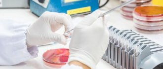
Laboratory examination of urine
A change in the composition and color of urine indicates the development of pathologies in the body. By assessing the symptoms and test results, the doctor will be able to diagnose the disease.
How to properly prepare for testing
To obtain accurate results when examining urine, it is important to properly prepare for the study:
- It is necessary not to eat brightly colored fruits and vegetables the day before the test, as they can change the color of the urine.
- It is prohibited to drink alcoholic beverages, take diuretic medications and biological supplements before taking tests.
- The doctor should know about the medications that were taken before the urine collection.
- It is not recommended to conduct research during a hypertensive crisis, during menstruation, or during infectious diseases.
Physical activity is also prohibited on the eve of the study.
How to properly collect urine
Basically, morning urine that has accumulated in the urinary tract overnight is suitable for research.
To obtain more accurate results, you should adhere to the following basic rules:
- In the morning, before collecting the material, you should wash your genitals.
- The container in which urine will be collected must be sterile.
- Part of the first portion of urine should be flushed down the toilet, and then, without stopping, fill the container. This is recommended to ensure that the number of bacteria in urine is optimal.
Collected urine is suitable for analysis within 2 hours if stored at a low temperature.
External definition
The presence of fat in urine can be determined even visually: fat shimmers in the light and resembles small droplets.
The presence of lipids in urine is determined using a Nicolas prism. In addition, microscopic examination allows you to see fatty acids in the urine.
Reasons for the development of lipuria
In a healthy person, a liter of urine contains about 2 mg of fat. In patients with severe diabetes mellitus, as a result of impaired carbohydrate metabolism, the kidneys secrete excessive amounts of lipids.
As a result of consuming very high-calorie foods with low energy expenditure, the amount of different fats in the human body increases. Excess carbohydrates are converted to fats.
The main reasons for the development of morbid obesity:
- excessive secretion of biologically active substances by the pancreas;
- deficiency of certain hormones.
Increased accumulation of various fats in tissue cells provokes their destruction and the development of dystrophic changes. Pathologies of fat metabolism cause the formation of many diseases in the body.
Fatty spots in urine may indicate the development of the following diseases:
- Pulmonary tuberculosis, which occurs in severe form.
- Complicated bone fracture. In such situations, fat cells penetrate into the blood from the bone marrow in large quantities. As a complication, fat embolism can develop, in which lipids completely clog the vessel.
- Diabetes.
- Pyelonephritis, kidney inflammation. With these pathologies, the kidneys function slowly and poorly, allowing fat cells to pass into the ureter.
- Urolithiasis disease.
- Pancreatitis, cholecystitis.
- Excess weight.
- Inflammatory and malignant processes occurring in the urinary organs.
- Cardiovascular pathologies.
Bougienage of the urinary system will help determine the reasons for the appearance of fat in the urine. For examination, a special instrument, a bougie, is inserted into the urethral cavity. To reduce injury to the organ, the bougie is pre-lubricated with sterile glycerin.
This procedure allows you to diagnose the following pathologies:
- narrowing of the urethra, its location and degree;
- localization of stones.
Having correctly determined the cause of the appearance of greasy spots in the urine, you can prescribe treatment, after which the greasy mark will disappear.
How to get rid of pathologies of fat metabolism
If lipuria is caused by physiological obesity, then getting rid of it is not very difficult.
The main method of treatment is drawing up a correct and complete menu. You should limit your carbohydrate intake. Food must contain the required amount of vitamins and fats, organic acids, and proteins.
If disorders of fat metabolism are caused by some pathological processes, the doctor prescribes medications that contain lipid-lowering components (statins). The mechanism of action of these drugs is to suppress the activity of an enzyme involved in the conversion of fats.
The most effective lipid-lowering drugs in recent years are:
- Pravastatin. This drug reduces the risk of cardiovascular pathologies and cholesterol concentrations. Well tolerated. In some situations, skin rashes may occur.
- Simvastatin is prescribed for high cholesterol levels. Reduces the risk of atherosclerosis and other pathologies of the cardiac system.
- Fluvastatin. It is effectively prescribed to prevent myocardial infarction, the formation of angina pectoris and other pathologies. The medication has a gentle effect on the liver.
- Rosuvastin. It is considered the most effective drug for lowering lipid concentrations.
- Atorvastatin. Helps to quickly reduce the amount of lipids. In addition, it normalizes cholesterol levels in patients with coronary heart disease.
The appearance of fat spots in urine is a rather serious sign that requires treatment. Lipuria develops as a result of an increase in undigested lipids in the blood. To find out the root causes of this condition, you should consult a doctor and undergo a comprehensive examination, consisting of laboratory and instrumental diagnostic methods. Lipuria is not often found in medical practice, but it indicates the development of a dangerous pathology in the human body.
nefrol.ru
Piuria
Pyuria - pus in the urine - is a general name that includes different quantitative gradations of a pathological increase in the number of leukocytes in the urine (leukocyturia). With significant pyuria, the urine is cloudy, with sediment quickly forming. Pyuria is easily distinguished microscopically from other causes of cloudy urine.
Active leukocytes are living leukocytes that have retained the biological activity of their membranes. They are easily identified by their ability to pass water and dye through the shell in a natural or created hypo-osmotic environment, for example, when adding distilled water or coloring ingredients. Then the leukocytes become visible under a microscope as round-shaped cells with a multilobed nucleus, enlarged 2-3 times. In their protoplasm, granularity is noted in a state of Brownian motion.
When examining urine with a small amount of urine, active leukocytes can be seen without special staining or by adding a drop of methylene blue solution to a drop of urine directly on a glass slide. It has been proven that such cells cannot be a consequence of inflammation of the urothelium and that their appearance with a high degree of certainty indicates a disease of the kidneys or accessory gonads.
Diseases of the genitourinary organs, most often accompanied by pyuria
A small number of leukocytes are found in the urine of absolutely healthy women. The presence of 2000 leukocytes in 1 ml of urine is considered acceptable. An increase in their number is considered a pathological phenomenon. To localize the source of pyuria, a two- or three-glass test is performed.
Even more graduated multi-glass tests have been proposed and used in the history of urology, but they have not received widespread acceptance and are now rarely used. Their main difference from classical tests is the retrograde lavage of the urethra during the collection of individual portions of urine. The non-physiological nature and direct danger of these intraurethral manipulations have limited the use of complicated beaker tests.
For differential diagnosis of the inflammatory process of the upper bladder, kidneys and mainly the lower urinary tract, it is sufficient to use a two-glass test. In this case, the patient is asked to urinate so that the first portion of urine (5-10 ml), i.e. almost washed off from the urethra, it was collected in the 1st glass, and all the rest of the urine - in the 2nd.
If an inflammatory process in the prostate gland is suspected, they resort to a three-glass test, when the patient is asked to urinate in three glasses so that the first and last portions of urine fall into the 1st and 3rd glasses, and the main part of it is released into the 2nd cup. If leukocyturia is higher in the 1st glass, we can assume inflammation of the urethra, if in the 3rd – in the prostate gland, if approximately the same in all 3 servings – inflammation can occur in the bladder, and in the bladder, and kidneys.
A two-glass test is more accurate in diagnosing the inflammatory process in the prostate gland and seminal vesicles, if you examine one portion of urine before and another after massage of the prostate gland and seminal vesicles. By the intensity of pyuria, one can additionally judge the severity of the inflammatory process and its transition to a purulent form.
Fat in the urine of a two-year-old child. We don't feed fatty foods. urine is clean and transparent. What is this connected with?
Once I was inserting a Viferon candle into my candle, and I also noticed grease, got scared, washed the pot, then the same grease again, only then it dawned on me that it was from the candles. Maybe you missed something too?
There is no fat in urine! It has too large molecules that cannot penetrate the membranes of the kidney glomeruli, tubules, etc.!!
A greasy jar or lid was used for testing.
Maybe you treated your child’s perineum with oil or baby cream. Well, there can’t be fat in the urine!!!
touch.otvet.mail.ru
Myoglobinuria
Myoglobinuria - the excretion of myoglobin in the urine - is accompanied by a red-brown color of the urine. Muscle hemoglobin, the red muscle pigment, is a chromoprotein. Myoglobin enters the blood in large quantities when striated muscles are damaged with crushing or crushing. Injuries accompanied by massive muscle damage are observed during earthquakes, military operations, when people fall under the rubble of buildings, etc.
Often, a few hours after rescue and liberation from rubble and rubble, people who seemed to have no life-threatening injuries developed a red-brown urine color with a sharp decrease in its amount, up to oligo- and anuria. This occurs as a result of some thickening of the blood accompanying the crush syndrome (crash syndrome), against the background of which a large amount of myoglobin enters the blood.
During microscopic examination of urine, myoglobin is detected as a brownish-brown pigment. Gradually, the kidneys are freed from myoglobin, but during this period the patient needs effective therapy and, in particular, several sessions of hemodialysis in specialized centers, where patients should be sent for anuria associated with crush syndrome.
Lipuria (fat in urine) - causes, diagnosis and treatment
Content:
Lipuria - what is it
Lipuria (from the Greek lipos - fat, uron - urine), or lipuria - the presence of fat in the urine. Normally, lipids are absent in the urine, or their amount does not exceed 2 mg/l. That is why lipuria is a pathological condition. Fats in the urine can be presented as grains, granular balls, lipoid casts, or clusters of lipoid grains that are visible only under a microscope.
Lipids are a general name for substances of various classes. These include
- Fats (triacylglycerides)
- Phospholipids (component of the cell membrane)
- Steroids (cholesterol, cholesterol derivatives, steroid hormones)
All these compounds are very important for the body, so disturbances in the metabolism of fats, fatty acids and cholesterol lead to dire consequences.
Causes
Urine is an indicator of the functioning of the kidneys, and the kidneys, in turn, are an indicator of the functioning of the body as a whole. Urine can be used to determine the function of many organs and their systems.
The appearance of fatty droplets in the urine floating on the surface is not an independent disease. Rather, this is a private manifestation of renal dysfunction (possibly against the background of a disease in another organ system), so it is important to diagnose and identify the cause of the deviation.
The symptom of lipuria is most often observed in nephrotic syndrome. Nephrotic syndrome is a concept that combines clinical symptoms and laboratory results, the leading of which are edema and proteinuria (protein in the urine). Some doctors identify it as an independent pathology, while others classify it as a concomitant syndrome. Its cause can be diseases in which the renal glomerulus, the cellular filter of the kidney, is damaged. Some of them:
- Acute and chronic glomerulonephritis.
- Pyelonephritis.
- Kidney amyloidosis.
In this case, lipuria is observed at the height of the disease, disappearing with its transition to the stage of irreversible chronic renal disease.
Fat in the urine can be detected when a long bone is fractured. When bones break, a structural change occurs in the surrounding tissue, after which liquid fat from the bone marrow enters the bloodstream. This also increases the level of lipids in the blood. A complication may occur in the form of fat embolism - complete blockage of the lumen of the vessel with fatty inclusions.
Surgical interventions for various reasons can lead to the release of fat in the urine, especially with extensive excision of subcutaneous fat. People who are obese are more susceptible to this.
Also, fat in the urine can be detected in diabetes mellitus. Diabetic nephropathy is a syndrome in which the kidney tubules and glomeruli are affected; manifests itself as a result of disturbances in the metabolism of proteins, fats, fatty acids and cholesterol.
Symptoms
A person may not notice abnormalities in urine, since fats hardly change the appearance of urine. It may become a little cloudy, but this may not be noticeable when urinating.
The symptoms are not related to lipuria per se, but to the disease that caused the condition.
In nephrotic syndrome, an edematous symptom is pronounced. Edema is general. First, the eyelids, face, ankles, and lumbar region swell, then the entire body swells. Swelling of the entire subcutaneous tissue of the body - anasarca - often accompanies this syndrome. In addition, before anasarca, swelling spreads to the body cavities, causing conditions such as ascites (fluid accumulation in the abdominal cavity), hydrothorax (fluid in the chest cavity).
In addition to edema, patients with nephrotic syndrome are concerned about skin changes - peeling, dryness, pallor. These disorders occur due to impaired skin trophism during edema.
As mentioned above, nephrotic syndrome can be an independent disease, or it can serve as a sign of other diseases. The doctor’s task is to conduct a differential diagnosis between pathologies of the excretory and other systems that can cause the syndrome.
Diagnostics
Urinalysis is one of the most important methods for diagnosing various diseases of organs and their systems. It can show abnormalities under various conditions.
In nephrotic syndrome, proteinuria is detected - protein in the urine. Normally, the protein content in urine is no more than 50 mg/l per day. With pathology, the amount of protein can reach more than 2000 mg/l per day.
Acute glomerulonephritis is characterized by hematuria - the presence of red blood cells in the urine. Urine takes on the appearance of “meat slop.”
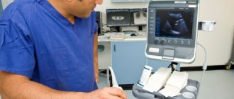
When diagnosing diseases that are the cause of lipuria, a general blood test and biochemical analysis are indispensable. If the doctor reveals abnormalities in the test results, then instrumental diagnostic methods will be used, for example, ultrasound, x-rays.
Treatment
The symptom of fat in the urine itself is not an indication for treatment. So, if lipuria occurs as a result of a fracture of a large bone or surgery, then no therapy is required.
If fat in the urine is a symptom of a disease, then the underlying disease must be treated. The choice of non-drug and drug therapy is made by a general practitioner or other specialist.
In any case, with lipuria it makes sense to reduce the fat content in food. If the patient is overweight, then sometimes lipid-lowering drugs are used. They reduce the concentration of fats in the blood. There are several groups of lipid-lowering drugs; Of these, statins are most often used - Rosuvastatin (Rozulip, Rozistark), Simvastatin (Vasalip), Atorvastatin (Atoris). All of them reduce the risk of cardiovascular complications associated with hyperlipidemia (high blood lipid concentrations) and high blood cholesterol.
For nephrotic syndrome, diuretics are indicated. Furosemide, hypothiazide, and a group of spironolactones (veroshpiron) are used. They are prescribed only for severe edematous syndrome.
If nephrotic syndrome is caused by glomerulonephritis, then the first drug prescribed is prednisolone, in the form of tablets 1-2 mg/kg in 2-4 doses per day.
Similar articles:
pochkizdorov.ru
Proteinuria
Proteinuria - the presence of protein in the urine - can be caused, on the one hand, by increased permeability of the glomerular filter, on the other hand, by inadequate protein resorption, which is mainly carried out by the proximal nephron. A healthy person excretes from 50 to 150 mg of protein per day in urine, which corresponds to 0.033% according to a general urine test.
True (renal) proteinuria depends on the content in the urine of protein passing through the nephron as a result of degenerative changes in the renal, primarily glomerular membranes, in particular the renal glomeruli and Bowman Shumlyansky's capsule. With glomerulonephritis and nephrosis, proteinuria can reach high levels.
False (extrarenal) proteinuria depends on the presence of a large number of erythrocytes or leukocytes in the urine, i.e. protein containing cells. When they are destroyed in concentrated urine, the amount of detectable protein increases, and in some cases, when protein and pus in the urine are the result of the same disease (pyelonephritis), the differential diagnosis of false and true proteinuria turns out to be difficult and requires the use of highly accurate immunochemical techniques.
Oily film on the surface of urine
The content of the article
A sign of phosphaturia is a white film on the urine
Have you been trying to cure your KIDNEYS for many years?
Head of the Institute of Nephrology: “You will be amazed at how easy it is to heal your kidneys just by taking it every day...
Read more "
Normally, fresh urine is clear. If turbidity or a white film appears on the urine (on the surface and at the point of contact with the vessel), this may be a sign of the presence of phosphate salts in it. Their appearance is associated with a violation of the metabolism of calcium and phosphorus in the body. If these insoluble salts are found in several urine tests, we can talk about phosphaturia, which can be an independent symptom or indicate the presence of kidney phosphate stones. They are formed in the kidneys (in 50% of cases), urinary tract and bladder. They grow especially quickly in the presence of pyelonephritis. You can recognize phosphaturia at home in this way: if you heat cloudy urine with a white film and add acetic acid to it, it will become transparent.
Accurate diagnosis
To obtain detailed data on the functioning of the urinary system, a daily urine test is performed, since it is the daily excretion of salts by the kidneys that is limited. Since its chemical composition directly depends on the food consumed, spicy, smoked, sweet, carbonated and alcoholic drinks and canned food are prohibited a week before the procedure. Urine is collected throughout the day in one container, starting at 6 a.m., each time the container is tightly closed with a lid to prevent oxidation, and it is put away in a dark, cool place. The last collection of material is done no later than 6 a.m. of the next day, after which the urine is shaken, 100 ml is poured into another clean jar and sent to the laboratory as quickly as possible. The accompanying note should indicate the total volume of daily urine.
OUR READERS RECOMMEND!
Our readers successfully use Renon Duo to treat kidneys. Seeing how popular this product is, we decided to bring it to your attention. Read more here...
If phosphates were detected in a random analysis, the doctor is obliged to inform the patient about the dangers of salts in the urine. Phosphorus is necessary for the body for normal metabolism, muscle and brain function. Its level increases with frequent consumption of foods containing phosphates. With their excess, the body poorly absorbs magnesium, calcium and iron. Phosphaturia therapy is the prevention of stone formation and chronic inflammation of the urinary system. To prevent the precipitation of insoluble salts, a special diet is prescribed, which restores the acidity of urine, and in an acidic environment, phosphorus-calcium sediment does not form.
Treatment with diet No. 14
The essence of the diet is to reduce the consumption of foods that contain calcium and have an alkalizing effect, and increase the amount of those that oxidize the body.
If you have phosphaturia, you should completely exclude the following from your diet:
- fatty fish and meats;
- milk soups;
- pickles;
- smoked meats;
- spices;
- baked goods;
- cocoa and chocolate;
- alcohol.
Drinking strong tea and coffee is not recommended, as they irritate the kidneys. The consumption of all dairy products, egg yolks, peanuts, mushrooms, potatoes and other vegetables is significantly limited. The basis of the diet is meat, poultry and fish, prepared in any way, and various cereals. Healthy dishes include seafood, green peas, Brussels sprouts, pumpkins, as well as sour apples, sauerkraut, cranberries and lingonberries. Meals should be fractional. Bread - dried, made from wholemeal flour. Refractory fats are prohibited.
There is no need to eat very dry food. So that the urine is not concentrated and salts are not detected in it, you need to drink as much liquid as possible, in the absence of restrictions - up to 2.5 liters per day: bread kvass, fruit drink, jelly. For phosphaturia, it is useful to drink a glass of slightly alkaline non-carbonated mineral water on an empty stomach.
Since the diet of diet No. 14 is not meager, it can be practiced for as long as treatment requires, and it is advisable to take vitamin complexes (A, B, D2). Urinalysis should be done periodically to check the salt concentration.
Folk recipes
The effect of herbal infusions for phosphaturia is that, along with excess fluid, they remove salts and toxins. Plants that dissolve phosphates:
- corn silk;
- bearberry;
- horsetail;
- wintergreen round-leaved;
- tricolor violet.
The following recipe is effective: mix flax seeds, sage leaves, madder and wheatgrass roots in equal quantities, then 4 tbsp. l. pour into a thermos and add 1 liter of water. Take 200 ml 1 hour before meals.
During the season, it is necessary to arrange watermelon days, flushing the urinary system. Rosehip decoction and apple compote (with peel) are very useful for oxidizing urine.
Self-massage and physical exercises help to cope with the disease: bending the body, rotating, walking. You need to move a lot. To treat phosphaturia, the doctor may prescribe benzoic acid orally (0.3 g 3-6 times a day) and antibiotics (the dosage is selected individually). If you strictly follow all the doctor’s recommendations, the disease will recede, but even then you should adhere to a healthy lifestyle.
1pochki-med.ru
Hematuria
Hematuria is blood in the urine. In the urine of a healthy person, a small number of red blood cells are usually determined (up to 1000 in 1 ml of urine). The presence of blood in the urine, invisible to the naked eye and detectable only by microscopic examination, is called microhematuria. This is what is most often called erythrocyturia, in contrast to microhematuria, when an admixture of blood turns the urine red of varying intensity. Gross hematuria can be a symptom of many diseases and requires a urological examination.
First of all, it is important to determine the source of the bleeding. Since the anterior urethra is separated from the posterior urethra by the striated muscular sphincter of the pelvic diaphragm, bleeding from the anterior urethra will be constant, regardless of urination - urethrorrhagia. Urethrorrhagia must be clearly distinguished from gross hematuria.
If the urine is more heavily stained with blood in the first glass (in the first portion), hematuria is called initial, or initial. The source of such hematuria, as a rule, is located in the posterior urethra: irrigation, cancer, severe inflammatory process, damage. If the blood stains the urine more strongly in the 3rd glass (the last portion of urine), hematuria is called final, or terminal. It is characteristic of diseases of the bladder neck - inflammation, tumors, stones, varicose veins.
Diseases of the genitourinary organs, most often accompanied by hematuria
It can be a manifestation of bleeding from the kidney parenchyma, renal pelvis, ureter, bladder (polycystic kidney disease, tuberculosis, pyelonephritis, urolithiasis, necrosis of the renal papillae, hemorrhagic cystitis, bladder ulcers, benign prostatic hyperplasia, endometriosis and schistosomiasis of the bladder, etc. .).
With total hematuria, the most dangerous, it is necessary to look for additional manifestations of the disease that caused it. First of all, you should pay attention to the shape of the clots. Shapeless blood clots usually form in the bladder; the possibility of their formation cannot be excluded during massive bleeding from the kidney.
If pain occurs after the onset of hematuria, it can be assumed that the ureter is occluded by a blood clot with the development of renal colic on the side of the disease, accompanied by bleeding from the kidney. When pain precedes hematuria, the cause of ureteral occlusion is hematuria, as happens with urolithiasis and, in particular, with associated renal colic.
Total painless hematuria does not allow one to accurately determine the level of bleeding and its side. Considering the seriousness of possible diseases accompanied by total painless macrohematuria, in particular tumor hematuria, emergency cystoscopy is necessary to visualize the bladder disease or at least identify the side of the lesion by bloody urine released from the ureter. This will significantly help further diagnosis by narrowing the targeted search area.
It should be borne in mind that red urine may not only be due to blood. Some medications (for example, phenolphthalein, or purgen) and foods (beets) can cause urine color to change. In these cases, diagnosis is helped by a careful questioning of the patient and microscopic examination of urine.
Hemoglobin can also give a bloody color to urine. Hemoglobinuria can be observed with certain blood diseases, poisoning, with extensive burns, after transfusions of incompatible blood. Urine with hemoglobinuria can be colored red of any intensity, but when examined in transmitted light, one can be convinced of its transparency, and, most importantly, during a microscopic analysis of urine, red blood cells are not found in it, but with spectral analysis, hemoglobin, methemoglobin, and oxyhemoglobin are determined.
It has been established that exceeding the concentration of hemoglobin in plasma 1.0-1.4 g/l already causes hemoglobinuria. All oxidizing agents, when ingested or inhaled, lead to the conversion of hemoglobin to methemoglobin, which entails hemolysis and, consequently, hemoglobinuria. Nitro compounds, phenacetin preparations, and sulfonamides in large doses promote hemolysis as a result of changes in the enzyme systems of the red blood cells themselves. Increased breakdown of red blood cells can be caused by a large intake of fatty foods or as a result of heavy physical activity.
A particularly striking symptom complex accompanies hemoglobinuric fever, a disease characterized by acute massive intravascular hemolysis under the influence of various drugs and poisons, as well as under the influence of other causes, such as cooling or a septic state. Free hemoglobin circulating in the blood is captured by the reticular endothelial cells of the liver, spleen, and lymph nodes and is converted into bilirubin, which can manifest as yellowness of the skin and mucous membranes of varying intensity.
Hemoglobinuria may appear in the absence of hemoglobinemia as a result of the breakdown of blood in the kidneys (indirect or false hemoglobinuria). In the urine sediment in these cases, lumps and yellow crumb-like masses of amorphous hemoglobin, often hyaline and granular cylinders, are always found. Urine has the color of black beer or red wine, which is due to the content of oxyhemoglobin in freshly released urine, and the presence of methemoglobin in standing urine.
Pneumaturia
Pneumaturia—excretion of air or gas in the urine—is quite rare, but is of some interest as a symptom. It is easily explained in patients after endoscopic manipulations, examinations with the introduction of oxygen into the bladder, for example, during X-ray examinations, with entero-urinary and entero-genital fistulas.
The appearance of pneumaturia outside of these situations indicates significant bacteriuria and the presence of intestinal and paraintestinal flora. This flora promotes the fermentation of glucose and the release of carbon dioxide bubbles (odorless) during urination. Then pneumaturia suggests diabetes mellitus, and with incomplete emptying of the bladder, the presence of pyelonephritis.
The release of gas with a pungent ammonia odor indirectly confirms a significant infection of the urinary tract by the same flora, which, when urine remains in the bladder for long periods of time, for example, with chronic urinary retention, “manages” to bring the process of metabolism of urea in urine to the formation of ammonia in the form of gas. Therefore, each case of pneumaturia requires careful clinical interpretation.
Other impurities in urine reflect both general and local processes.
Bacteriuria
Bacteriuria - the release of bacteria in the urine - is usually accompanied by leukocyturia and indicates the presence of an inflammatory process in the organs of the urinary system and the male reproductive system. The urine of a healthy person is sterile. There is no bacteriuria without an inflammatory process at some level, since numerous studies have proven the impossibility of microorganisms passing through the kidney filter.
With the expansion of ideas about the causative agents of inflammatory processes, with the establishment of the role of Trichomonas, fungi, mycoplasmas, chlamydia, viruses, and their immunological relationships with the macroorganism, it becomes clear that each infection has its own specifics, so that the previous division of them has lost the meaning previously attached to this definition.
Some saprophytic flora enters the urine due to contamination during urine collection for testing. Colony counting allows one to distinguish such bacteriuria from true bacteriuria. When the microbial count is 105 in 1 ml or more, we can talk about the presence of an infectious inflammatory process.
To clarify the nature of the microflora in inflammatory diseases of the male reproductive system, the secretions of the prostate gland, seminal vesicles or ejaculate are subjected to bacteriological examination. Bacteriological examination of ejaculate is a valuable method for identifying the inflammatory process of the entire vas deferens system with its accessory glands.
Possible diseases that contribute to the release of sticky urine in humans
Most often, glucose levels increase for the following reasons:
Diabetes
Excess sugar content gives the biofluid viscosity, and this most often happens during diabetes. Glucose levels can increase in people of different genders and age groups. It was noted that for a long period additional symptoms did not bother.
For this reason, you must always monitor your own health. When a person’s weight begins to change, he constantly feels thirsty, wounds take a long time to heal, resistance to physical stress disappears, you need to consult an endocrinologist who will prescribe a urine sample for a general analysis. In particularly severe cases, loss of consciousness may occur.
Kidney diseases
The main symptom of this pathology is swelling. When it is accompanied by poor appetite, general weakness, changes in pressure in the arteries, pain in the lumbar region, then the likelihood of diseases of the paired organ arises.
Kidney problems also arise if diabetes progresses in the body. Severe complications can only be prevented by regular examinations. liver diseases.
With an increased level of sugar in biological fluid, a person may experience certain symptoms in the form of nausea, belching, a specific and not entirely pleasant smell of sweat, a yellowish color of the skin on the face, diarrhea and changes in the color of stool.
Other diseases
Deviations from normal sugar levels can occur in other diseases. These include:
- acute pancreatitis;
- infectious meningitis;
- brain oncology;
- concussion or stroke.
How to eliminate metabolic disorders
The problem can be eliminated using an integrated approach, which includes nutritional correction and the use of medications. Only a doctor can prescribe medications, taking into account the individual characteristics of the patient. If fat appears in the urine due to a chronic disease, it is recommended to take medications to treat the specific pathology.
Medicines
To correct the condition, medications are most often prescribed that suppress the activity of an enzyme that is involved in lipid conversion:
- Pravastatin. Reduces the risk of pathologies of the heart and blood vessels, normalizes the amount of cholesterol in the blood.
- Simvastatin. Normalizes cholesterol levels. Prevents atherosclerosis and other cardiovascular diseases.
- Fluvastatin. Reduces the likelihood of heart attacks, angina pectoris, and has a gentle effect on the liver.
- Rosuvastatin is the most effective drug that reduces the amount of fat.
- Atorvastatin. Reduces the amount of fat and cholesterol levels in patients suffering from coronary heart disease.
Diet
If the cause of lipuria is physiological obesity, then solving the problem will not be easy. The main method of therapy is choosing the right diet. The patient is recommended:
- limit the amount of fats and carbohydrates in the diet;
- eat small and balanced meals;
- Avoid eating fried, smoked foods and alcohol.
Fat in a urine test is a symptom that should prompt you to see a doctor. This disorder is rarely encountered in medical practice, but it always indicates serious health problems. Self-medication is excluded. Only a specialist should prescribe medications and select a treatment regimen after urinalysis and other studies.
Diseases
There are many diseases that are caused by urine. The reasons for its appearance, in addition to the already mentioned kidney pathologies, lie in such an ailment as diabetes mellitus. This phenomenon occurs especially often in the insulin-dependent form of the disease. At the same time, the level of sugar in a person’s blood is also high. Another reason for increased glucose levels may be an attack of acute pancreatitis.
It would seem, what does the state of the brain have to do with urine analysis? But as it turns out, there is a relationship, and it is quite close. A number of brain injuries can cause high sugar in the urine. The reasons for its appearance are an infectious disease (meningitis, encephalitis), traumatic brain injury, hemorrhagic stroke. And in this case, they talk about the occurrence of glucosuria of central origin.
In addition, disturbances in the functioning of the endocrine system can provoke an increase in sugar levels. An increase in hormone levels - the release of adrenaline, thyroxine or glucocorticoids into the blood - may well provoke glucosuria. And then it will be called endocrine.
The causes of toxic glucosuria are extremely unusual in everyday life. It develops from poisoning with substances such as morphine, strychnine, chloroform or phosphorus. In any case, whatever the reasons for such an imbalance of substances in the urine, if you detect high sugar levels, you should definitely consult a doctor and undergo additional tests in order to prevent the development of serious diseases.
Hiluria
Hiluria is an admixture of lymph in the urine, when the urine has the color and consistency of thick milk, usually due to the formation of a message between large lymphatic vessels and the urinary tract. This occurs most often in the fornix zone of the renal calyces or its pelvis. The formation of such fistulas is usually associated with inflammation, tumor processes, and injuries, as a result of which the thoracic duct is compressed and the intraductal lymph pressure increases.
It should be remembered that chyluria often occurs with filariasis, in which filariae primarily affect both the lymphatic and urinary tracts. Despite the obviousness of the diagnosis, it can be quite difficult to establish and eliminate the cause of chyluria. Since patients lose most of their energy reserves, they need enhanced nutrition, including parenteral nutrition.
What test do you need to take?
If fatty spots are found in the urine of a child or adult, it is necessary to undergo a general urine test. To obtain a reliable result, you need to prepare for the study:
- do not eat vegetables and fruits the day before the test;
- exclude alcohol, diuretics and dietary supplements;
- notify the doctor about the medications you are taking during this period;
- before collecting urine, wash the genitals, sterilize the container or purchase a special one at the pharmacy;
- take only a middle portion of urine for analysis.
A visual sign that the urine contains lipids is opacity. The cause of turbidity is determined by examining urine sediment under a microscope, or using chemical analysis. The fact that lipids are present in the biomaterial is indicated by the disappearance of turbidity when ether is added.
A liter of urine should normally contain no more than 2 ml of lipids.
The presence of fat in the urine alone is not enough to make a diagnosis. A biochemical urine test is also prescribed to identify the following indicators:
- diastasis;
- bilirubin and other liver enzymes.
Additionally, you need to donate blood for biochemistry. If there are deviations in the tests, the kidneys, pancreas and gall bladder are examined using instrumental methods.
If you are obese, you need to take an additional blood test for hormones.