About the structure of the kidney
The structure of the kidneys is considered one of the most complex in the human body. The pelvis and calyces are designed to connect them with the urinary ducts. The largest pelvises include several large calyces, each of which consists of smaller calyces. There are several types of pelvis in the fetal kidneys:
- embryonic - small in size, attached to one larger pelvis;
- fetal - there are no pelvic structures, instead there are cups leading into the ureter;
- mature - exactly like an adult.
Indications for ultrasound are limited
Like any other screening test, a kidney ultrasound should be performed annually.
Indications for an extraordinary ultrasound are:
- presence of pain in the lumbar region;
- detection of changes in urine analysis;
- urinary incontinence;
- paroxysmal colic;
- lack of urination;
- the presence of painful and frequent emptying of the bladder;
- suspicion of a tumor process in the kidneys;
- inflammatory processes in the genital organs;
- traumatic injury to the lumbar region;
- change in the amount of urine.
What size should the fetal renal pelvis be normally?
There are several types of abnormal changes in the size of the fetal pelvis. Primary dilatation (hydronephrosis) is caused by pathological processes in the body of the baby and mother. In this case, the outflow of urine is difficult. The reduction of structures is called hypoplasia. In some cases, the pelvis may be absent. Their expansion due to overflow with urine is called pyeloectasia. In this case, the pelvis cannot cope with the incoming volume of secreted fluid: they are enlarged because they are stretched.
At the 32nd week of gestation, the normal size of the pelvis should not exceed 5 mm. By the 36th week they should increase to 7 mm. An enlargement of the renal pelvis in an unborn baby to 10 mm or more is pyelectasis.
The pathology is found in 2% of pregnant women, and boys are 3 times more likely to suffer from it. The diagnosis is made based on ultrasound results from the 16th week, taking into account the fact that the kidneys enlarge as the fetus grows. To distinguish normality from pathology, limits of acceptable values have been established. The sonologist takes them into account during the ultrasound examination.
Up to the 32nd week of gestation, the size of the renal pelvis is in the range of 4–7 mm. Exceeding the norm by 1 mm requires regular monitoring. Often it does not lead to pathologies in the future; the situation evens out as the fetus grows. With an increase of 2 mm or more, the likelihood of kidney abnormalities after birth increases. In this case, medical correction is required.
Prevention of discharge pyramidal syndrome in the kidneys
Few people know about the echogenicity of their organs, and even fewer think about it. Meanwhile, the hyperechogenicity of the kidneys indicates the presence of dangerous inclusions, which indicate the syndrome of isolated pyramids developing in the body.
Sharp outlines or white spots in an ultrasound image of the kidneys are signals of ongoing pathologies and even insufficient body weight. In any case, everyone needs to know about the condition of their kidneys.
Briefly about the syndrome
Echogenicity is characterized by the degree of reflection of sound from the tissues and fluid of internal organs, and the appearance of strong reflections on ultrasound signals the presence of various foreign inclusions.
Hyperechoic kidneys on ultrasonogram
Most often, hyperechoic formations form in the renal pyramids and parenchyma.
Pyramids are the triangle-shaped areas of the kidneys through which, after filtration, urine exits into the pyelocaliceal system, and parenchyma refers to the tissue that fills the organ from bone and cortex.
Any extra element of the structure, being a consequence of the pathology occurring in the organ, introduces dissonance into its functioning, and often developing on one, soon affects the second kidney.
All excess elements are divided into calcium stones, stones, sand and neoplasms. The last group includes formations of various types:
- Small formations appear as white dots during ultrasound. The absence of an acoustic shadow indicates their safety;
- The following type of inclusions is characterized by a large size, which can be either benign or malignant, cancerous tumors. They are extremely rare and require constant monitoring by doctors;
- Large light spots on ultrasound indicate cancerous inclusions, which, unlike other formations, necessarily have an acoustic shadow, areas of sclerosis, and also contain calcifications and psammoma bodies.
8 main causes of hyperechogenicity
Hyperechoic renal pyramid syndrome is not an independent disease, but an accompanying ailment, as it is a consequence of a pathology developing in the body. The cause of their appearance may be the following pathological renal processes:
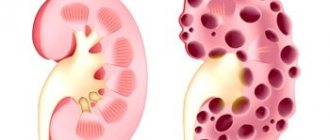
- Polycystic disease;
- Sclerosis of blood vessels;
- Injuries;
- Bleeding;
- Purulent inflammation of tissues;
- Accumulation of several abscesses and boils in one place;
- Oncological processes;
- Fatty formations in the pyramids of the kidney.
Hyperechoic inclusions on ultrasound
Symptoms and diagnosis
Hyperechoic pyramid syndrome can be suspected by the following characteristic clinical manifestations that occur against a general background of weakness and fatigue: high temperature (up to 39°C), dark brown or red urine, stabbing pain in the kidney area; spasms in the abdomen and groin, vomiting, nausea, nagging pain in the groin.
The symptoms of the syndrome are characteristic of many renal diseases, however, with the help of ultrasound, a specialist will immediately diagnose the presence of hyperechoic inclusions, and will also assess the condition of the kidney parenchyma against the background of prominent pyramids.
When a diagnosis of “hyperechoic pyramid syndrome” is made during diagnosis, additional procedures are prescribed to identify the root causes of the process and prescribe appropriate therapy. Thus, general blood, urine, and stool tests are required.
Diagnosis of hyperechogenicity
Possible causes of enlarged renal pelvis
The cause of congenital pyelectasia is a genetic factor and illnesses that the mother suffered while carrying the baby (especially pyelonephritis, urinary tract infections). Each case is individual, but it has been proven that pathology is provoked by:
- congenital malformations of paired organs of the urinary apparatus;
- compression of the ureters by vessels or fetal organs when they are not formed correctly;
- narrowing of the urinary tract;
- incorrect functioning of the urinary system due to weakened fetal muscles;
- intrauterine intoxication due to the fact that the expectant mother abuses alcohol, leads an unhealthy lifestyle, smokes, and eats poorly;
- regular intake of more fluid than necessary into the fetus’s body.
What is the risk of pyeloectasia for a baby?
The reasons that caused fetal pyeloectasis should be identified in a timely manner. In the future, they need to be eliminated or their harmful effects minimized. Otherwise, the pathology will worsen, which will lead to compression of the kidney tissue and subsequent atrophy of the organ. After the birth of a child suffering from pyeelectasis, sooner or later a chronic inflammatory process will form in the kidney, caused by the penetration of bacterial microflora. This leads to hardening of the organ.
Another danger of pyeloectasia is prolapse of the diseased kidney. This is the result of an increase in the mass of the kidney due to the fact that more urine accumulates in the dilated pelvis. The problem can be aggravated by ureteral prolapse - prolapse of structures in the urethra and vagina.
Types of inclusions and possible diseases
Sclerosis is a pathological replacement of healthy functional elements of an organ with connective tissue, followed by disruption of its functions and death.
If a single formation is found inside the kidney that does not cast an acoustic shadow, it may be a signal:
- cystic cavity filled with fluid or empty;
- sclerosis of kidney vessels;
- small, not yet hardened concretions (stones);
- sand;
- inflammatory process: carbuncle or abscess;
- fatty compaction in the kidney tissue;
- hemorrhages with the presence of hematomas;
- development of tumors, the nature of which needs to be clarified.
If hyperechoic formations are small (0.05-0.5 cm3), reflected on the screen with bright sparkles, and there is no acoustic shadow, these are echoes of psammoma bodies or calcifications, which often, but not always, indicate malignant tumors.
Psammoma (psammotic) bodies are layered formations of rounded forms of protein-fat composition, encrusted with calcium salts. Found in vascular connections, meninges, and some types of tumors.
Calcifications are calcium salts deposited into soft tissues affected by chronic inflammation.
Is there a treatment for intrauterine pyelectasis and is it possible to prevent the pathology?
An anomaly detected in the prenatal period requires observation. The doctor evaluates the enlargement of the fetal pelvis over time and assigns one of three degrees to the pathology. In mild and moderate forms of the pathology, the prognosis is favorable: the pelvis returns to normal during the first year of life as the baby grows. In severe cases of bilateral progressive pyelectasis, surgery is indicated. It is done in 30% of cases.
The intervention is performed under anesthesia using laparoscopy, which eliminates large incisions. Miniature surgical instruments are inserted through the urethra and manipulations are performed under ultrasound guidance. The doctor’s goal is to restore the normal flow of urine and eliminate its backflow into the pelvis. After surgery, the baby is prescribed anti-inflammatory drugs.
The operation does not guarantee a 100% recovery, and the little patient must be observed by a urologist until school, and then until puberty. During periods when the child is actively growing, relapse of the pathology is possible.
There are no special measures that can prevent pyeloectasis. Experts recommend that women plan pregnancy, treat genitourinary infections and kidney diseases before conception, and prevent relapses of chronic pathologies. For this purpose, before and after pregnancy you need to follow a diet, drinking regime and personal hygiene.
During pregnancy, it is important to avoid exposure to toxins and stay in environmentally polluted places. As recommended by your doctor, you should take diuretics to improve urine flow and prevent swelling. If a pathology is discovered in the fetus, it is important to adjust the diet and drinking regimen and carefully follow the doctor’s recommendations. Moderate physical activity of the mother in this situation will have a positive effect on the condition of the fetus.
What is pyelectasis
The size of the fetal pelvis in the second trimester of pregnancy is 5 millimeters, in the third – 7 mm. If there is an enlargement of the renal pelvis in the fetus, the doctor will find out about this at 16-20 weeks with an ultrasound examination. The baby is constantly monitored, since this phenomenon may be temporary.
A temporary pathology that manifests itself during fetal development is associated with a heavy load on a woman’s kidneys. Because of this, part of it passes to the fetal urinary organs. Due to the structural features of the urinary system, pathology is more often observed in boys.
Causes and consequences of pyelectasis
The most common reason for an enlarged renal pelvis in the fetus is a genetic predisposition. Therefore, if a pregnant woman has relatives in her family with this disease, it is imperative to inform the doctor and the specialist performing the ultrasound about this.
The second reason is the abnormal development of the kidney structure. Urine has the ability to return to the organ in the absence of direct exit. Under pressure in the kidneys, the pelvis expands.
There is also a list of reasons that provoke the disease:
- compression of the ureters by blood vessels or other organs due to improper formation of fetal organs;
- abnormal intrauterine development of the child (the urinary organs do not work normally due to weakening of the muscles);
- congenital kidney defects, obstruction of urine outflow;
- narrowing of the lumen of the urinary tract.
If the fetal renal pelvis is dilated, complications may develop that are life-threatening to the baby. It is important to diagnose the disease in a timely manner, because otherwise, under strong pressure in the bladder, the ureters will enlarge and spasms will occur at the bottom of the urethra. Complications of pyelectasis include the development of urethrocele, severe pyelonephritis and cystitis, and atrophic changes in the urinary organ.
With constant inflammation, the renal structural unit, the nephron, dies. Prolapse of the kidney may occur due to the filling of the renal pelvis with urine, as a result of which the mass of the urinary organ increases. Expansion of the renal pelvis in the fetus leads to prolapse of the ureters with prolapse of these structures into the vagina and urethra.
Changes in the kidney parenchyma
To maintain normal functioning, the body needs to carry out metabolism. In order for the body to receive everything it needs from the environment, there must be a continuous cycle between the person and the external environment.
During metabolic processes, metabolic products are formed in our body, which must be excreted from the body. This includes urea, carbon dioxide, ammonia, etc.
Substances and excess water are removed, as well as mineral salts, organic substances and toxins that enter the body with food or other routes.
The elimination process occurs through the excretory system, namely the kidneys.
The kidney is a paired parenchymal organ, bean-shaped . The kidneys are located in the abdominal cavity, in the lumbar region, retroperitoneally.
Normal kidney values:
- length 10-12 cm,
- width – 5-6 cm,
- thickness from 3 to 4 cm;
- the weight of one kidney is 150-200 g.
The structure of the kidney also includes the main tissue - parenchyma .
What is renal parenchyma?
The term “parnechyma” itself is defined as a collection of cells that perform an organ-specific function. Parenchyma is the tissue that fills the organ.
The parenchyma of the kidney consists of the medulla and cortex, which are located in the capsule. It is responsible for all functions performed by the organ, including the most important one – urine excretion .
Examining the structure of the parenchyma using light microscopy, you can see the smallest cells densely intertwined with blood vessels.
Normally, the thickness of the kidney parenchyma of a healthy person ranges from 14 to 26 mm, but can become thinner with age.
For example,
in elderly people, the normal size of the kidney parenchyma is no more than 10-11 mm.
Interestingly, kidney tissue has the ability to regenerate and restore its functions. This is a big plus in the treatment of various diseases.
Many people do not know where their kidneys are, so sometimes they do not even realize that they may have impaired kidney function.
Kidney pain can indicate various diseases. Read our article about how kidneys hurt in various pathologies.
Increased echogenicity of the renal parenchyma - is it dangerous?
According to statistics, today, against the background of general morbidity, people more often suffer from problems of the urinary system. Pathological processes in the kidneys cannot always be observed; more often they occur hidden.
Echogenicity of the kidneys can be diagnosed using ultrasound.
The technique is invasive, is completely painless and has a big advantage : using ultrasound, you can detect the slightest pathological changes even in the early stages.
This will increase the patient's chances of recovery. The diagnostic process itself takes no more than 20-25 minutes, during which time you can find out such parameters as:
- the size of the organ itself,
- its location,
- neoplasms, if any.
Increased echogenicity of the kidneys may indicate:
- diabetic nephropathy (enlarged kidneys, but the pyramids located in the medulla have reduced echogenicity);
- glomerulonephritis , which occurs in severe form, and the renal parenchyma itself diffusely increases its echogenicity.
- increased echogenicity of the renal sinus indicates that there are inflammatory processes, metabolic and endocrine disorders .
Kidneys whose tissue is healthy have normal echogenicity; it is homogeneous on ultrasound.
Diffuse changes in the renal parenchyma
A serious signal for a detailed study of the kidneys are changes in their parenchyma. The reasons for changes in organ size can be different:
- development of urolithiasis
- inflammation of the glomeruli or tubules
- diseases affecting the urinary system
- formation of fatty plaques near the pyramids
- diseases leading to inflammation of the kidney vessels and adipose tissue
Renal parenchyma cyst
This disease arises and develops when fluid is retained in the nephrons of the kidney and develops from the parenchyma. A cyst can occur on both the parenchyma of the right and left kidney.
The cyst is characterized by an oval or round shape and measures 8-10 cm .
Sometimes the size of the cyst reaches rather large sizes (fluid accumulates up to 10 liters), thereby squeezing the structures lying nearby.
A cyst removed in time is the key to not just a quick recovery, but to saving the kidney. is diagnosed using ultrasound.
The symptoms are not difficult to identify. This may include muted pain in the hypochondrium and lower back, increased blood pressure and the presence of blood in the urine.
Unfortunately, symptoms do not always appear, and the disease occurs in a latent form.
In such cases, the disease is detected in late stages, when the only treatment method is surgery.
Thinning of the kidney parenchyma
The reasons for the appearance of this pathology may be different. For example, incorrect selection of treatment method or infectious disease .
It must be remembered that the kidney parenchyma can decrease with age, but sometimes shrinkage is observed in chronic diseases.
If you feel discomfort in the lower back or pain when urinating, seek help from specialists, do not treat yourself.
This will not only save your time, but also improve your health.
In utero detection of pyelectasis
The pathological phenomenon is diagnosed starting from the 16th week of pregnancy. If the pelvis is dilated by no more than one millimeter, there is a high probability that the deviation will go away on its own. When they increase by more than 10 millimeters, there is a risk of developing complications, which is extremely dangerous for the health of the unborn baby.
The disease is detected using ultrasound. If the pathology has not been eliminated during the period of intrauterine development, the baby is observed after birth by doctors. Additional diagnostics are carried out by intravenous urography and cystography.
When sick, a newborn’s body temperature rises and he eats poorly. Pain is observed in the lower back and abdomen. Signs of gastrointestinal distress appear: diarrhea, nausea, vomiting.
Increased echogenicity
So, what is the basic structure of the parenchyma, you can imagine. But a rare patient, having received the result of an ultrasound examination, does not try to decipher it on his own. It is often written in the conclusion that there is increased echogenicity of the parenchyma. First, let's look at the term echogenicity.
Examination using sound waves is based on the ability of tissues to reflect them. Dense, liquid and bone tissues have different echogenicity. If the fabric density is high, the image on the monitor looks light, the image of fabrics with low density appears darker. This phenomenon is called echogenicity.
The echogenicity of the renal tissue is always homogeneous. This is the norm. Moreover, both in children and adult patients. If during the examination the structure of the image is heterogeneous and has light inclusions, then the doctor says that the renal tissue has increased echogenicity.
With increased echogenicity of the parenchyma, the doctor may suspect the following ailments:
Pyelonephritis. Amyloidosis. Diabetic nephropathy Glomerulonephritis. Sclerotic changes in the organ.
A limited area of increased echogenicity of the kidneys in children and adults may indicate the presence of a neoplasm.
Methods for eliminating the pathological condition
Treatment depends on the degree of development of the disease, that is, on how enlarged the fetal pelvis is. There are three degrees of development of pathology: mild, moderate and severe. During the first, the organ of the unborn child is monitored. The development of the second stage may also have a favorable outcome. The disease goes away on its own with further development of the urinary channel. But the period of observation with ultrasound increases.
In severe cases of the disease, surgery is performed. The instruments used for this type of surgery are very small to avoid tissue damage. The use of anti-inflammatory drugs is necessary to minimize the risks of complications. With the help of the operation, the outflow of urine is restored, stopping its reflux into the bladder.
After surgery, a special diet is prescribed, in which food that irritates the ureteral mucosa is excluded. Drug therapy is ineffective if the renal pelvis is dilated.
It is difficult to predict further developments. The child may experience relapses, especially at the age of 6 years and during puberty. Therefore, patients are monitored at the dispensary throughout their lives.
Signs of pyelonephritis on kidney ultrasound
Ultrasound examination of the kidneys is currently most widespread for the diagnosis of any form of pyelonephritis. Due to :
low invasiveness; high diagnostic significance; absence of contraindications to the study.
Evaluation of the results should be carried out by a specialist in the field.
ultrasound has better specificity in detecting pyelonephritis compared to urine tests, but lower resolution (seeing small details) compared to NMR or CT examination of the kidneys.
This aspect is compensated by the comparatively lower cost of the ultrasound method and the absence of radiation exposure. As a result, ultrasound is the method of choice for pregnant women and children .
In the screening diagnosis of renal diseases or examination of persons at risk (arterial hypertension, diabetes mellitus), the method takes leading importance . In pregnant women, ultrasound is especially useful throughout all trimesters of pregnancy to assess the structure and function of a woman’s kidneys and monitor treatment.
Doctors perform ultrasound diagnostics in several positions of the sensor and the patient (polypositional). This is due to the anatomical location of the kidneys. The study is carried out at the height of inspiration or during deep breathing. This achieves the most complete picture.
The main parameters of the kidneys assessed by ultrasound are :
contour; dimensions; echogenicity of the parenchyma; homogeneity; mobility; structure of the pyelocaliceal system; presence of stones or inclusions.
In a healthy person, the normal length of the kidney is 7.5–12 cm, width about 4.5–6.5 cm, thickness 3.5–5 cm, parenchyma from 1.5–2 cm. Ultrasound examination of the kidneys is used to diagnose any form pyelonephritis. The expansion of the pyelocaliceal system indicates the obstructive nature of the disease.
For pyelonephritis:
Uneven contour of the kidneys. Indicates infiltration of renal tissue. Dimensions. With unilateral lesions, size asymmetry is noted due to inflammatory edema. When both organs are involved, their sizes significantly exceed normal values.
The density of kidney tissue and homogeneity in an acute process can be unevenly reduced due to focal or diffuse inflammation of the tissue; in a chronic process, on the contrary, an increase in echogenicity is observed.
Deterioration in kidney mobility , as well as a combined enlargement of the organ, is a significant sign of acute pyelonephritis according to ultrasound data.
The condition of the parenchyma , expansion of the pyelocaliceal system or its deformation indicates the obstructive nature of the disease, but can also occur in other diseases (hydronephrosis, congenital anomalies). Restricted respiratory mobility indicates swelling of the perinephric tissue.
The most common conclusion based on renal ultrasound is: asymmetry in the size of the kidneys, diffuse acoustic heterogeneity of the renal parenchyma, expansion and deformation of the renal parenchyma, shadows in the renal pelvis, compaction of the renal papillae, uneven contour of the kidneys or an increase in the thickness of the parenchyma.
In acute pyelonephritis, the ultrasound picture changes depending on the stage of development of the pathological process and the degree of obstacles to the outflow of urine.
Acute primary (without obstruction) pyelonephritis, especially at the beginning of the disease, in the phase of serous inflammation, can give a normal ultrasound picture on the echogram. As the pathological inflammatory process develops and interstitial edema increases, the echogenicity of the organ tissue increases. Its cortical layer and the structure of the pyramids become better visible.
In secondary (complicated or obstructive) forms of the disease, it is possible to identify only signs of blockage of the urinary tract (such as dilation of the calyces and pelvis, an increase in the size of the kidney). With apostematous nephritis, ultrasound results may be the same as with serous inflammation.
Other signs: the mobility of the organ is usually reduced or absent, the cortical and medulla layers are less distinguishable, the boundaries of the kidney lose clarity, sometimes shapeless structures with heterogeneous echogenicity are found. With a carbuncle, bulging of the external contour of the organ is often noted, lack of differentiation between the cortical and medulla layers, heterogeneous hypoechoic structures .
When an abscess forms at the site of destruction, anechoic formations are revealed, and sometimes a fluid level and an abscess capsule are observed. When paranephritis forms or an abscess breaks through the boundaries of the fibrous capsule of the organ, a picture of a heterogeneous structure with a predominance of echo-negative structures is observed. The external contours of the kidneys are clear and uneven.
With various obstructions (stones, tumors, strictures, congenital obstructions, etc.), in the area of the upper urinary tract there is an expansion of the calyces, pelvis, up to the upper third of the ureter.
Read further:
Exacerbation of pyelonephritisDiet for pyelonephritisRules for collecting and evaluating urine analysis for pyelonephritis
An experienced doctor will immediately notice signs of pyelonephritis on an ultrasound. The disease is common. Occurs due to infection, inflammation in the renal collecting system.
In the chronic form, there are exacerbations with remissions. The reason for the transition to a chronic form is poor treatment of the disease at the acute stage. The kidney tissues degenerate and do not perform their functions; the kidneys work much worse. This can lead to severe complications.
Doctors often see the disease on ultrasound. It affects older and younger people. Most of them are women. The kidneys usually get sick directly, and not through inflammation of the lower or upper urinary tract. The disease occurs in two types: in patches or in a diffuse state.
With focal pyelonephritis in the parenchyma zone, the local expansion is anechoic or echohomogeneous. The contours of the kidney sometimes bulge. After treatment and recovery, no traces of the disease remain.
Ultrasound diagnosis of the kidneys will be difficult if the organ has a current or, for example, three-day hematoma, acute inflammation of the cavity (also fresh), an acute carbuncle, or other formations that look similar on an echogram in the acute stage.
"Advice. For diagnosis, look for an experienced specialist. Only an ultrasound specialist who has worked for a sufficient amount of time in a hospital and has seen many ultrasound screenshots will correctly decipher the data.”
Foci of inflammation in the kidneys can only be diagnosed using ultrasound; doctors do not use any other diagnostic method. This one is safe and informative.
When pyelonephritis is diffuse in the acute stage, the kidney becomes larger, capturing the area of the parenchyma. It expands and has low echogenicity. If the disease is at an early stage, then the kidney on ultrasound will have clear contours. And with severe swelling of the parenchyma, the specialist will see on the screen that the contours are blurred and the capsule located near the kidneys and consisting of fat is inflamed.
Pyelonephritis in the emphysematous form is extremely rare. With this disease, gas bubbles form in the area of the renal pelvis. They are black, round and highly echogenic. They leave an acoustic shadow.
An ultrasound helps determine whether the kidneys are asymmetrical and will show their volume. To do this, use the formula for calculating epilepsoid. You will need given - the largest dimensions: transverse with longitudinal. These data are also used to establish the diagnosis of an abscess in the lower or upper urinary tract.
The apparent reasons are varied. If you have chronic pyelonephritis, you may not know about it for some time (before diagnosis). Pain is felt in the lumbar region. Aching or dull and weak.
When it's cold or damp outside, they get worse. Women experience frequent urination and even urinary incontinence. Blood pressure increases in patients. Women feel pain when urinating.
How intense will the disease manifest itself? It depends on whether it is 1 kidney or both and how long ago? If a woman has chronic pyelonephritis, then during the period of remission she will not feel much pain and will decide that she is healthy. Painful sensations will become noticeable during the acute stage of the disease.
What causes the exacerbation? Visible reasons: people have weak immunity. It happens after eating spicy foods, if you often drink alcohol in any form, or if you get hypothermic somewhere. Symptoms of the disease:
Your temperature is above +38 °C; You feel a nagging pain in your lower back. There are also pains in the peritoneal area, but less often. If you stand somewhere for a long time or play sports, they will remind you of themselves.
You get tired faster than usual and often feel weak; Headache; Muscle pain is felt; You feel sick; The face and limbs swell; Urination becomes more frequent, persistent frequent urge; You feel pain when urinating; Urine is cloudy; There was blood in the urine.
Source: https://no-gepatit.ru/2017/10/14/priznaki-pielonefrita-na-uzi-pochek/
Preventive measures during pregnancy
There are no specific preventative measures. But, planning to become a mother, a woman must undergo a full medical examination, and if diseases of the genitourinary system are detected, they should be treated immediately. If the expectant mother has already suffered from pyeloectasia, during pregnancy you should protect yourself from increased physical activity, reduce the daily volume of fluid consumed and fully follow the doctor’s advice.
It is difficult to protect yourself and the fetus from all possible diseases. But if you take responsibility for planning your pregnancy, lead a healthy lifestyle and do not ignore visits to doctors, you can significantly reduce the risk of developing pathologies in your unborn baby.


