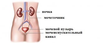
Radiation studies of the organs of the urinary and reproductive system usually begin with a survey X-ray of the kidneys and all urinary tracts. However, this method provides doctors with only part of the necessary information about the condition of these organs and their ability to perform their function. Many more precise methods are used, for example, x-rays of the bladder, which allow one to study a specific area of the urinary tract.
X-ray examination of the bladder using radiocontrast
To do this, they use the ability of the kidneys to take up contrast agents from the blood, which then enter the urine, making it possible to examine structures such as the renal calyces, pelvis system, ureters and bladder. This study is called excretory (infusion) urography.
general information
The procedure involves the introduction of a radiopaque contrast agent into the cavity of the bladder. Doctors fill the cavity with a solution (from 50 to 200 ml depending on the age of the child) containing 10-30% Urografin, Iodamine, Triombrast.
At the beginning of the procedure and after filling all areas with a contrast agent, the doctor takes x-rays to assess the condition of the genitourinary system. A special preparation stains the tissue, all affected areas stand out clearly in the photographs. According to the results of the study, a clear radiographic picture of the bladder cavity is visible. The procedure, depending on the variety, takes from 30 to 60 minutes, with preparation - up to two hours.
Features and types of research
Cystography is carried out using two main methods:
- bottom-up method.
An X-ray contrast agent is injected into the bladder through the urethra using a catheter. To reduce discomfort, use Katedzhel gel with an analgesic effect; - top-down method.
The composition is injected into a vein, then the blood carries the substance throughout the body, after a certain time the drug enters the bladder and stains the tissue. From this point on, x-rays can be taken. The method is less painful, but the penetration of contrast into the blood increases the risk of allergic reactions.
On a note:
- with the development of non-contrast and small tumors, another type of study is required - pneumocystography. The main difference is the introduction into the cavity of the bladder not of a liquid form of the drug, but of gas. For the procedure, oxygen, carbon dioxide or nitrous oxide is used;
- Sometimes doctors combine gas and liquid to diagnose complex cases of diseases, abnormalities of the urinary tract and tumor process. Lacunar cystography is a highly informative method;
- If a child has problems with urination, the urologist may prescribe voiding cystography. The study is carried out directly during urine excretion. The technique provides accurate data on areas of X-ray contrast agent leakage. An unpleasant moment is psychological discomfort that is difficult for a child to cope with.
Diagnostic options
A urological examination is performed to identify possible pathologies of the urinary system and determine their stage. Diagnostics refers to x-ray methods with preliminary introduction of a contrast agent into the body. The contrast can be in gaseous or liquid form. Its distribution in the examined area makes it possible to visualize the affected parts of the genitourinary area on an x-ray.
Cystography options are classified according to the method of contrast administration and procedure technique. Main types: retrograde cystography, otherwise ascending, excretory (descending), voiding cystography. The decision on the choice of examination option is made by the urologist, depending on the individual indications of the patient.
This option is based on the introduction of a contrast agent into the blood. During natural circulation, the drug is delivered through the bloodstream to the kidneys, from where it enters the bladder with urine. The patient is given an intravenous injection of contrast, and x-rays are taken at hourly intervals. Descending cystography is prescribed less frequently than others due to the time duration of the study. This type of examination is recommended when, in addition to the ureters and bladder, it is necessary to evaluate the shape, outline and anatomical location of the kidneys.
The retrograde method is more informative, while the diagnostic time is shorter. This effect is achieved because the contrast agent is injected directly into the bladder through the opening in the urethra. Bypassing the bloodstream, the contrast is quickly distributed throughout the urinary organs and gives a clear picture on the x-ray.
This type of examination takes place in three stages. Contrast is initially administered through the urethra, as in ascending endoscopy. X-rays are taken during the process of emptying the bladder, this allows you to evaluate the performance of the organ. Next, a control image of an empty bladder is taken. The technique of voiding cystography is the most complex, but it allows us to identify pathological changes in organs at the initial stage of development.8
Advantages and disadvantages
Diagnostic studies using contrast have both strengths and weaknesses. Despite the high information content of cystography, the method has some negative aspects that are important to draw the attention of parents to.
The doctor should explain how to prepare the child for the procedure to reduce the risk of complications and inaccurate results. Doctors must prevent negative consequences, strictly follow diagnostic rules, take into account the patient’s age, and the condition of the patient’s problematic organs.
Advantages:
- All deviations that are difficult to recognize with and are visible on x-rays;
- for the most complete picture in complex cases, doctors can choose the optimal method: lacunar or pneumocystography;
- the result of the study is ready a short period of time after the procedure. The doctor evaluates tissue damage, gives a preliminary conclusion, and refers to a pediatric urologist;
- new X-ray machines with digital resolution make it possible to study the entire process: from filling the bladder to removing urine. With dynamic cystography, radiation exposure is significantly reduced, which is especially important when examining children.
Flaws:
- psychological discomfort;
- the child does not always understand the doctor’s instructions;
- Painful sensations appear during catheter insertion;
- complications are possible in the form of allergic reactions to the drug, nephrotoxic effect. With retrograde cystoscopy, the listed complications are absent, but other problems are possible: acute urine retention due to sphincter spasm and injury to the urethral mucosa (more often in elderly patients);
- When the walls of the bladder rupture, the penetration of the contrast agent into the bloodstream can provoke sepsis.
Symptoms
Symptoms of bladder cancer depend on the stage of the disease, type of tumor, malignancy, location, and complications present. Symptoms include hematuria and dysuria.
Hematuria
(blood in the urine) is a symptom that occurs most often in patients with bladder cancer and is one of the earliest. The amount of blood in the urine does not depend on the size of the tumor, since even the smallest tumor can cause severe bleeding, and a large tumor may not bleed.
Bleeding may occur at varying frequencies. There are cases when a month or a year passes after the first bleeding, which can delay the process of identifying the disease. In the later stages of the disease and with disintegrating tumors, bleeding occurs constantly, it can be severe and require surgical intervention.
Sometimes hematuria cannot be detected without a detailed urine test.
Dysuria
– urination becomes frequent and painful. Older people may experience urinary retention, which is mainly associated with the presence of prostate adenoma. If the tumor disintegrates, the urine may contain pus, particles of disintegrating tissue, and also have a strong odor.
Pain in the suprapubic region in patients is usually not associated with the process of urination, but intensifies at this time. Pain is most often associated with the transfer of pathogenic cells to surrounding organs. The spread of pain to the perineal area, genitals, sacrum and thigh is associated with damage to nerve endings.
Pain in the lumbar region appears due to the pressure of the tumor on the orifices of the ureters, as well as the development of hydronephrosis and pyelonephritis. During an exacerbation of pyelonephritis, patients experience fever and chills. Hydronephrosis and pyelonephritis can lead to kidney failure, which in turn causes constant thirst, nausea, dry mouth, loss of appetite and other symptoms. Bladder cancer can be significantly complicated by chronic pyelonephritis, which ranks first among the causes of mortality in this disease.
Indications for diagnostics
A complex diagnostic study is prescribed after a preliminary examination of a small patient. It is important to collect blood tests, urine tests, do an ultrasound, and conduct.
The introduction of a radiopaque substance into the bladder cavity in childhood is undesirable; the child and parents are often afraid of the procedure, and conditions are created for the development of stress. If, based on the results of other types of examination, there is no accurate data on the causes of negative symptoms, localization, and severity of the pathological process in the urinary tract, then cystography has to be performed.
Main indications:
- pathologies of the bean-shaped organs and bladder;
- bladder rupture or reflux;
- detection of a tumor of unknown etiology;
- accumulation of salt stones;
- developmental abnormalities of the genitourinary system.
Contraindications
Ascending cystography is not performed in the following cases:
- inflammation in the scrotum, urethra, bladder;
- excretion of blood clots in the urine or massive hematuria.
On a note!
Descending cystography has the same limitations as excretory urography: severe pathologies of the liver and kidneys, allergies to iodine preparations, renal failure, thyroid diseases, poor blood clotting.
In most cases, doctors avoid cystography and use other diagnostic methods. If descending or ascending cystography is unavoidable, parents will have to explain to the child how the study will be carried out and why they will have to be patient a little during the procedure.
Psychological preparation, an accessible explanation of the essence of the method and the importance of the examination are important. The less parents panic, the lower the risk of developing fear in their son or daughter.
To obtain reliable results, you will have to change the diet of the young patient:
- For a week, exclude from the menu all items that provoke increased gas formation in the intestines. Do not give legumes, carbonated drinks, baked goods, fresh milk;
- if the child suffers from constipation, then for two weeks before the procedure the little patient takes a mild laxative;
- children receive tea that prevents the accumulation of gases or dill water;
- two days before the bladder examination, the doctor categorically prohibits foods that cause flatulence;
- On the day of the study, an enema must be given to maximize the removal of food debris and gases from the intestines.
How to treat ? View a selection of effective treatment options.
About how excretory urography of the kidneys is performed using a contrast agent is written on the page.
How is the procedure performed?
All types of cystography cause psychological discomfort to the patient; with the ascending method of examination, pain is felt when inserting a urethral catheter. It is important to follow all medical commands to get the most accurate result. After the procedure, the urine often changes color: a radiopaque substance comes out.
Study information:
- After preparation, the patient lies down on the X-ray machine (“supine” position). During the first phase you need to lie still. The doctor takes a general photograph of the organs of the genitourinary system;
- Then the doctor proceeds to the most unpleasant part - he inserts a catheter through which the cavity of the bladder is filled with liquid with a radiopaque substance or gas. Up to 12 years of age, 50-100 ml of the drug is sufficient; for adolescents, the adult norm is 200-300 ml;
- During the administration of contrast and during examination, the doctor presses the catheter, which provokes pain and the urge to urinate. This step cannot be avoided - it is important to retain the contrast inside the cavity so that the substance does not leak out of the bubble during the procedure;
- After filling the bladder with a special substance, the radiologist takes several pictures in different projections: on the side, from the stomach and from the back. A small patient should raise his legs to 90 degrees and additionally raise his shoulders. In this position, the pictures are the clearest and most informative;
- at the end of the procedure, the doctor removes the catheter and takes a photo of the empty bladder;
- After the examination, the doctor examines the finished images and analyzes the image. If fistulas or ruptures of the walls of the urethra or bladder are detected, hospitalization of the child is required to prevent sepsis: through the zones of ruptures, contrast enters the blood;
- After the procedure, if there are no complications, a hospital stay is not required; most often, cystography is performed on an outpatient basis. Observation by doctors of the urology department is necessary if a small patient was previously admitted to a medical institution with severe pathologies of the urinary tract, and cystography is one of the routine examination methods.
Quantitative diagnostic methods
If there is a suspicion that there is an obstruction to the outflow of urine from the bladder, doctors radiometrically determine the volume of remaining urine. This is because in such cases, after urination, some urine still remains inside the bladder.
To measure its amount, a radiopharmaceutical is injected into the patient, and after one and a half or two hours the radiation intensity is measured over the bladder. After the patient is asked to empty it, the radiation level is measured again and the volume of residual urine is calculated. The most common and informative alternative to this study is ultrasound sonography. However, sometimes these methods are used together, as this makes it possible to obtain a complete picture of the pathological process.
Diagnostic results
What does cystography show? The procedure allows you to accurately determine the localization zone of the pathological focus, the severity of the changes and the spread of inflammation, the type of tumor process or the size of the stones. In the first image, the radiologist sees the exact location of the kidneys and other organs of the urinary system. After filling the cavities with an X-ray contrast agent, it is possible to identify pathologies that are difficult to recognize on ultrasound and cystoscopy.
Deviations are clearly visible in the colored organs:
- congenital defects of the bladder and bean-shaped organs;
- and renal-ureteral reflux;
- stones in the urinary tract;
- change in the structure and thickness of the walls of the bubble;
- fistulas, traumatic injuries to the walls of the bladder and other parts of the urinary system;
- tumors and stones of any size;
- inflammatory processes in natural filters, bladder, ureters, other pathologies.
With proper preparation and following the instructions of a radiologist, cystography in children provides accurate data on the nature, stage, and localization of pathological foci in the urinary tract. Parents must properly prepare the child psychologically and change the diet to eliminate interference during the study.
X-ray (cystography) of the bladder is an endoscopic diagnosis that is performed to identify pathological changes in the genitourinary system. The procedure is prescribed for both adults and children. An X-ray examination is done with the addition of a contrast agent, which allows one to study the integrity, condition and structure of the internal organ. As a result of the study, it is possible to identify serious diseases, make an accurate diagnosis and begin immediate treatment.
What can be seen in the photographs?
Normal urethrocystogram (pictures taken in different projections)
One of the main pathologies that are detected during X-ray examination are tumors. Cysts or neoplasms can develop covertly for a long time, without disrupting the functioning of body systems. The most informative method used to diagnose bladder tumors is computed tomography.
Doctors can make an accurate diagnosis of cancer only after cystoscopy with a biopsy, but there are a number of factors that make radiation examination necessary. Firstly, malignancy of papillomas most often occurs deep in the wall of the organ, and this cannot be determined by examining a biopsy specimen. Secondly, cystoscopy does not provide information about tumor growth into the wall and neighboring organs, nor does it show the presence of metastases in regional lymph nodes.
Therefore, if there is a suspicion of cancer, it is better to start the diagnostic search with radiation diagnostics. When an artifact is discovered, one cannot say whether it is benign or malignant. A reliable sign of cancer on radiographs is its germination deep into the wall of the bladder and invasion into the paravesical tissue.
Tomography is valuable because it can accurately visualize tumors in the fundus and apex of the bladder. Cystography is also capable of visualizing the process, however, for this, a double contrast procedure must be performed.
Lacunar cystogram of a patient with prostate adenoma, papillary tumor and bladder stones
Another use of radiography in bladder examination is to diagnose congenital anomalies.
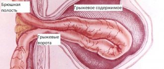
The most common of these is diverticulum. It is a cavity formation that is connected to the bladder through a thin neck. It is best determined by cystography. A ureterocele may also be present. At first glance, it may be similar to a diverticulum, however, its main difference is that it does not lie away from the organs, but is, in fact, a hernia of the ureter.
Features of bladder cystography
An X-ray of the bladder is highly informative and is prescribed by a urologist or surgeon. Cystography is carried out with the addition of a solution, which is administered in 2 ways: the first is ascending (using a catheter, 150-200 milliliters of the substance is administered through the urethra), the second is descending (intravenously). After intravenous administration, you should wait 30-45 minutes for the bladder to fill.
The following drugs can be used as contrast: urografin, iodamide, triomblast. X-ray with contrast helps to detect serious diseases: vesicoureteral reflux, fistulas, pathological narrowings, chronic cystitis (inflammation of the bladder), malignant and benign tumors, bladder diverticula, neoplasms, stones.
Contrast x-rays are prescribed to assess the condition of the bladder and urinary tract. Using the procedure, you can identify the cause of urine incontinence and analyze the excretory function of the kidneys. 25-30 minutes after the administration of the contrast agent, with normal kidney function, only remnants of the contrast will be visible in the pelvis and calyces. If a large amount of the substance remains in the pelvis and calyces, this will indicate slow excretion of urine.
X-ray examination of the bladder
Radiation studies of the organs of the urinary and reproductive system usually begin with a survey X-ray of the kidneys and all urinary tracts. However, this method provides doctors with only part of the necessary information about the condition of these organs and their ability to perform their function. Many more precise methods are used, for example, x-rays of the bladder, which allow one to study a specific area of the urinary tract.
X-ray examination of the bladder using radiocontrast
To do this, they use the ability of the kidneys to take up contrast agents from the blood, which then enter the urine, making it possible to examine structures such as the renal calyces, pelvis system, ureters and bladder. This study is called excretory (infusion) urography.
Indications for the procedure
The bladder appears on an x-ray as a shadow in the form of a transverse oval, the lower edge of which coincides with the upper edge of the pubic bone. In the case of urography, its shadow has medium intensity and an even contour. The advantages of urography when examining the bladder are:
- The procedure is available to all patients.
- The research has a low cost.
- It is non-invasive (no penetration).
- The doctor has the opportunity to study the structure of the kidneys, ureters and bladder in one study.
- Possibility of diagnosing calcifications at different parts of the urinary tract.
However, urography also has a number of disadvantages.
It provides only limited information about the structural features of the renal parenchyma, and cannot completely replace other methods for their study. With its help, it is impossible to obtain data on the functional state of urination, and it is not possible to perform a study with reduced renal filtration. It is contraindicated in case of heart, liver, kidney failure and intolerance to iodine preparations.
Other studies
An alternative research option is the introduction of a contrast agent through the urethra itself. In this case, it is assumed that cystography has been done, or an x-ray of the bladder itself. Since the concentration of contrast in this case is much higher, you will get a shadow of high intensity that stands out against the background of the bones.
In the case of a normal x-ray picture, the shadow of the bladder is homogeneous, its contour is even and regular. If there are stones or a tumor inside, then either the uniformity of the shadow or the clarity and evenness of the contours changes.
Indications for cystography are:
- Traumatic extravasation.
- Postoperative extravasation.
- Suspicion of bladder diverticula.
- Suspicion of vesicoureteral reflux.
Also, this procedure can be prescribed after abdominal injuries, when there is no accurate information about the condition of the bladder. It is also carried out when there is a violation of the innervation of this organ, which can manifest itself in various types of urinary outflow disorders. It is also convenient in diagnosing urolithiasis and recurrent cystitis. In the latter case, cystography can also prevent the development of neoplasms, since in the case of a chronic course, hypertrophy of the bladder walls develops, from which cancer can develop.
When urinating, the contrast agent moves from the bladder into the urethral space. The filming carried out at this time is called voiding cystography. It allows you to obtain an image of the initial parts of the urethra and evaluate the functioning of the bladder sphincters.
Quantitative diagnostic methods
If there is a suspicion that there is an obstruction to the outflow of urine from the bladder, doctors radiometrically determine the volume of remaining urine. This is because in such cases, after urination, some urine still remains inside the bladder.
To measure its amount, a radiopharmaceutical is injected into the patient, and after one and a half or two hours the radiation intensity is measured over the bladder. After the patient is asked to empty it, the radiation level is measured again and the volume of residual urine is calculated. The most common and informative alternative to this study is ultrasound sonography. However, sometimes these methods are used together, as this makes it possible to obtain a complete picture of the pathological process.
What can be seen in the photographs?
Normal urethrocystogram (pictures taken in different projections)
One of the main pathologies that are detected during X-ray examination are tumors. Cysts or neoplasms can develop covertly for a long time, without disrupting the functioning of body systems. The most informative method used to diagnose bladder tumors is computed tomography.
Doctors can make an accurate diagnosis of cancer only after cystoscopy with a biopsy, but there are a number of factors that make radiation examination necessary. Firstly, malignancy of papillomas most often occurs deep in the wall of the organ, and this cannot be determined by examining a biopsy specimen. Secondly, cystoscopy does not provide information about tumor growth into the wall and neighboring organs, nor does it show the presence of metastases in regional lymph nodes.
Therefore, if there is a suspicion of cancer, it is better to start the diagnostic search with radiation diagnostics. When an artifact is discovered, one cannot say whether it is benign or malignant. A reliable sign of cancer on radiographs is its germination deep into the wall of the bladder and invasion into the paravesical tissue.
Tomography is valuable because it can accurately visualize tumors in the fundus and apex of the bladder. Cystography is also capable of visualizing the process, however, for this, a double contrast procedure must be performed.
Lacunar cystogram of a patient with prostate adenoma, papillary tumor and bladder stones
Another use of radiography in bladder examination is to diagnose congenital anomalies.
The most common of these is diverticulum. It is a cavity formation that is connected to the bladder through a thin neck. It is best determined by cystography. A ureterocele may also be present. At first glance, it may be similar to a diverticulum, however, its main difference is that it does not lie away from the organs, but is, in fact, a hernia of the ureter.
Methodology of the procedure
The study is quite safe and can be performed on all patients except pregnant women, since the influence of X-rays on the fetus cannot be allowed. Cystography, in which a contrast agent is injected into the bladder through the urethra, is not recommended for people with acute infectious diseases, as well as for weakened patients. This procedure should not be performed if a traumatic rupture of the organ is suspected; in this case, excretory urography should be prescribed.
Schematic representation of ascending urethrocystography
All studies are carried out on an empty stomach. The patient must be warned in advance about the procedure; the doctor must collect a detailed medical history, clarify the tolerance of iodine and drugs containing it, if urography is planned. The patient should cleanse the intestines the night before, and the procedure is repeated in the morning on the day of the study. A person must come to the radiology room on an empty stomach. The patient is then placed in a supine position and a plain radiograph is taken. Then they begin additional research methods.
diagnostinfo.ru
Indications and contraindications
Indications include: ruptures and reflux, congenital pathologies, complications after infectious and inflammatory diseases, enuresis, sand and stones, tuberculosis of the internal organs of the genitourinary system, suspected tumor, trauma to internal organs, diseases of the bladder and kidneys.
Although the diagnosis is considered informative and reliable, it has contraindications. Cystography should not be performed on pregnant women; patients who have an inflammatory process in the bladder or ureters (the catheter tube can injure the urethra, which leads to aggravation of the inflammatory process); people who pass urine containing blood clots (clots can distort X-ray images and cause incorrect test results).
The origin and development of bladder cancer
The causes of neoplasms can be the following factors:
- carcinogenic substances and chemical elements that fill the external environment;
- age and gender of the patient;
- chronic inflammation of the bladder;
- hormonal disorders;
- diseases caused by parasites;
- vehicle exhaust gases, smoking.
There are many cases of bladder cancer caused by carcinogens that affect DNA, damaging it and leading to mutations.
The development of bladder tumors in older people is explained by the fact that carcinogens have a negative effect on the mucous membrane of the bladder for a longer period of time. Irreversible age-related changes also occur, for example, hormonal imbalance, decreased immunity, and changes in the structure of the bladder mucosa.
Alternative research methods
If there are contraindications, the patient may undergo alternative diagnostics, for example, magnetic resonance imaging or pneumocystography. The peculiarities of pneumocystography are that gas is injected instead of a contrast solution. Sometimes lacunar cystography is prescribed - a combined procedure, during which a specialist injects 15-20 milliliters of contrast and 180-200 cubic centimeters of gas.
Another effective diagnostic method is voiding cystography, which is performed during urination. During the procedure, the work of the urinary canals is monitored, and areas of leakage of the contrast agent can be detected.
Cystography and other manipulations are needed to determine in detail the parameters and location of the bladder, identify pathologies and anomalies, stones and sand, ruptures, thickening of the walls of the internal organ, disruption of the urinary system and other dysfunctions.
An X-ray of the bladder is performed if ultrasound and cystoscopy fail to help make the diagnosis. The procedure is not recommended for the purpose of preventing the disease, since it is painful and the patient often feels discomfort during it.
Preparation
Preparation is also important for cancer patients. Yes, suspicion of bladder cancer is very serious, but this stage should still not be neglected. The fact is that the procedure always involves the use of a contrast agent, and a study using it will be informative only if you follow some rules in advance:
- 3 days before the study, you must begin to follow a special diet (limit any foods that increase gas formation in the intestines);
- take activated charcoal one day in advance, which will help with cleansing;
- in the evening before the examination, as well as in the morning immediately before it, it is necessary to do a cleansing enema;
- You need to come for diagnostics on an empty stomach; you cannot eat 6-8 hours before.
Bladder tuberculosis.
X-raying of the bladder for children
X-rays are performed on children from the age of five months. Parents are afraid of such an examination, since the child’s body is exposed to radiation and a special solution is injected. But if the attending physician insists on performing the procedure, you should prepare your child for the upcoming diagnosis.
For 1-2 weeks, the child should not drink carbonated water or eat food that can cause excessive gas formation and flatulence. In the morning, before cystography, it is recommended to give the child a cleansing enema. To avoid an allergic reaction to the medications that will be administered, it is advisable to conduct an allergy test.
Features of bladder cystography of a child:
- a catheter tube will be inserted through the urethra through which contrast is injected;
- the drug can be prescribed before and after urination;
- small children are given anesthesia to make the procedure painless;
- you should not eat or drink 4-6 hours before the test;
- the examination takes no more than 10-15 minutes;
- after the x-ray, the child is under the supervision of medical staff for 2 hours (during this time the bladder should empty).
After the procedure, the baby should take a furagin tablet to prevent the occurrence of an inflammatory process in the genitourinary system. Adverse reactions occur rarely. The child needs to drink a lot of fluids throughout the day so that the medication is eliminated faster.
Methodology for examining children
An infant's bladder can be x-rayed no earlier than five months of age. Many parents are not ready to expose their child to radiation, and are also wary of injecting contrast into the bladder. If there is no other way out, then you should carefully prepare for the procedure.
The child should receive dietary food that does not cause bloating for about a week. On the day of the procedure, an enema is indicated to cleanse the intestines. A drug test should be carried out.
If there are compelling reasons to examine the bladder, an x-ray is performed on the child as follows:
- a contrast agent is injected into the bladder through the urethra;
- contrast is administered both before and after urine output;
- so that young children can endure the procedure, they are given anesthesia;
- the child should not eat or drink 6 hours before the procedure;
The duration of the examination is no more than 15 minutes.
X-ray of a child's bladder. Source: dcenergo.ru
After the x-ray, the little patient remains under the supervision of medical staff for 2 hours. It is important to empty your bladder during this time so that all the contrast material comes out. It will take about a day for complete removal. At this time, an increased drinking regime is indicated.
How is cystography performed on adult patients?
You should prepare in advance for x-rays. 4-5 days before the test, you must stop eating foods that cause gas and flatulence. It is forbidden to drink strong coffee and tea, sparkling water. You should refrain from consuming fermented milk products, whole milk, corn, cabbage, grapes and legumes. Before the procedure, you need to do a cleansing enema.
With the ascending diagnostic method, the patient takes a horizontal position, then the specialist injects 200 milliliters of a special solution into his bladder. After the substance is completely injected, the doctor clamps the catheter so that the injected liquid does not flow back out. Next, you need to listen to the specialist’s commands so that the pictures are detailed and accurate.
Images are taken in different positions: lying on your back, on your side, during and after urination. When the bladder is empty, comparison pictures will be taken. A diagnosis will be made based on the x-ray images.
Descending diagnosis is carried out 30-60 minutes after administration of the drug into a vein. During this time, the contrast fills the bladder and provides detailed visualization of the contours of the internal organ. Next, photographs are taken.
The medical staff warns the patient in advance that the examination may be accompanied by pain and unpleasant discomfort. If the patient has a high pain threshold, for example a child, the examination will be carried out with simultaneous anesthesia. Before cystography, it is recommended to consult with a nephrologist, urologist and radiologist. They will be able to give recommendations on how to carry out the manipulation.
To protect yourself from side effects, after the x-ray, the patient should remain in bed for 24 hours and drink plenty of fluids.
Methodology for examining adults
Patients must prepare for the study. All foods that cause signs of flatulence are excluded from the diet. The following drinks are prohibited: tea and strongly brewed coffee, carbonated water.
The patient needs to lie down. Then he is injected with 200 ml of a solution obtained by diluting the contrast agent.
When all the fluid has been introduced, the catheter is clamped. Otherwise, the solution will flow back. The specialist will manage the process, so all his commands must be followed. As a result, the images will reflect the health status of the organ as accurately as possible.
X-ray of the bladder is performed in several positions. The patient will have to turn from back to side. Pictures are also taken after the bladder is empty. By comparing the images that were taken when the bladder was full and when it was empty, a reliable diagnosis can be made.
Diagnostics using the descending type are carried out 30 minutes after the administration of contrast. Or even in an hour. This pause is necessary to provide more detailed visualization of the contours of the bubble. And after this, the pictures will show whether there are pathological changes.
Doctors always warn patients that some aspects of the procedure may cause discomfort and pain. If such inconveniences are unpleasant due to an increased pain threshold, anesthesia is given.
Cystography requires preliminary consultation with a urologist, nephrologist and radiologist. It is important to discuss all the nuances of the study with these specialists in order to carry out the procedure with maximum benefit and comfort as possible.
Examination of the bladder - x-ray with contrast injection - requires bed rest after the procedure. The patient needs to drink a lot of clean water to cleanse the body of the solution.
Decoding images
On images, a normal bladder should have a pyramidal or oblong shape with round or oval contours. Women most often have an oval bladder; X-rays show a concavity of the outer contour. The pear-shaped bladder is characteristic of children. When diagnosing with contrast, you can monitor the condition of the organ and identify diverticula, calculi (stones) and adenoma (benign or malignant). In men, the internal organ has a triangular shape.
If the organs of the genitourinary system are asymmetrical and have a strange structure, the radiologist sees this as a pathology and writes it down in his report. During interpretation of cystography, pathological displacements of the bladder, called cystoceles, can be detected.
A qualified radiologist will be able to decipher images and describe them in 15-30 minutes. Next, the patient is sent with a conclusion to the attending physician, who referred him for diagnosis. With a correct diagnosis, serious illnesses can be prevented and effective treatment can begin.
Bladder x-ray is a type of contrast study. The study allows you to detect changes in shape and pathological formations (X-ray “minus-shadow” syndrome). Has a high degree of reliability. Used in urology and surgery.
An alternative replacement for this X-ray diagnosis can only be magnetic resonance angiography. These research methods have different purposes and purposes, so they should not be compared.
What does a contrast x-ray of the urinary system show?
X-ray of the bladder and kidneys with contrast agent
Contrast X-ray of the urinary system shows the following diseases:
- stones;
- foreign bodies;
- tumors;
- diverticula;
- chronic cystitis;
- pathological narrowing;
- fistulas (artificial passages between organs);
- Vesicoureteral reflux.
Contrast radiography of the bladder is performed to evaluate the causes of urinary incontinence. It allows you to analyze the excretory function of the kidneys. 24 minutes after the administration of contrast, physiologically, during normal functioning of the renal excretory system, only remnants of iodide radiopaque urografin can be traced in the projection of the pelvis and calyces. If a lot of the substance is retained, slow urine excretion can be suspected.
Application area
The cystographic technique is most often used to confirm the existing diagnosis. The indications for it are very extensive. It is prescribed to infants to identify developmental anomalies of the urinary tract (double bladder, atresia). For an adult patient, cystography is prescribed in a situation where the doctor suspects tumor diseases, tuberculosis of the genitourinary organs, bladder diverticula and other diseases of the urinary system. The presence of stones and sand, nephrotic syndrome are also indications for such a study. Cystography shows the presence of reflux, rupture, various complications after infections, tuberculosis, prostate cancer. It is also used for urinary incontinence. Cystography is mandatory for traumatic injuries, for fistulous openings between the intestine and the bladder.
Radionuclide cystography is used to detect vesicoureteral reflux; it is based on the introduction of labeled proteins injected with contrast
Should a child be afraid of cystography with contrast?
Cystography is prescribed for children starting from the 5th month. Some parents are afraid of the procedure, as it involves radiation and contrast injection.
Cystography is prescribed to identify serious pathology, and therefore is necessary if recommended by a doctor.
In order not to be afraid, we describe to parents the stages of the procedure:
- A urinary catheter (contrast tube) is inserted through the urethra.
- A contrast agent is given before and after urination.
- If x-rays are performed on small children, anesthesia is administered.
- Preparation requires fasting and stopping drinking liquids 3 hours before the examination.
- The duration of cystography is 10 minutes.
- After 20 minutes, the child can be given food and drink (when he wakes up from anesthesia).
Doctors observe the baby for 2 hours after the X-ray. During this time, the bladder should be emptied and the urinary contrast material should be removed.
At home, the child should be given furagin to drink to prevent inflammation.
Obviously there is nothing to be afraid of, as side effects are rare.
Factors influencing the result of the study
- Presence of gas, stool, contrast agent or foreign bodies in the intestines (poor image quality).
- The presence of calcified fibromatous nodes of the uterus or space-occupying formations of the ovaries.
- Obesity or ascites (poor image quality).
BH Titova
“General radiography of the urinary system” and others
In order to identify pathologies of the urinary system, the patient is often prescribed cystography of the bladder, with the help of which a detailed examination is carried out. This procedure helps make a diagnosis. X-ray diagnostics is carried out using various methods, which necessarily require careful preparation. This type of study is also carried out in newborns if there is a suspicion of the development of urological problems.
How to do an x-ray of the urinary system
X-ray of the urinary system and bladder may be:
- ascending;
- descending.
An ascending x-ray is done after contrasting the genitourinary tract. For these purposes, 150-200 ml of the substance is sufficient. The patient is advised to bend his legs at the knees and hip joints.
The vertical direction of the central beam during exposure allows clear visualization of the bladder. To prevent shadows of the pubic bones from superimposing, it is necessary to conduct research in three intersecting planes: right and left oblique, axial.
Ascending is carried out using a urinary catheter (as in the example described above).
Descending urography is carried out simultaneously with excretory urography 30 minutes - 1 hour after the contrast enters the bloodstream. By this time, the substance densely fills the bladder and provides clear visualization of the contours of the organ.
Descending examination is used when, for certain reasons, a catheter must be inserted into the bladder, showing the structure of the urethra, bladder, and the location of the prostate gland.
What does the bladder look like on a photo?
In the image, a normal bladder is characterized by the following indicators:
- pyramidal or oblong shape;
- oval or rounded contours.
What does the genitourinary system look like on x-ray in women, men and children:
- In women, the bladder shape is oval. Its transverse size is larger than its longitudinal one. The cystogram visualizes the concavity of the outer contour.
- In children, the organ is pear-shaped. It faces the narrowed part towards the pubic bones. Physiologically, the shadow of the bladder is located at the level of the pubic symphysis.
Cystography with liquid contrast clearly shows pelvic tumors (adenoma, stones, diverticula). On an axial radiograph in men, a triangular shape of the organ can be seen. In this case, the bag-like protrusions look like areas of clearing against the background of an intense metallic shadow.
Fibroids and an increase in the size of the uterus take on a saddle shape when contrasted. Depending on the position of the uterus, the compression may be located in the middle part of the bladder.
Asymmetrical deformation causes the most bizarre shapes, which the radiologist classifies as a sign of pathological changes.
Cystocele is a pathological displacement of the bladder in which the lower contour is located below the pubic symphysis. X-rays for this disease are valuable before planning surgery.
Contraindications to x-ray diagnostics of the urinary tract
Contraindications to x-ray diagnostics of the urinary tract are:
- Acute inflammatory processes of the urethra.
- Diseases of the seminal vesicles and prostate gland.
The conditions described above are not contraindicated for ascending cystography.
In conclusion, I would like to note that radiography of the urinary tract of a child is performed in case of emergency, since it has a radiation load on the pelvic organs. Cystography for adults is carried out on the basis of research.
X-ray studies of the urinary system remain the highest quality and most promising. Over the long period of existence of this diagnostic method, one can note its gradual development.
One type of contrast study is an x-ray of the bladder. The study facilitates the accurate detection of pathological formations and has a significant level of accuracy.
What pathologies can an x-ray of the bladder with a contrast agent detect?
Formations in the form of stones
Objects that came from outside.Types of tumor neoplasms
Changes by which cystitis can be identified.
Fistulas formed as a result of inflammatory processes.
Often, an x-ray of the bladder with a contrast agent is performed in the presence of urinary incontinence, since this type of x-ray allows an in-depth study of the excretory potential of the kidneys. Twenty-four minutes after the administration of the contrast agent, provided that the renal excretory system works smoothly, only minimal traces of the contrast agent, which is the iodide radiopaque urografin, are detected - this indicates a slowdown in the process of urine excretion.
Is it safe to use a bladder x-ray with contrast (cystography) for a child?
Many parents are afraid of carrying out this type of examination on their children, since it involves radiation and contrast injection. To allay concerns, it is worth knowing the following nuances:
Inserting a catheter into the urethra
Injecting contrast before and after urination.If the patient is young, anesthesia can be used
Stop eating and drinking three hours before the procedure.
The approximate duration of the procedure is ten minutes.
Twenty minutes after it ends, you can resume eating and drinking water.
Within two hours after diagnosis, doctors need to monitor the child’s condition. During this period, the contrast agent should be completely eliminated from the body. Further, in order to avoid inflammation of the urethra, it is prescribed to use the drug furagin. And, in fact, there is nothing to fear, since the risk of developing side effects is almost zero.
Progress of the cystography procedure
Cystography is divided into ascending and descending types. The first is performed with catheterization of the urethra and requires less contrast agent than the second, ascending type of urography. The second is carried out at the same time as urography, aimed at determining the excretory capacity of the excretory system after an hour, which makes it possible to achieve dense filling of the bladder and the appearance of clear boundaries of the organs being studied in the image.
The need for a descending study occurs when catheterization is required in order to obtain information about the bladder, prostate gland and urethra.
Bladder X-ray
Based on the results of the image, the bladder may be given characteristics such as pyramidality or elongation, as well as roundness. In organisms that differ in structure, the shape of the bladder varies accordingly. For example, the female bladder is oval in shape, and its transverse part is wider than the longitudinal one. The child's bladder is pear-shaped, with the narrow part facing the pubic bone.
Bladder X-rays with contrast are particularly effective in detecting tumors in the pelvic area, such as adenomas. Such formations are clearly visible as a light area in the metallic shadow of the image. Pathological displacements of the bladder are also detected in this way and contribute to the effective collection of information before surgery.
How is an x-ray of the urethra and bladder done using a contrast agent?
Radiation examination methods are used in urology to identify problems with the urinary system. They give a clear picture of the condition of the bladder and urethra. These methods include x-rays of the bladder - cystography and urethra - urethrography using a contrast agent.
What are the types of radiation methods for urological examination, and in what cases are they prescribed?
Both bladder cystography and urethrography are types of x-ray examinations. In this case, urethrography is an examination of the urethra, and cystography is an examination of the bladder.
There are ascending and descending urethrography.
Ascending examination is performed on men.
The patient is placed in a horizontal position and a radiopaque liquid is injected into the urethra. At the moment of maximum filling of the urethra, the penis is raised and a picture is taken. Carrying out such an examination for women is very problematic (due to the peculiarities of the anatomy).
Descending (mictional) urethrography is often combined with cystography.
A large amount of contrast fluid is injected into the urethra (so that the bladder is also full). The patient is then asked to urinate and a picture is taken of the bowel movement.
Other types of urological X-ray examinations:
- Urography (kidney examination);
- Retrograde ureteropyelography (ureters are examined);
- Pyelography (examination of the kidney cavities);
- Pneumorenus (assessment of the external contours of the kidneys and adrenal glands).
The purpose of all of the above procedures is to obtain a clear image of the necessary part of the genitourinary system using contrast material.
X-ray images will help in diagnosing various diseases and selecting the optimal treatment method. Urethrography in men is usually performed to identify the causes of decreased urine flow, benign prostate tumors, strictures, and urethral cysts.
Cystography is performed on both men and women to detect stones in the bladder and identify the causes of hematuria. X-ray examinations are also carried out if there is a suspicion of a rupture of the bladder wall.
Preparation for the procedure and procedure for urethro-cystography
Cystography and urethrography are often performed simultaneously, since the procedure is almost identical. A comprehensive examination of the bladder and urethra is called urethrocystography.
Recommendations for preparing for the study depend on the suspected disease. If the purpose of the procedure is to detect stones, then the patient must refuse to eat or drink much fluid for at least 8 to 12 hours. Otherwise, you can live your daily life, sticking to your normal diet.
To stretch the bladder, the patient is given approximately 400-500 ml to drink an hour before the procedure. ordinary clean water. It is forbidden to urinate during this hour.
The examination is carried out in an X-ray room. The patient is put on a robe without metal fasteners, the piercings are removed from the genital area and navel, and then a catheter is inserted into the urethra.
Adult women and men are examined without anesthesia, and local anesthesia may be used for the procedure for children. A contrast agent is injected through the catheter (usually a liquid that darkens on an x-ray, but sometimes a special gas is also used).
Depending on the type of procedure, the picture is taken at the moment of maximum filling of the bladder (if retrograde cystography is prescribed), or during urination (if descending urethrography is necessary). At the end of the examination, the therapist will lubricate the urethral opening with a soothing gel with an antibacterial effect.
Contraindications for procedures
Before taking an X-ray of the patient’s bladder, the urologist will definitely examine the medical record for any serious abnormalities in the patient’s health. In particular, the procedure will have to be postponed if:
- Radiotherapy was recently performed;
- Acute renal failure was diagnosed;
- There is a suspicion of the presence of acute inflammatory processes in the genitourinary system;
- Urethrorrhagia was detected.
Also, urethrocystography is postponed if a woman is carrying a child. The procedure can be performed in the first trimester of pregnancy, but only if the potential benefits of the examination significantly outweigh the risk of harm to the fetus.
What side effects may occur during urethrocystography?
One of the most common side effects of urethrocystography is an allergy to the contrast agent. To determine how the body reacts, the radiologist may administer a small amount of gadolinium beforehand.
Damage to the urethra may also occur. Most often, male patients are susceptible to such complications (especially if the procedure is performed on an emergency basis). Trying to insert a catheter into an already injured urethra can lead to stretching of the wound.
If a non-sterile catheter is used, urinary tract infection may occur. After the procedure is carried out in violation of the rules of asepsis, minor discomfort in the urethra usually occurs. Then a strong burning sensation will develop when urinating. If unpleasant symptoms do not go away within 24 hours, the doctor will prescribe antibiotics.
Other side effects that usually resolve within 24 hours without additional medical intervention include:
- Increased urge to urinate;
- Slight increase in temperature (up to 37º);
- Chills;
- Trembling in the lower extremities;
- Discharge of blood from the urethra.
The above complications usually do not pose any threat, but if the temperature rises above 38°, you need to see a doctor again.
Cystourethrography is performed quite often, so there is no need to worry before the procedure. You need to know information about side effects in order to independently recognize a deterioration in your health condition and promptly seek help.
cistitus.ru