Kidney surgeries
Surgical interventions on the kidney are carried out using various approaches: extraperitoneal, transperitoneal or transthoracic. The most commonly used extraperitoneal approach through exposure of the retroperitoneal space is lumbotomy. The incision for lumbotomy is made in the lumbar region.
Rice. 3. Operative approaches to the kidney: 1 — Fedorov’s incision; 2 - Simon's section; 3 — Bergmann-Israel section; 4 — Cherny section; 5 - Pean's section.
The most common are the Fedorov section and the Bergmann-Israel section (Fig. 3).
For large kidney tumors, when approach to the upper pole of the kidney is difficult, combined approaches are used: thoraco-abdominal extraperitoneal and abdominal transperitoneal-extraperitoneal.
Most kidney operations are currently performed under endotracheal anesthesia with the use of muscle relaxants, which greatly facilitates access to the retroperitoneal space to the kidneys.
The patient is placed on the operating table on the healthy side. The nature of the operation is dictated by the disease and condition of the patient.
In case of paranephritis, the retroperitoneal space and purulent focus are opened, followed by drainage with rubber drainage and gauze swabs. In acute pyelonephritis and apostematous nephritis, the kidney is decapsulated. Often this operation is accompanied by another operation - nephrostomy (see) or pyelostomy.
In case of multiple kidney stones or coral stones, nephrotomy is performed, combining it with pyelolithotomy (an operation of dissecting the pelvis to remove stones). More often, an incision is made along the posterior or lower surface of the renal pelvis, to which access is freer and safer. With a posterior pyelotomy, it is dissected in the longitudinal direction, after which the stone is removed from the incision of the pelvis with a special tool. If the kidney stone was infected, then the operation is completed by draining the pelvis with rubber drainage - pyelostomy; if there was no secondary pyelonephritis, then several catgut sutures can be placed on the incision of the pelvis and the perinephric space can be drained.
In case of kidney tumors or large degenerative changes in the kidney, the kidney is removed - nephrectomy.
In case of pathological mobility of the kidney, it is fixed in the renal bed - nephropexy; in case of hydronephrosis, plastic surgery is often performed on the renal pelvis and ureteropelvic anastomosis.
In case of kidney tuberculosis, in case of traumatic injury, in case of kidney stones, part of the kidney is removed, usually one of the poles - kidney resection.
In case of polycystic kidney disease, patients undergo an operation that combines the emptying of kidney cysts with improving its blood supply by sewing an omentum into the renal parenchyma - omentorenopexy. In case of hypertension caused by narrowing of the renal artery, plastic surgery is performed on it.
Nursing care - see Postoperative period.
Surgical approaches to the kidney, its exposure and interventions on it are carried out using various approaches: extraperitoneal, transperitoneal (abdominal) and transthoracic (thoracic). In some cases, for example, with large renal tumors, a combined approach to the kidney is used—thoracoabdominal. Most kidney surgeries are performed from an extraperitoneal approach by exposing the retroperitoneal space - lumbotomy. A lumbotomy can be performed through various incisions in the lumbar region. The most common are the Fedorov and Bergmann-Israel incisions; the Simon, Pean, and Cherny incisions are used less frequently (Fig. 35).
For very large kidney tumors and adrenal tumors, a Nagamatsu incision is used: a vertical incision along the edge of the rectus dorsi muscles with continuation to the anterior abdominal wall along the lower edge of the XII rib with subperiosteal resection near the vertebrae of the X, XI and XII ribs.
During kidney operations, the patient is placed on the operating table on the side opposite to the surgical intervention, and a cushion is placed, which facilitates access to the kidney. After cutting the skin and tissue, the muscles are dissected and the retroperitoneal space is exposed. The peritoneal sac is pushed inward. The renal capsule is opened behind and the kidneys are isolated from the perinephric tissue. Intermuscular approaches to the kidney without dissecting the muscles, by moving them apart along the fibers, have become possible with the use of modern types of pain relief using muscle relaxants.
Kidney decapsulation - removal of the fibrous capsule - is used for acute pyelonephritis, perinephritis, and sometimes for renal failure. As a result of this operation, it is possible to reduce increased intrarenal pressure and improve blood and lymph circulation in the kidneys. After exposing the kidney, a longitudinal incision of the fibrous capsule is made along its lateral edge (Fig. 36). A grooved probe is inserted into the incision and, lifting the capsule, it is dissected along it. Then the edges of the capsule are peeled off from the renal parenchyma up to the renal hilum. There is no need to excise the fibrous capsule. Kidney decapsulation is often combined with other operations, such as nephrostomy.
Nephrotomy - an incision in the renal parenchyma - is performed to remove stones and foreign bodies from the kidney, to perform nephrostomy, and sometimes for diagnostic purposes. Longitudinal sectional nephrotomy is used to remove large coral stones. An incision of the renal parenchyma is made longitudinally along the Tsondeka line, 0.5 cm posterior to the convex edge of the kidney. Sectional nephrotomy should be performed after preliminary temporary compression of the vascular pedicle of the kidney. To do this, it is necessary to mobilize the kidney and apply a soft clamp to the vascular pedicle. In addition to sectional longitudinal nephrotomy, transverse nephrotomy is used. The time of cessation of blood circulation in the kidneys should not exceed 30 minutes. Local hypothermia - cooling the kidney to a temperature of 14-16° - allows you to turn off blood circulation in the kidney for a longer time, reduce bleeding from the renal wound and improve wound healing and the course of the postoperative period. More often it is necessary to use partial nephrotomy in renal surgery by dissecting the renal parenchyma above the calyx or pole of the kidney to remove the stone. After nephrotomy, bleeding is stopped by applying catgut sutures to the wound (Fig. 37). The application of large mattress, U-shaped tightening sutures to the renal parenchyma is not justified, since this results in renal ischemia with the development of infarctions and secondary bleeding. Extensive nephrotomy must be combined with nephrostomy.
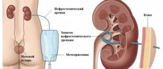
Nephrostomy - renal fistula; provides drainage of the collecting-pelvis system, is performed in case of acute purulent-inflammatory processes of the kidneys, hydronephrosis, calculous anuria, etc. In the case of thinned renal parenchyma, it is enough to make a nephrotomy 2 cm long and insert a rubber drainage into the pelvis through the renal wound. If there is a large amount of renal parenchyma, for the correct installation of renal drainage, it is necessary to dissect the pelvis (pyelotomy), then perforate the parenchyma through one of the renal calyces using a Fedorov clamp and, grasping the drainage tube with it, insert it into the pelvis. The drainage should be fixed to the fibrous capsule of the kidneys with a catgut suture (Fig. 38).
Pyelostomy is the placement of a fistula on the renal pelvis. This operation is performed less frequently than nephrostomy. After dissecting the renal pelvis, a rubber drainage tube is inserted into it, which is fixed with a catgut suture to the edges of the renal pelvis wound (Fig. 39). Nephrostomy with ring-shaped drainage, which consists in passing a drainage tube through wounds of the pelvis and kidney, has not been justified, as it causes severe complications up to the cutting of the drainage tube through the entire renal parenchyma.
Pyelotomy is a dissection of the renal pelvis. This operation is usually used to remove stones from the pelvis and calyces. Depending on the location of the incision of the pelvis, anterior, lower, posterior and upper pyelotomy are distinguished. Anterior pyelotomy is dangerous due to possible damage to the renal vessels; it is used in the presence of a large extrarenal pelvis, and more often on abnormal kidneys (horseshoe-shaped, dystopic kidney), in which the pelvis is located in front, away from large vessels. Much more often it is necessary to perform a posterior pyelotomy (Fig. 40).
After isolating the posterior surface of the kidney from the pelvic fat, the posterior wall of the pelvis is released and dissected. With an intrarenal type of pelvis, you should first move the posterior lip of the kidney outward with a finger or a hook, which is usually easy with a well-developed renal sinus. The renal lip is lifted with a hook, as a result of which the posterior surface of the pelvis is exposed (Fig. 41). On the inner surface of the renal lip, and sometimes along its lower edge, there are a. et v. retropyelica, wounding of which should be avoided. The wall of the renal pelvis is dissected in the longitudinal or transverse directions. When removing large and especially staghorn stones, it is necessary to dissect not only the pelvis, but also the calyx; This operation is called calicotomy.
With the intrarenal type of pelvis, if stones are located in the pelvis and lower calyx, a lower pyelotomy is used. After isolating the lower pole of the kidney and the upper third of the ureter, the ureteropelvic segment, usually covered by the renal parenchyma, is mobilized. The lower pole of the kidney is peeled off from the pelvis and moved outward with a hook; as a result of this, the lower surface of the pelvis is exposed, which is cut longitudinally. When performing a lower pyelotomy, you should not damage the ureteropelvic segment itself, since if it is injured, a stricture may subsequently develop.
To remove large stones from the upper calyx, an upper pyelotomy is used. The technique of this operation is similar to lower pyelotomy, but is more difficult and requires preliminary mobilization of the entire kidney with its dislocation into the wound. After pyelotomy, it is advisable to suture the wound of the pelvis with thin catgut sutures. If this is technically impossible, then it is sufficient to drain the renal wound well.
All kidney operations involving opening of the urinary tract require good wound drainage. For this purpose, rubber-gauze, cellophane-gauze graduates or thin drainage tubes are used, inserted to the site of opening of the pelvis and cups and into the lower corner of the wound.
Kidney resection - removal of part of the kidney - is performed in case of tuberculosis, traumatic injuries, hydrocalycosis, nephrolithiasis, solitary cyst, fornical-calyceal canal, manifested by bleeding, and occasionally with a tumor of a single kidney. More often, one of the poles of the kidneys is resected, less often its middle part. It is first necessary to mobilize the vascular pedicle in order, if necessary, to apply a soft clamp to the vessels and thereby perform the operation bloodlessly. Sometimes this can be achieved by compressing the kidney itself with fingers or a soft clamp applied centrally to the resected renal segment. If there is a separate arterial vessel to the pole of the kidney to be resected, it is ligated, which greatly facilitates the operation. After excision of the pole of the kidney, the renal calyx and pelvis are ligated and sutured (Fig. 42).
Vessels that are crossed and bleeding in the renal wound are sutured with catgut sutures and bandaged. The renal wound is sutured with interrupted catgut sutures. Tamponade of a kidney wound during resection with muscle or fat tissue is currently not performed. If during resection of the kidney there was a wide opening of the pyelocaliceal system, then it is necessary to perform a nephrostomy. When one of the halves of the abnormal double kidneys is affected, heminephrectomy is used, which is not much different from kidney resection. Abnormally located vessels are ligated in close proximity to the part of the kidney being removed.
For cavernous tuberculosis, along with kidney resection, cavernotomy is used. An incision along the outer surface of the kidney opens the purulent cavity located in the parenchyma, and then removes the caseous masses and sometimes the pyogenic membrane.
For certain indications, cavernotomy has the advantage over partial nephrectomy in that it preserves most of the functioning parenchyma.
Nephrectomy - removal of the kidney - is performed in case of malignant tumors, hydronephrosis, large damage to the kidney, in advanced stages of inflammatory (purulent) kidney diseases, with nephrogenic hypertension, etc. The kidney is isolated from the surrounding tissues, the vascular pedicle is mobilized, on which catgut ligatures are applied. The vascular pedicle is ligated closer to the main vessels. Above (central) ligatures are applied to the pedicle with a Fedorov clamp, after which the vascular pedicle is crossed. The ureter is ligated in its upper third and crossed between two ligatures. When removing a kidney, the vascular pedicle central to the clamp applied to it is additionally sutured and bandaged with catgut (Fig. 43).
For extensive sclerotic peri- and paranephritis, pyonephrosis, when it is impossible to isolate the kidney from the surrounding tissues, subcapsular nephrectomy according to Fedorov is used (Fig. 44).
During this operation, the sclerotic perinephric tissue and along with it the fibrous capsule are dissected. The kidney is decapsulated. In front and behind the renal hilum, semilunar bordering incisions are made through the fibrous capsule detached from the renal parenchyma and the sclerotic perirenal fatty tissue fused to it. Through these incisions it is possible to mobilize and ligate the renal vascular pedicle.
For papillary tumors of the renal pelvis, along with nephrectomy, you should always perform a ureterectomy (see Ureter, operations) with resection of the bladder according to the location of the ureteric orifice of the same side. It is more expedient to perform such an operation from two incisions of the abdominal wall: from the lumbar - nephrectomy and from the inguinal-iliac - ureterectomy with resection of the bladder.
Nephropexy , a kidney fixation operation, is indicated for nephroptosis. Many proposed operations for the treatment of nephroptosis are performed using homo- or alloplastic materials. Various methods of nephropexy using synthetic materials have not proven successful. With these methods, the kidney, fixed to the ribs, is deprived of its physiological mobility, and as a result, renal hemodynamics are significantly impaired. Nephropexy according to Fedorov and its various modifications of fixing the kidney by the fibrous capsule suffer from the same drawback. The Gorash operation (suturing the ruptured layers of the renal fascia) does not provide sufficient fixation of the kidney.
Recently, nephropexy methods using a muscle flap taken on a pedicle from the psoas muscle have become most widespread. The most commonly used procedure is the Rivoire operation, which involves isolating a pedunculated muscle flap from the psoas muscle, placing it under the fibrous capsule of the kidney and fixing the flap to the free end of the 12th rib. This operation, like those mentioned above, significantly limits the physiological mobility of the kidney. The best results are obtained by nephropexy according to Rivoire as modified by the urological clinic of the 2nd Moscow Medical Institute. The muscle flap is passed subcapsularly along the posterior surface of the kidney and, going around its lower pole, the end of the flap is fixed along the anterior surface of the organ. This achieves the elevation of the kidney into its bed in a normal position while maintaining its physiological longitudinal axis. The muscle flap of the psoas muscle is fixed with interrupted silk sutures to the fibrous capsule of the kidney (Fig. 45). This operation allows you to preserve the physiological respiratory mobility of the kidney and prevents it from falling below its normal position.
Enterorevascularization of the kidney aims to create a roundabout flow of blood to the kidneys and is used for nephrogenic hypertension caused by chronic pyelonephritis. The resected segment of the jejunum with the mesentery feeding it is dissected along the length, the mucous and submucosal membranes are excised, and the flap is sutured to the kidney along its entire surface, devoid of a fibrous capsule (Fig. 46).
For renovascular hypertension caused by stenotic lesions of the renal artery and its branches, plastic and reconstructive operations on the renal artery are used: resection of a narrowed segment of the artery with an end-to-end connection of the ends of the artery, endarterectomy (excision of atheromatous plaques from the artery wall with restoration of its lumen), artery bypass surgery with the aorta, splenorenal arterial anastomosis, etc.
Kidney surgery includes a kidney biopsy. The biopsy can be performed percutaneously or by lumbotomy. For a renal biopsy, a special needle is used to obtain a piece of tissue from the kidney parenchyma for histological examination.
Plastic surgeries used in the treatment of hydronephrosis—see Hydronephrosis.
Rice. 35. Operative approaches to the kidney: 1 — Fedorov’s incision; 2 - Simon's section; 3 — Bergmann-Israel section; 4 — Cherny section; 5 - Pean's section. Rice. 36. Kidney decapsulation (stages 1 and 2 of the operation). Rice. 37. Sectional nephrotomy. After removing the calculus, mattress sutures are applied, in the loops of which free pieces of muscle tissue are fixed. Rice. 38. Nephrostomy: 1 - a pyelotomy was previously performed and, using a probe inserted into the pelvis and then into the lower calyx, a drainage tube was inserted into the pelvis; 2 - the drainage tube is fixed to the fibrous capsule of the kidney. Rice. 39. Pyelostomy. The drainage tube is inserted into the pelvis. Sutures are placed on the edges of the pelvic wound to secure the tube. Rice. 40. Posterior pyelotomy. Rice. 41. Exposure of the renal sinus with hooks. Rice. 42. Resection of the upper pole of the kidney. Rice. 43. Nephrectomy: 1 - the ureter is ligated and crossed; applying a general ligature to the renal vessels - the renal pedicle; 2 — applying clamps to the renal pedicle; 3 — the kidney is removed, the renal vessels are sutured for subsequent ligation. Rice. 44. Subcapsular nephrectomy according to Fedorov: 1 - isolation of the kidney from the thickened fibrous capsule; 2 - the kidney is isolated, the renal pedicle is covered with an inverted capsule, in the area of the renal hilum the capsule is cut circularly; 3 - the capsule is shifted, the renal pedicle is exposed. Rice. 45. Nephropexy according to Rivoire (scheme). The lower pole of the kidney is fixed with a flap cut from the muscle. The distal end of the muscle flap is fixed to the free end of the XII rib. Rice. 46. Enterorevascularization of the kidney (scheme): 1—4—consecutive stages of the operation.
Brief structure of the urinary system
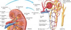
The main function - blood filtration and urine formation - is performed by the kidneys, or more precisely by their structural functional cells, nephrons.
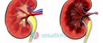
On the outside, the kidney is covered with a protective two-layer capsule, which consists of two membranes of adipose and connective tissue. Nephrons are located in the parenchyma, which is located immediately under the protective capsule.
And the calyces and pelvis form the so-called renal collecting system.
From the pelvis, urine flows through the ureters into the bladder. In normal condition, the length of the ureter in an adult is about 25–30 cm. Its internal diameter is on average 4 - 5 mm.
But in some places the ureter narrows: at the point of exit from the renal pelvis, at the mouth in the bladder and at the intersection with the iliac vessels.
The wall of the ureter consists of three layers. On the outside it is covered with a tube, which is formed from connective tissue. From the inside, its surface is a mucous membrane of transitional epithelium.
The middle layer consists of longitudinal and transverse smooth muscle fibers. Thanks to their involuntary contractions, urine from the kidney enters the bladder. As it accumulates, it is discharged through the urethra.
Diseases of the urinary system are very common. Unfortunately, many people are familiar with the diagnoses of cystitis, urolithiasis, and pyelonephritis.
However, only a few pathologies necessitate plastic surgery of the ureteral wall. Let's look at them in a little more detail.
Pyelotomy
The main indication for pyelotomy is the presence of stones in the renal pelvis. The technique consists of cutting the skin and step-by-step direction to the organ. Next, the kidney is freed from fatty tissue and rotated in such a way as to facilitate access to the stones. The main difficulty is to avoid damaging the kidney vessels. After removing all the stones, the surgeon sutures all the tissues one by one. If necessary, a catheter is inserted. After the operation, slight swelling is possible, which subsides after 1-2 days.
Factors that make intervention impossible
Despite the positive prognosis, the operation has contraindications, the presence of which is the main factor for the procedure.
It is prohibited to perform surgery in the following cases:
- If a patient has a blood disease (clotting disorder), it can be either congenital or acquired during life.
- The presence of infection in the body.
- Long gestation period.
- Serious inflammation.
Problems can also occur if there is scarring in the abdominal cavity. They greatly complicate the process of isolating the kidney and performing minimally invasive interventions.
Urethrourethroanastomosis and ureterocystoanastomy
Plastic surgery during urethro-urethroanastomosis is performed by suturing the ends of both ureters together. In order to increase the diameter of the lumen and reduce the risk of scar development, oblique incisions are made.
The ureter is sutured onto a catheter, which is removed a few weeks after the plastic surgery.
Urethrocystoanastomy is performed when the lower part of the ureter is damaged. Plastic surgery involves its direct attachment to the bladder.
Suturing is carried out using a thin catheter, which remains until the surface of the organ heals - for 8 - 10 days.
Before performing these types of operations, a special Foley catheter is placed in the bladder, which remains there for several days after the plastic surgery.
Pyeloplasty
An operation called “pyeloplasty” should be performed in patients with impaired functioning of the renal pelvis. In preparation for surgery, it is necessary to undergo laboratory tests and consult with the attending physician. Depending on indicators such as age, general health and severity of the disease, the doctor will determine the type of pyeloplasty required. During the operation, an incision is made and the ureter is disconnected from the damaged area of the pelvis. Next, the doctor removes the blocked part. At the end of the procedure, the doctor connects the ureter to a healthy pelvis. For the duration of the postoperative period, a catheter or stent is placed in the person’s ureter, and drainage may be performed. In the absence of adverse reactions and complications, the drainage is removed for 3-4 days.
Recovery period
In the first month after kidney surgery, you will need to take medications, treat wounds, avoid exercise, and follow a diet.
The first week after surgery will be the most difficult, so during this time it is important to be under constant medical supervision. The patient will experience pain in the kidneys after surgery, and a bacterial infection may occur. Therefore, a drug therapy regimen is prescribed, during which painkillers, antibiotics, vitamins and other auxiliary drugs are used. Also, in the first six months after treatment, it is important not to put stress on the organ, eat right, give up bad habits, and lead a healthy lifestyle. After surgery, it is important to undergo regular medical examinations, take all tests and monitor your condition.
Preoperative diagnosis
In order for the operation to proceed without complications, and for the patient to avoid postoperative negative consequences, it is important to undergo a number of diagnostic procedures the day before, which will help eliminate contraindications and take adequate measures in a timely manner. Therefore, the doctor will give directions for the following activities:
- consultation of highly specialized specialists;
- submitting blood and urine samples for general clinical analysis;
- ECG of the heart;
- Ultrasound of the kidneys and bladder;
- MRI or CT scan of the urinary system;
- FGS;
- X-ray.
Hydroureteronephrosis
Unlike hydronephrosis, this disease, in addition to the renal collecting system, also affects the ureter. This process occurs when it is compressed or blocked, which is caused by many factors.
Moreover, the lower the ureteral obstruction is localized, the larger the area of its damage.
The most common causes of hydroureteronephrosis are:
- scars that form after surgical interventions;
- damage to the ureter as a result of medical error during operations on the abdominal organs;
- compression of the ureter by tumors of nearby organs, such as the uterus, ovary, rectum;
- narrowing of the lumen of the ureter due to various inflammatory and infectious diseases;
- congenital abnormalities of the structure or location of the urinary tract;
- radiation therapy for cancer.
The mechanism of development of this disease is the same as with hydronephrosis, with the only difference being that the ureter is also involved in the pathological process.
Hydroureteronephrosis is manifested by pain in the lumbar region on the side of the affected kidney. In most cases, the disease affects only one ureter; a bilateral process can be observed as a complication of radiation therapy.
Complications
In order for the operated kidney to recover faster, it is important to follow all the doctor’s recommendations, then the risk of negative consequences is reduced. The most common complications after surgical treatment are:
- infection of internal tissues;
- injuries to neighboring organs, veins or arteries;
- bleeding;
- thromboembolism;
- relapse of the oncological process;
- the formation of adhesions that negatively affect the kidney and its function.