Indications for installation of a urinary catheter
Installation of a urinary catheter is indicated for the following conditions:
- Urinary retention as a result of infections and surgical interventions.
- Unconscious patient with uncontrolled outflow of urine.
- Acute inflammatory diseases of the urinary organs, requiring lavage and administration of medications into the bladder.
- Injury to the urethra, swelling, scars.
- General anesthesia and postoperative period.
- Spinal injuries, paralysis, temporary incapacity.
- Severe cerebral circulatory disorders.
- Tumors and cysts of the urinary organs.
Catheterization is also carried out if it is necessary to collect urine from the urinary bladder.
To prevent catheterization from harming the body, you should read the instructions for its implementation. The steps for inserting a catheter are different for men, women, and children.
Installation of a urological catheter in women is performed as follows:
- The patient takes a horizontal position: lies on her back, bends her knees, spreads them apart. A diaper is placed under the patient’s buttocks.
- The labia are washed, treated with an antiseptic and pulled apart.
- The entrance to the urethra is treated with a solution of furatsilin.
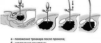
- A tube soaked in Vaseline is inserted into the urethra using tweezers.
- When the device is inserted 7 cm deep, urine begins to flow through the tube. The second end of the catheter is fixed in the urinal bag.
Depending on the purpose of the procedure, it can end at this point, or continue with rinsing, administering medications and further removing the device.
Due to physiological characteristics, women tolerate this procedure much easier than men.
Placement of a urethral catheter for men is carried out as follows:
- The patient lies in a horizontal position on his back. Legs are bent at the knees and spread apart. Oilcloth is placed under the buttocks.
- The penis is wrapped in a napkin, the urethra is treated with furatsilin solution and wiped.
- The catheter is taken with tweezers and inserted into the urethral canal. The penis is slowly and gently pulled onto the tube until it moves towards the external sphincter.
- The device is slowly lowered into the scrotum until the obstacle is overcome.
- The second end of the catheter is fixed in the urine bag. The specialist waits for the outflow of urine from the bladder.
Further instructions depend on how long the catheter is placed. For short-term use, the device is removed after urine drainage or medication administration. For long-term use, catheterization is completed after insertion.
If the procedure was carried out correctly, there is no pain.
The general algorithm for installing a catheter in children does not differ from adult instructions.
There are important features when performing the procedure in children:
- The urethral catheter for children should have a small diameter so as not to damage the child’s genitourinary organs.
- The device is placed on a full bladder. You can check the fullness of the organ using ultrasound.
- Treatment with medications and strong antibacterial compounds is prohibited.
- In girls, you need to spread the labia carefully so as not to damage the frenulum.
- The insertion of the tube should be gentle, slow, without applying force.
- The catheter must be removed as soon as possible so as not to provoke inflammation.
DETAILS: Urological diseases in adults
The procedure in children, especially infants, should be performed by a urologist with pediatric training.
Catheter installation technique and complications
A. Thermodilution
Method of introducing saline solution into the right atrium, in an amount of 2.5; 5 or 10 ml, low temperature makes it possible to change the temperature of the blood that is in contact with the thermistor located in the pulmonary artery. The degree of temperature fluctuation is inversely proportional to the amount of cardiac output. The change in blood temperature during such activities is small when cardiac output is high, and drops sharply when cardiac output is low. When graphically depicting the dependence of temperature fluctuations, it is expressed by a thermodilution curve
Cardiac blood output is determined by a computer program that integrates the area of the thermodilution curve. To better measure cardiac output, it is necessary to quickly and uniformly administer the solution, the temperature and volume of which must be strictly fixed by entering the calibration factors of the solution into the computer. These factors consist of the volume and temperature of the solution and the type of catheter, as well as cardiac output data during operation of the electrocautery.
Malfunction of the tricuspid valve, as well as intracardiac shunts, significantly reduce the reliability of the results obtained, since it is really possible to measure only the output of the right ventricle, which in this case does not correspond to the data of blood output from the left ventricle. Occasionally, a rapid infusion of a cold (ice) solution provokes the appearance of arrhythmia. Complications that arise when measuring cardiac blood output coincide with the manifestation of complications when installing catheters in the pulmonary artery and central veins.
The new thermodilution technique makes it possible to continuously monitor cardiac output. In such cases, special monitors and catheters containing thermofilaments are used, which generate low-intensity heat pulses into the blood of the proximal pulmonary valve. In addition, these catheters are equipped with thermisters that measure fluctuations in blood temperature in the pulmonary artery. The monitor determines cardiac blood output by cross-correlating heat and changes in blood temperature.
Clinical Opportunities
Cardiac blood output makes it possible to determine indices that reflect the functioning of the circulatory system. Pulmonary artery blood pressure measurements are difficult to compare with cardiac output data. An example of this is data from normal blood pressure and pressure when the pulmonary artery is “jammed”. In this case, the perfusion of important organs of the body is reduced due to low cardiac output and high peripheral vascular resistance. Without measuring cardiac output, effective pharmacological effects on myocardial contractility, preload and afterload are impossible.
Permanent or temporary urinary catheter
The use of catheters, depending on the situation, can be either temporary (if necessary) or permanent. The constant use of a urinary catheter is necessary for chronic diseases that cannot be radically cured with medications or surgery. Often these are neurological patients.
While women often have a Foley catheter installed in the urethra, this option is not acceptable for men. Why? Yes, for the reason that the male urethra communicates not only with the bladder, but also with the prostate, testicles, and seminal vesicles. And a foreign body in the urethra will sooner or later lead to complications, such as acute prostatitis or epididymitis...
That is why a permanent urinary catheter is more often used in women, and in men it is also used, but after performing an operation - epicystostomy with the formation of a suprapubic vesical fistula. It is into this fistula that a permanent urinary catheter is installed in men. In this embodiment, it is practically safe and does not cause complications.
Features of catheterization
Regardless of the type and reason for use, all catheters require mandatory fixation. The tube is secured to the skin using medical tape or suture material. Modern models are initially equipped with special clamps, which greatly facilitates the catheterization process. Additionally, it is necessary to set the position of the tube inside the cavity; most often, the instrument has a device that allows you to quickly change its shape after insertion into the hollow organ.
The most widely used system is the Pigtail system - the tip of the catheter made of polyvinyl material looks very similar to a pig's tail, hence the name. During production, this device is placed in a special stylet or conductor, and after installation it is released and, twisting, prevents the tube from falling out of the organ. This type of fixation system is recognized as the safest and easiest to implement.
For a more rigid fixation, a loop is used, which is tightened with a fishing line previously placed in the catheter cavity.
Types of catheterization
Based on the duration of the procedure, catheterization is divided into long-term and short-term. In the first case, the catheter is installed on a permanent basis, in the second - for several hours or days in a hospital setting.
Depending on the organ undergoing the procedure, the following types of catheterization are distinguished:
- urethral;
- ureteral;
- renal pelvis;
- vesical.
Catheterization can also be divided into male, female and pediatric.
The procedure does not require special preparation. Before catheterization, the patient should wash himself and, if necessary, shave the hair in the intimate area.
The nurse or attending physician must sterilize and prepare the necessary instruments for use. The catheterization kit includes the following:
- sterile tray for instruments;
- diaper or oilcloth;
- disposable rubber gloves;
- antiseptic for rubber processing;
- gauze napkins;
- Vaseline or glycerin;
- tweezers;
- Janet syringe;
- furatsilin solution;
- 2 new catheters.
You may also need a container to collect urine for analysis.
Before performing the procedure, the specialist thoroughly washes his hands, puts on disposable gloves and treats them with an antiseptic. The tip of the selected device is lubricated with Vaseline or glycerin.
After discharge from the hospital, the relatives of the patient with a catheter are left alone with this problem. And many simply do not know how to properly flush the bladder through a urinary catheter. You should adhere to the following rules:
- Rinse the urinary catheter at least 1 time a day, preferably 2 times (if necessary, you can rinse the bladder through a urinary catheter 5 or even 10 times a day);
- Before rinsing, be sure to disconnect the urinal and tubes extending the catheter. Flush the bladder directly through the catheter;
- Use special solutions to rinse the bladder. It is strictly not recommended to rinse with plain water because... this will inevitably lead to the development of inflammation in the bladder and may result in the development of acute ascending pyelonephritis;
DETAILS: Total and free PSA values are normal
It must be admitted that relatives of patients with a catheter do not always invite medical specialists to replace the catheter and change it themselves. If the procedure for replacing the Foley and especially Petzer catheter is entrusted to a urologist, then they dare to insert the catheter into the woman’s bladder themselves. In such situations, you must adhere to the following rules:
- Before catheterization, it is necessary to treat the area of the external urethral opening with an antiseptic;
- Wash your hands thoroughly twice with soap and treat them with alcohol;
- When inserting the catheter, hold it with sterile tweezers. Otherwise, you will get an infection in the bladder;
- It is advisable to use a special Zhanne syringe or a disposable alternative with a volume of 60 - 150 ml for rinsing.
Although it may seem difficult to insert a catheter into the bladder at first glance, we urge you to entrust this procedure to qualified medical professionals. Remember that improper catheterization can injure the bladder or urethra, which will ultimately result in emergency surgery.
Indications for pulmonary artery catheterization.
Heart diseases:
- Heart defects.
- IHD with left ventricular dysfunction or recent myocardial infarction.
- Heart failure (eg, cardiomyopathy, cardiac tamponade, cor pulmonale).
Lung diseases:
- Acute respiratory failure (eg, adult respiratory distress syndrome).
- Severe chronic obstructive pulmonary disease.
Infusion therapy:
- Sepsis.
- Shock.
- Burns (acute period).
- Hemorrhagic pancreatitis.
- Acute renal failure.
Surgical interventions:
- Valve replacement.
- Coronary artery bypass surgery.
- Clamping of the aorta (for example, during operations for an aortic aneurysm).
- Pericardiectomy.
- Portal vein bypass.
- Operations on the posterior cranial fossa with the patient in a sitting position.
Complicated pregnancy:
- Placental abruption.
- Severe toxicosis.
Despite the fact that the effectiveness of pulmonary artery catheter monitoring in many groups of surgical patients remains largely unproven, it is nevertheless well established that thanks to information about important hemodynamic parameters, it is possible to significantly reduce the risk of developing certain perioperative complications (to for example, heart failure, pulmonary edema, renal failure, myocardial ischemia).
In contrast to clinical testing, monitoring cardiac output and pulmonary artery pressure provides the most accurate information about the circulatory system during critical illness. Catheterization of the pulmonary artery is performed only in cases where it is necessary to obtain information about preload, cardiac indices, degree of oxygenation of mixed venous blood or bcc. This information is of particular value for patients whose risk of hemodynamic disorders (for example, myocardial infarction) is extremely high, or during operations that are characterized by a high risk of complications with blood circulation (for example, intervention due to a thoracic aortic aneurysm).
Contraindications to pulmonary artery catheterization.
Blockade of the His bundle, especially its left bundle, makes it impossible to insert a catheter into the pulmonary artery, as there may be a threat of complete AV block. The risk category also includes concomitant diseases, such as Ebstein's anomaly, which can provoke an attack of tachyarrhythmia. In cases of severe Wolff-Parkinson-White syndrome, caution should also be exercised, since a floating catheter can become a source of infection, and in case of hypercoagulation, provoke the formation of a blood clot. For such diseases, it is best to use catheters with built-in pacemakers.
Methodology and complications.
There are different types of catheter that are inserted into the pulmonary artery. In practice, the most commonly used catheter is a four-channel polyvinyl chloride catheter of size 7F, its length is 110 cm. Inside the thermistor channel with which the catheter is equipped, a wire is passed that connects the thermistor to a device that helps regulate the rate of cardiac output of blood. It is also equipped with an air channel designed for pumping air into the cylinder.
The percutaneous introducer (system for percutaneous introduction of the Swan-Ganz catheter) consists of a vasodilator and a sheath catheter, which are passed over a guide wire.
The proximal port of the catheter has an outlet into a channel intended for the introduction of infusion solutions, a sensor for measuring cardiac blood output and pressure in the right atrium of the myocardium. The distal port is connected to a channel designed for collecting venous blood samples, as well as monitoring pressure in the right pulmonary artery.
When inserting a catheter, one should be guided by the Seldinger technique, when catheterization is carried out into the central blood vein. In such cases, a vasodilator is used, as well as a special sheath catheter, which are guided through a special wire guide. A floating catheter is inserted into the lumen of the sheath catheter, and the vasodilator and guidewire are removed.
Floating catheter with a balloon for catheterization of the pulmonary artery (Swan-Ganz catheter).
Before the procedure for installing a floating catheter, it is checked, the balloon is inflated and emptied, and the catheter is flushed with heparinized sodium chloride saline solution. After this, the port is connected to the sensors, and it is installed at the level of the mid-axillary line, passing the floating catheter to the internal jugular vein, to the 15 centimeter mark. In this case, the tip of the catheter will be inserted into the right atrium of the myocardium. On a rehabilitation medicine monitor showing a venous pressure curve, the minus waves of the myocardium will coincide with the respiratory cycle curve. The balloon is filled with air to a volume of 1.5 ml, which is recommended by the catheter manufacturer. Increased systolic blood pressure indicates a correctly performed procedure. When the catheter is inserted to a depth of 35-40 cm, its tip enters the pulmonary artery, and this is what provokes an increase in diastolic pressure.
When installing a catheter into the right atrium, ensure that the balloon being advanced is filled with air. This will prevent injury to the endocardium by the catheter and cause it to migrate with the blood flow. When removing the catheter, the balloon must be free of air.
Strict ECG monitoring is necessary during catheter insertion to prevent the occurrence of arrhythmia, as this phenomenon occasionally occurs frequently due to irritation of the right ventricular endocardium by the catheter tip or balloon . In such cases, intravenous administration of lidocaine is not required. If there is no change in blood pressure as the catheter is advanced, the balloon should be emptied and the catheter removed immediately to prevent knot formation.
Normal pressure values and waveform as the Swan-Ganz catheter moves from the right atrium to wedge in the pulmonary artery.
In emergency cases, which are accompanied by low cardiac output of blood, pulmonary hypertension, congenital heart disease, advancement of the catheter with blood flow can be carried out by increasing the tidal volume, that is, the patient is recommended to take a full breath or lower the lower part of the operating table, lay the patient who is undergoing manipulation, on the right side. It is also recommended to administer a cold isotonic solution (sodium chloride) through the paroxysmal port. Such manipulation will increase the rigidity of the catheter walls, but may provoke perforation. You can also administer a small dose of an inotropic drug intravenously. This will increase cardiac output of blood.
The amplitude of the blood pressure curve decreases sharply if, when a catheter with an inflated balloon enters the pulmonary artery, it is moved forward a small distance. If at this time the air is pumped out of the balloon, a curved line will again appear on the monitor, indicating the pressure in the pulmonary artery. This “jamming” of pressure can also occur when the catheter tip migrates too distally. In this case, you should empty the balloon and pull out the catheter a little.
With such manipulations, an overinflated balloon can lead to rupture of the pulmonary artery , which causes 50-70% of deaths. To prevent this from happening, during catheter insertion it is necessary to constantly monitor the pressure in the pulmonary artery, avoiding “jamming” pressure. It should be noted that when the proximal right ventricular outlet is open and the tip of the catheter is 10 cm from it, when the latter is displaced in the distal direction, the blood pressure curve will show data similar to blood pressure in the pulmonary artery. Correct insertion of the catheter should be monitored using a lateral chest x-ray.
During the manipulation, the catheter can move to the right and in a caudal direction, and it can also migrate forward to the vena cava. In this case, the pressure in the pulmonary capillaries may be reduced compared to the pressure in the alveoli, and during positive pressure ventilation it shows a false increase in values.
Installation of a catheter can be complicated by the same symptoms as installation through a central vein. There may also be a risk of endocarditis, bacteremia, blood clots, pulmonary infarction, rupture of the pulmonary artery, which most often occurs in patients taking coagulants, elderly patients and women suffering from pulmonary hypertension. Risk factors also include catheter nodulation, heart rhythm disturbances, conduction disturbances, and damage to the pulmonary valve.
When to combine a catheter with a urine bag?
The constant use of a catheter for urine drainage forces us to solve the problem of urine collection. After all, with a permanent catheter, the patient is not always bedridden. Many lead relatively active lifestyles. The most practical option is to use a catheter with a urine bag. The urine bag is a plastic bag with a tube for collecting urine, which is connected to a catheter and a second tube with a valve to drain accumulated urine. A catheter with a urinal is used both in the option of urinary diversion through a catheter in the urethra and through a suprapubic fistula.
| It should be remembered that any permanent catheter must be replaced with a new one at least once a month. When replacing the catheter, be sure to also change the urine bag, because In an old catheter and urinal, colonies of bacteria form over time, which will constantly provoke inflammation in the bladder. |
Clinical Opportunities
The floating Swan-Ganz catheter made it possible to monitor the patient's condition during surgery. By penetrating the pulmonary artery, it makes it possible to calculate preload on the left ventricle more accurately than insertion of a catheter through a central vein or physical examination. With its use, it becomes possible to collect venous blood, diagnose air embolism, and myocardial ischemia. The ability to calculate hemodynamic parameters is provided by catheters equipped with a thermistor. It measures the cardiac output of venous blood.
Hemodynamic parameters calculated from data obtained during pulmonary artery catheterization .
Index | Formula | Norm | Unit |
| Cardiac index | Cardiac output (l/min) Body surface area (m2) | 2,8-4,2 | l/(min x m2) |
| Total peripheral vascular resistance | (BPmean - CVP) x 80 Cardiac output (l/min) | 1200-1500 | din x c x cm-5 |
| Pulmonary vascular resistance | (DLAav. - DZLA) x 80 Cardiac output (l/min) | 100-300 | din x c x cm-5 |
| Stroke volume | Cardiac output (l/min) x 1000 heart rate (min-1) | 60-90 | ml/stroke |
| Impact index (SI) | Stroke volume (ml/stroke) Body surface area (m2) | 30-65 | ml/stroke/m2 |
| Right ventricular stroke index | 0.0136 (DLAav. - CVP) x UI | 5-10 | g-m/blow/m2 |
| Left ventricular shock index | 0.0136 (ADav. - DZLA) x UI | 45-60 | g-m/blow/m2 |
Note. DLAsr. - average pressure in the pulmonary artery; PAWP—pulmonary artery wedge pressure; gm - gram-meter.
To provide assistance to patients, models of catheters with built-in electrodes have been developed that allow intracavitary ECG and cardiac pacing. In some catheter models, the fiber optic bundle makes it possible to monitor blood oxygen saturation.
According to Starling, there is a dependence of the function of the left ventricle of the heart on the length of its muscle fibers in diastole, which corresponds to its end-diastolic volume. This pressure reflects the length of the fibers, which can be changed due to decreased compliance acquired during cardiac ischemia or tamponade. In the absence of damage to the mitral valve, during its opening (diastole), blood enters the left ventricle, its pressure is equal to the blood pressure in the left ventricle.
The left atrium is connected by pulmonary vessels. A properly “wedged” catheter isolates the distal lumen with a filled balloon and allows for an analysis of arterialized blood; “wedging” pressure is applied to the distal outlet of the catheter. In the absence of increased airway pressure or pulmonary vascular disease, the wedge pressure becomes equal to the atrial pressure.
Thus, we can conclude: the pressure in the artery corresponds to measurements of the length of muscle fibers and left ventricular function.
Inserting a catheter into the central vein makes it possible to more accurately determine the functioning of the right ventricle of the heart. Insertion of a catheter into the pulmonary artery is indicated when dysfunction of both ventricles occurs, which leads to hemodynamic disorder between the circulation. CVP does not always correspond to pulmonary capillary pressure when the ejection fraction is less than 0.50. And the “jamming” pressure of the pulmonary artery may not always correspond to the end-diastolic pressure data in the left ventricle.
How to rinse a urinary catheter?
When a patient is discharged from a urology hospital, the attending physician recommends specific rinsing solutions. But if you don’t know how to flush the urinary catheter, first carefully study the recommendations in the discharge summary that the patient received upon discharge from the hospital. There, the attending physician is simply obliged to indicate how and with what to rinse the bladder through the catheter.
For the last 30-40 years, urologists have recommended flushing the catheter to remove urine with a solution of furacillin 1:5000. But the high resistance of urinary infections to this solution forced doctors over time to abandon furacillin in favor of a solution of potassium permanganate or the more modern Betadine and Vokadin.
DETAILS: What injections are prescribed for prostatitis, names of drugs, prostatilen || Injections for urological diseases
| Pay special attention to the fact that solutions must be prepared before use, i.e. dilute according to the attached instructions. It describes how to properly prepare a solution for washing a urinary catheter. |
Urinary catheter care
An indwelling urinary catheter should be carefully cared for to avoid urinary tract infections. The algorithm for processing it looks like this:
- Place the patient on his back, place an oilcloth or bedpan under the buttocks. Drain the drainage fluid and carefully remove the device.
- Drain the urine from the drainage bag, rinse it with water, treat it with an antiseptic: Chlorhexidine, Miramistin, Dioxidine, boric acid solution.
- Flush the catheter using a 50 or 100 mg syringe. Pour an antiseptic into it, and then rinse with running water.
- For inflammatory processes of the urinary tract, treat the catheter with a solution of furatsilin, diluting 1 tablet in a glass of hot water.
The urine bag must be emptied 5-6 times a day and washed with antiseptics at least once a day. The catheter should be cleaned no more than 1-2 times a week.
In addition, it is necessary to thoroughly wash the patient's genitals.
How to change a catheter yourself at home?
Replacing a catheter at home is a dangerous procedure that can cause serious injury to the urinary organs. Carrying out the procedure yourself is only permissible for a soft urethral device, and if there is a serious need.
To replace the device, you must remove the old catheter:
- Empty the urine bag. Wash your hands with soap and wear gloves.
- Lie in a horizontal position, bend and spread your legs to the sides.
- Wash the device tube and genitals with an antiseptic or saline solution.
- Locate the device's cylinder opening. This is the second hole, not used for draining urine and flushing the bladder.
- Empty the balloon using a 10 ml syringe. Insert it into the hole and pump out the water until the syringe is completely filled.
- Gently pull the tube out of the urethra.
After removing the device, a new one is inserted into the urethra, according to the instructions given above for representatives of different sexes.