The bladder begins to develop from the embryonic petal from the 25th day from the moment of conception and is finally formed by the 22nd week of pregnancy. By this time, the bladder reaches a size of 8 mm. With a transvaginal ultrasound, the organ is visible already at 11 weeks, with a transabdominal ultrasound - from the 16th week.
Anomalies of the genitourinary system are extremely rare. They are often associated with chromosomal abnormalities and are accompanied by a whole range of disorders and syndromes.
Ultrasound diagnostics in the 2nd trimester reveals 85% of pathologies. The most common bladder abnormalities are:
Fetal size by week of pregnancy
During the nine months of pregnancy, your baby goes through a journey that took humanity millions of years to complete. His growth in the mother's womb is an accelerated version of human evolution, and watching the baby grow is very interesting. Today, when examination methods such as ultrasound diagnostics have appeared, many aspects of the baby’s intrauterine life, previously hidden from the eyes of others, have become obvious, and therefore it has become possible to find out how the size of the fetus changes throughout the entire gestation period.
How does a child grow?
The baby's growth begins 12 hours after conception. During the first 12 hours, the first cell of his body is formed, which will give rise to everything else. It is called a zygote. Maternal and paternal reproductive cells (egg and sperm) take part in its formation. They merge, exchange genetic information, give up 23 chromosomes, and after 12 hours, just imagine, a completely new organism settles inside the woman.
Already at this stage, the cell contains all the information about the future person - it is known what gender the baby will be born, what kind of eyes and ears he will have, whether he will be tall or short, fat or thin. The cell contains information about what diseases he inherited from his ancestors, where his moles will be located, whether he will have talent for music, sports, science, what his psychotype will be.
And in the first days after conception, the zygote is intensively fragmented without increasing in size. That is, the number of cells within a single cell increases, but the overall size of the cell does not. About a week after conception, the baby becomes a blastocyst and attaches to the wall of the uterus, where it will spend the next nine months.
It should be noted that in the first trimester all embryos grow at approximately the same rate.
During this period, all the forces of the tiny organism are devoted not to growth as such, but to the formation of internal organs. Embryogenesis processes take place - the heart, intestines, spine, brain, cerebellum, kidneys, stomach, genitals, and so on are formed. From 10 weeks, the baby turns from an embryo into a fetus and remains so until birth.
After embryogenesis ends, children grow differently. Everyone has their own genetic program - some are destined to be tall and others short, some will be chubby and others thin, influenced by race, ethnicity, parental genes and partly the gender of the child. Therefore, the size of the baby in the first trimester is the most informative in order to determine the exact duration of pregnancy.
From the second trimester, growth and weight gain become the main task for the baby in the womb. And the growth rate already indicates how well the baby feels - whether he has enough oxygen, nutrients, whether everything is fine with him.
Ultrasound standards are fetometry. Fetometric indicators are quite arbitrary, and in each specific case they may differ from the statistical average, and therefore it is not possible to fit all children to one standard. All sizes that we will talk about below are only average, typical for children, according to statistics.
Treatment of hypoplasia
While the fetus is in the womb, no treatment is required. After birth, the doctor is required to assess the condition of the newborn. If the second kidney is working normally and the tests are normal, then therapy is not indicated. If the child’s life is at risk, then specialists take urgent measures.
Hypoplasia of the fetal kidney is not a death sentence. The child will grow and develop normally. With a correct lifestyle that excludes excessive stress on one kidney, there may also be no problems in the future. The main thing is not to overcool and avoid pyelonephritis.
In addition, the most accurate ultrasound results are obtained after birth. Many diagnoses are eliminated.
What are the sizes?
The first size that doctors begin to determine, according to ultrasound diagnostics, is the average internal diameter of the gestational sac (AVD). After 8 weeks, doctors have a real opportunity to measure the child (embryo) itself, but before this period, the only informative indicator of its growth rate is the distance between the inner walls of the fetal membrane. At the same time, the size of the yolk sac is recorded.
The next size is the coccygeal-parietal (CTP). As the name suggests, this is the distance from the bottom of the tailbone to the top of the crown. It is this size that is considered the most important for establishing the exact gestational age. According to the clinical recommendations of the Ministry of Health, an error of only 3-4 days is considered acceptable.
, the biparietal head size (BDS) begins to be measured This is the distance between the internal points of the parietal bones of the skull in a plane. And almost simultaneously with this, the fronto-occipital size (FOR) begins to be determined - the distance from the frontal bone to the center of the occipital bone.
Osteoarticular system
Examination of the fetal osteoarticular system is of great importance for a more in-depth analysis of fetal development. Visualization of small parts of the body (upper and lower extremities) is possible from 12 weeks, however, differentiation of tubular bones (humerus, ulna, radius, femur, tibia and tibia) is possible only from 14 weeks, and from 24 weeks a more in-depth study of the entire bone structure is possible. articular system with its ossification centers.
It should be noted that sometimes it is very difficult to differentiate tubular bones that have a similar echographic picture ( femur and humerus, bones of the forearm and tibia, consisting of two long bones
). This requires a detailed scan of the length of the bones, identifying their distal and proximal ends, until a clear image of the joints is obtained. Small bones—the bones of the hands and feet—are quite easily differentiated.
There is a definite relationship between the length of the femur and other long bones and the gestational age of the fetus. Some authors claim that determining the age of the fetus based on the length of the femur is 20% more accurate than determining the fetal head, which can be used to diagnose hydrocephalus and microcephaly. But since it is not always possible to differentiate these bones ( they are especially difficult to differentiate when there is a small amount of water
), then the BDP of the head is the main and stable indicator for determining the duration of pregnancy.
Determination of gestational age by the length of tubular bones
Today I’ll tell you about one developmental defect that can manifest itself, which means it can be suspected already during screening in the 1st trimester.
Prune Belly
- a syndrome that includes a number of developmental anomalies, among which there are three main ones:
- weakness, underdevelopment of the anterior abdominal wall
- bilateral cryptorchidism
- abnormalities of the genitourinary tract
In my search for information about this syndrome, I came across so many variations of translations and pronunciations!
Both in the Italian manner of Prune Belli, and with the French accent of Prune Belli, giving me the impression that this is the name and surname of a certain scientist. As well as the “cut belly” syndrome and the “plum belly” syndrome, from Frölich and Obrinsky. Prune is an English word that translates as plum, although in this context another meaning was meant - prune, due to the characteristic wrinkled appearance of the anterior abdominal wall, belly is translated as belly. The term “prune belly” is not used in Russian-language literature, so in the future I will use its English version, I think it will be more correct. This type of abdominal wall defect was first described by Frölich in 1839, and the term Prune Belly was proposed by Osler in 1901.
Although the characteristic appearance of the baby's abdomen is the hallmark of this anomaly, which is usually the basis for the diagnosis of the newborn, the underlying genitourinary tract abnormalities are the most important factor determining overall survival. There is a wide spectrum of severity of manifestations within the syndrome. Some children with severe respiratory and renal impairment die in the neonatal period, while in others the changes are moderate and can be corrected. Severe renal dysplasia, oligohydramnios and, as a consequence, pulmonary hypoplasia in 20% of cases lead to antenatal fetal death and in 30% to progressive renal failure in the first two years of a child’s life. Urogenital tract abnormalities associated with Prune Belly include
- hydronephrosis
- tortuous dilated ureters
- varying degrees of renal dysplasia
- enlarged bladder
In addition, other systems may be involved: cardiovascular, musculoskeletal, respiratory, gastrointestinal.
In 95% of cases it occurs in boys, but similar changes, including the absence of the muscles of the anterior abdominal wall in combination with anomalies of the genitourinary system, are also described in girls.
As an illustration, I offer a story that I found freely available on one of the forums:
“ Girls, who are interested, I had a child with Prune Belli syndrome.
I’ll start my story by saying that neither I nor my husband are sick with anything, the pregnancy was going well, I didn’t sniff paint, I don’t drink, I don’t smoke, the reason for this situation has never been found, either here or in America. Yes, of course, we were upset by this situation, and if they had informed me about this at 12 weeks, I probably would have terminated the pregnancy, but now I have a wonderful child, whose urine tests periodically go bad, this greatly affects the kidneys, he will need surgery in the future on the abdominal wall, for cryptorchidism, as well as for a heart defect, he has flat feet, he used to have club feet, but from the age of 2 weeks we cast him using the Panseti method, his legs have improved, he behaves like an ordinary child, his teeth are also setting, gets up, walks, walks, well, in general, a wonderful baby, although he does all this with a delay of 3-4 months. I know a child with the same diagnosis as ours, he had an operation in England and collected about 4 million rubles and everything seems to be fine with him, ugh, ugh, ugh. 1. Our baby started peeing at about 18 weeks (we thought that his urinary tract had decreased because he started peeing, but that was not the case, a hole formed in the peritoneum from strong pressure, so the bladder returned to normal) we were immensely happy jumped and ran... not for long. 2. At week 20, they discovered problems with the kidneys (bilateral hydronephrosis), but immediately said that it can be treated, they say, don’t worry 3. At week 24, club feet were discovered (they said that everything would be corrected, they even say it suits boys) 4. At 28 week cryptorchidism... The set of all these indicators is Prune Belli Syndrome, but none of the ultrasound specialists said this.
So, I found information on the Internet that someone was already told at 12 weeks that the child had Prune Belli syndrome, the ultrasound specialist saw a thin wall of the child’s abdomen, but no one saw us .”
There are three main embryological theories of the occurrence of this syndrome:
Bladder outlet obstruction
This theory, proposed as early as 1903, was later substantiated by recreating the phenotypic changes characteristic of Prune Belly in urethral obstruction in fetal sheep at 43–45 days of gestation. According to this theory, all other changes are secondary. The absence of urine outflow from the bladder leads to its significant enlargement, stretching of the anterior abdominal wall, disruption of its blood supply and atrophy, and also disrupts the process of descent of the testicles into the scrotum and leads to the formation of hydronephrosis and dilation of the ureters. However, in humans, urethral obstruction with this syndrome occurs in 10-20% of cases. Some scientists believe that the obstruction may be transient, others argue that obstruction occurs in the most severe forms of Prune Belly. According to human fetal embryology, such changes should appear at 13-15 weeks, since by this time the urachus begins to close and urine production by the fetus increases significantly. While this theory is compelling, it does not explain all of the changes associated with Prune Belly.
Mesodermal developmental delay theory
It is hypothesized that urinary tract abnormalities may be explained by the abnormal development of mesonephros between the 6th and 10th weeks. Changes in the development of the Wolffian duct lead to prostatic hypoplasia, delayed development of the prostatic urethra and valve-like obstruction. However, this theory cannot include all the anomalies encountered in this syndrome.
Yolk sac theory
There is a hypothesis that in Prune Belly, excess volume of the yolk sac may lead to abnormalities in the development of the anterior abdominal wall, due to the involvement of a large part of the allantois in the formation of the urinary tract.
How to suspect Prune Belly syndrome during an ultrasound?
The first thing that catches your eye is megacystis, i.e. a significant increase in the size of the bladder, as well as a thin, stretched and protruding anterior abdominal wall. At 10-14 weeks of pregnancy, the diagnosis of megacystis is made when the longitudinal size of the bladder exceeds the conventional standard of 7 mm
. With sizes of 8-12 mm, in most cases, spontaneous normalization occurs, but dynamic monitoring is required once every 2 weeks. This appears to be due to the fact that the formation of smooth muscle and innervation of the bladder does not end by the 13th week of gestation and continues in the following days, which provides the basis for self-resolution of the problem in subsequent weeks of fetal development. Enlarged bladder, dilated ureters, and pyelectasia may occur with megacystis-megaureter syndrome and posterior urethral valve, but in these conditions the amount of amniotic fluid usually remains normal and there is no such thinning or bulging of the anterior abdominal wall. Progression of megacystis and oligohydramnios are prognostically unfavorable signs indicating obstruction of the bladder outlet.
It is not possible to definitely diagnose Prune Belly syndrome at 11-13 weeks; it can only be suspected, and the main diagnosis will be Megacystis
, which, as I already said, can occur in various conditions.
What to do?
This is your child and, as always, you have to make the choice. But sometimes, it is very important for someone to lift and take upon themselves this burden of responsibility, the burden of the choice that you must make with your own, and not only your life. It is very important to feel that only the best decisions will be made here and now. Here are the recommendations published in the article “Prenatal consultation by a pediatric urologist and decision-making tactics when diagnosing megacystis syndrome in the first trimester of gestation” https://www.lvrach.ru/2015/01/15436142/: “Given that “megacystis” in 25–40% it is combined with chromosomal pathology; the results of genetic studies play a significant role in making a decision on prolongation or termination of pregnancy. Most authors agree that posterior urethral valves, as well as prune-belle syndrome, are not a genetically inherited pathology, but this does not exclude the possibility of chromosomal damage. AW Liao states in 25% of cases the presence of trisomy on the 13th and 18th chromosomes with an increase in the size of the bladder from 7 to 15 mm in fetuses of 10–14 weeks of gestation.
The analysis of diagnostic measures and pregnancy outcomes allowed us to develop a diagnostic algorithm, which involves mandatory karyotyping of the fetal material in the presence of a bladder size within the range of 7–15 mm. If a karyotype abnormality is confirmed, it is advisable to terminate the pregnancy, otherwise, dynamic observation until convincing markers for the prognosis of the diagnosed condition appear.
A significant initial enlargement of the bladder ≥ 20–30 mm clearly indicates severe obstruction of the lower urinary tract and does not require dynamic monitoring; termination of pregnancy is advisable.
The presence of dilatation of the upper urinary tract does not always complement the “megacystis” syndrome in the early stages of gestation (11–13 weeks) and is most clearly defined in the second and third trimesters of gestation.
However, in all cases its presence is a factor aggravating the prognosis. Thus, summing up the analysis of literature data and the results of our own observations, several conclusions are obvious:
- The set of measures included in early prenatal screening of 11–14 weeks of gestation (molecular genetic and ultrasound markers of congenital malformation and CA) does not allow determining the likelihood of a fetus having congenital malformation of the urinary system. Identification of a risk group for fetuses at risk due to the presence of congenital pathology MBC occurs on the basis of the diagnosis of an increased longitudinal size of the bladder ≥ 7 mm, which is interpreted as fetal megacystis syndrome, and requires careful diagnostic measures to predict the outcome.
- “Fetal megacystis” syndrome is considered as a manifestation of severe disturbances in the urodynamics of the lower urinary tract of anatomical or functional origin, underlying the development of obstructive disorders of the upper urinary tract and dysplastic development of the renal parenchyma, including its cystic dysplasia, which predetermines decompensation of renal functions and an unfavorable outcome.
- A set of diagnostic procedures that clarify the prognosis when identifying a group of fetuses with megacystis syndrome includes karyotyping of fetal material (chorionic villi) in the absence of dilatation of the upper urinary tract and bladder size ≤ 20 mm and termination of pregnancy in cases of detected chromosomal pathology.
- The presence of bladder enlargement ≥ 20 mm, isolated or in the presence of dilatations of the upper urinary tract, is an indication for termination of pregnancy.
- Prolongation of pregnancy is recommended in all cases of moderate expansion of the bladder within 7-15 mm in the absence of expansion of the upper urinary tract, positive dynamics of observation for 2-3 weeks, restoration of bladder size.
- Carrying out diagnostic measures and choosing tactical decisions should be carried out with the participation of a pediatric urologist, including the ante- and postnatal stages of dynamic observation and necessary treatment.”
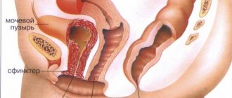
Some women may experience unpleasant symptoms during pregnancy, indicating that the uterus has begun to put pressure on the bladder. In this case, the patient visits the toilet very often and feels pain in the lower abdomen. Let's look at why this happens.
Collapse
Bladder pathologies in the fetus: what can be determined by screening ultrasound
The bladder begins to develop from the embryonic petal from the 25th day from the moment of conception and is finally formed by the 22nd week of pregnancy. By this time, the bladder reaches a size of 8 mm. With a transvaginal ultrasound, the organ is visible already at 11 weeks, with a transabdominal ultrasound - from the 16th week.
bladder pathology in the fetus
» data-medium-file=»https://i1.wp.com/medcentr-diana-spb.ru/wp-content/uploads/2018/05/patologii-mochevogo-puzyirya-u-ploda.jpg?fit= 450%2C300&ssl=1?v=1572898551″ data-large-file=”https://i1.wp.com/medcentr-diana-spb.ru/wp-content/uploads/2018/05/patologii-mochevogo-puzyirya -u-ploda.jpg?fit=825%2C550&ssl=1?v=1572898551″ class=”alignnone size-large wp-image-9236″ src=”https://i0.wp.com/medcentr-diana-spb .ru/wp-content/uploads/2018/05/patologii-mochevogo-puzyirya-u-ploda-825×550.jpg?resize=790%2C527″ alt=”bladder pathologies in the fetus” width=”790″ height =”527″ srcset=”https://i1.wp.com/medcentr-diana-spb.ru/wp-content/uploads/2018/05/patoloii-mochevogo-puzyirya-u-ploda.jpg?w=825&ssl =1 825w, https://i1.wp.com/medcentr-diana-spb.ru/wp-content/uploads/2018/05/patologii-mochevogo-puzyirya-u-ploda.jpg?w=450&ssl=1 450w , https://i1.wp.com/medcentr-diana-spb.ru/wp-content/uploads/2018/05/patologii-mochevogo-puzyirya-u-ploda.jpg?w=768&ssl=1 768w, https: //i1.wp.com/medcentr-diana-spb.ru/wp-content/uploads/2018/05/patologii-mochevogo-puzyirya-u-ploda.jpg?w=900&ssl=1 900w" sizes="(max -width: 790px) 100vw, 790px" data-recalc-dims="1″ />
Prevention
In medical practice, there are no preventive measures to prevent enlarged bladder syndrome in the fetus. Doctors advise women planning to conceive a child to fully prepare: undergo all tests, be examined by all specialists, and take a complex of vitamins. If any disease is detected, be sure to start treatment.
Pregnant women are advised to follow and comply with all doctor’s instructions in order to avoid serious complications.
In boys, during diagnostic ultrasound, a phenomenon such as a stream of turbulence into the amniotic fluid can be observed. Due to the overcrowded bladder of the fetus, it is easily mistaken for hydronephrosis, ovarian cyst, multicystic disease, or megacystis. Repeated examination provides an accurate description of the organ.
Pathologies and abnormalities of the bladder in the fetus
- Megacystis . This is an enlargement of the bladder over 8 mm in a longitudinal section on ultrasound. Pathology is detected at 10-15 weeks of pregnancy. Along with it, a violation of the relationship of the bladder to the coccygeal-parietal zone is usually detected (10.4% instead of 5.4%). Megacystis is of a chromosomal nature and is expressed in urodynamic disturbances due to blockage or fusion of the urethra.
- Detrusor obstruction . It is expressed in the absence of contractility of the muscular layer of the bladder, which is responsible for the expulsion of urine. On ultrasound, the bladder is pear-shaped, the walls are thin, and the organ itself is enlarged in size. If echographic signs of pathology are detected, the fetus is examined by vesicocentesis. Then karyotyping of the fetus is carried out, and if chromosomal abnormalities are confirmed, the woman is sent for an abortion. The same thing happens when the organ increases to 20-30 mm instead of the required 8 mm. There is a possibility of normalization after the 13th week of pregnancy.
- Exstrophy . This is the absence of the anterior wall of the bladder. On ultrasound, the bladder is completely absent, but the structure of the kidneys remains unchanged.
- Atresia. The absence of the urinary canal leads to an enlargement of the bladder to such a size that the volume of the fetus's tummy increases significantly. A woman is advised to terminate her pregnancy because the baby may be born with severe pulmonary hypoplasia or die in utero.
- Posterior urethral valve . This problem occurs only in boys. In girls, pmne-bUy syndrome occurs, which has similar symptoms. The anomaly is that due to an intrauterine disorder, the lower part of the urethra, which opens into the bladder, is too narrow, causing urine to flow back into the kidney. The result is hydronephrosis, an accumulation of excess fluid in the kidneys. On ultrasound, the fetus will have enlarged kidneys and a small bladder. The pathology is corrected immediately after the birth of the baby by excision of the site of pathological narrowing of the urethra. There is a high risk of infant death due to pulmonary hypoplasia due to renal failure.
- Vesicoureteral reflux. Normally, the ureter enters the bladder in such a way that the muscular wall of the organ serves as a valve that prevents the flow of urine back to the kidney.
If the ureter does not enter the bladder correctly, reflux occurs—urine flows back into the ureter. The anomaly is not a reason for termination of pregnancy, because it disappears by itself in the first 2 years of the baby’s life. In severe cases, surgical intervention is performed.
Bladder pathologies in the fetus are clearly visible on screening ultrasound, so such examination cannot be ignored. A screening ultrasound of the fetus should be performed only with the help of good equipment.
If you find an error, please select a piece of text and press Ctrl+Enter
Causes of pathology
Some researchers argue that if an enlarged bladder is detected in the fetus in the first trimester of pregnancy, it will not be possible to determine the true cause of its occurrence. Scientists identify two main etiological reasons for the development of megacystis in the fetus:
- Urodynamic disturbances due to urethral obstruction (fibrostenosis, strictures, in male fetuses it is expressed by posterior urethral valves, and in female fetuses - atresia).
- Functional insufficiency of urine outflow (manifested by the “megacystis-megaureter-microcolon” and Prune-Belly syndromes).
In the early stages of pregnancy, the diagnosis of an enlarged bladder can be established only by some echographic signs, because during this period there are still no specific indicators with which to differentiate intrauterine malformations.
The diagnosis of "megacystis-megaureter-microcolon" is more often diagnosed in female embryos (4:1), and urethral obstruction and Prune-Belly syndrome - in males.
Obstruction of the detrusor (the muscular membrane that expels urine) of the urinary tract is caused by blocking or complete absence of contractile function, which is characterized by degenerative processes, sparse smooth muscle, proliferation of fibrous connective tissue and hyaline deposits.
Diagnosis of the “keyhole” symptom, characteristic of valvular urethral obstruction, and dilation of the intestinal lumen most often occurs in the second half of pregnancy. These signs indicate the development of the “megacystis-megaureter-microcolon” syndrome.
A fetus with Prune-Belly syndrome is characterized by a thinned anterior peritoneal wall. Most researchers suggest that, although Rune-Belly syndrome and posterior urethral valve obstruction are not genetically determined anomalies, further development of chromosomal abnormalities cannot be ruled out.
Fetal bladder
Fetal kidney stones are a very rare pathology. In an adult, on scanograms they are defined as oval-shaped hyperechoic formations that give an acoustic shadow if their thickness exceeds 5 mm. In the fetus, due to the small size of the cameas, acoustic leakage is never observed. On scanograms of the fetus, they are defined as oval-shaped hyperechoic formations, the length of which is usually 3-5 mm, thickness - 2-3 mm.
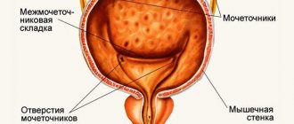
The fetal bladder begins to be detected on scanograms at 12-13 weeks of gestation. On transverse scanograms it is defined as round, and on longitudinal scans it is defined as an echo-negative oval-shaped formation with clear, even contours, completely devoid of internal echo structures.
The size of the bladder is subject to significant individual fluctuations and depends on the degree of its filling. Emptying of the bladder occurs completely or fractionally, i.e. in parts. In some cases, in the amniotic fluid at the location of the genital organs, the appearance of a turbulent flow can be observed, the occurrence of which is caused by emptying the bladder.
Anomalies of the bladder and urethra are rare. In the antenatal period, the following malformations are mainly observed: bladder exstrophy, ureterocele, urethral atresia, posterior urethral valve, syndrome .
Bladder exstrophy is a congenital disease characterized by a defect in the lower abdominal wall and the absence of the anterior wall of the bladder. This malformation is extremely rare - 1:45,000 newborns. In boys, this defect is often combined with total epispadias, and in girls, with anomalies of the uterus and vagina. The main echographic sign of bladder exstrophy is the absence of its image on scanograms, while the size and structure of the kidneys remain normal.
The amount of amniotic fluid is also unchanged. The diagnosis of exstrophy can be made as early as 16-18 weeks of pregnancy . Treatment is only surgical. Taking into account the large number of unsatisfactory long-term results, the question of the advisability of continuing pregnancy must be resolved together with specialists working in the field of pediatric urology.
Ureterocele is often detected only at the end of pregnancy and mainly with pronounced dilation of the ureter. Due to the fact that this pathology is almost always accompanied by pyelonephritis, urethritis and cystitis. in the immediate period after birth, the child must be sent to a specialized hospital for further examination and treatment.
conclusions
Bladder pathologies in the fetus are clearly visible on screening ultrasound, so such examination cannot be ignored. A screening ultrasound of the fetus should be performed only with the help of good equipment.
– functional disorders of bladder filling and emptying associated with disruption of nervous regulation mechanisms. Neurogenic bladder in children can manifest as uncontrollable, frequent or rare urination, urgency, urinary incontinence or retention, and urinary tract infections. The diagnosis of neurogenic bladder in children is made according to laboratory, ultrasound, radiological, endoscopic, radioisotope and urodynamic studies. Neurogenic bladder in children requires complex treatment, including drug therapy, physiotherapy, exercise therapy, and surgical correction.
Fetal bladder
Fetal kidney stones are a very rare pathology. In an adult, on scanograms they are defined as oval-shaped hyperechoic formations that give an acoustic shadow if their thickness exceeds 5 mm. In the fetus, due to the small size of the cameas, acoustic leakage is never observed. On scanograms of the fetus, they are defined as oval-shaped hyperechoic formations, the length of which is usually 3-5 mm, thickness - 2-3 mm.
The fetal bladder begins to be detected on scanograms at 12-13 weeks of gestation. On transverse scanograms it is defined as round, and on longitudinal scans it is defined as an echo-negative oval-shaped formation with clear, even contours, completely devoid of internal echo structures.
The size of the bladder is subject to significant individual fluctuations and depends on the degree of its filling. Emptying of the bladder occurs completely or fractionally, i.e. in parts. In some cases, in the amniotic fluid at the location of the genital organs, the appearance of a turbulent flow can be observed, the occurrence of which is caused by emptying the bladder.
Anomalies of the bladder and urethra are rare. In the antenatal period, the following malformations are mainly observed: bladder exstrophy, ureterocele, urethral atresia, posterior urethral valve, syndrome .
Bladder exstrophy is a congenital disease characterized by a defect in the lower abdominal wall and the absence of the anterior wall of the bladder. This malformation is extremely rare - 1:45,000 newborns. In boys, this defect is often combined with total epispadias, and in girls, with anomalies of the uterus and vagina. The main echographic sign of bladder exstrophy is the absence of its image on scanograms, while the size and structure of the kidneys remain normal.
The amount of amniotic fluid is also unchanged. The diagnosis of exstrophy can be made as early as 16-18 weeks of pregnancy . Treatment is only surgical. Taking into account the large number of unsatisfactory long-term results, the question of the advisability of continuing pregnancy must be resolved together with specialists working in the field of pediatric urology.
Ureterocele is often detected only at the end of pregnancy and mainly with pronounced dilation of the ureter. Due to the fact that this pathology is almost always accompanied by pyelonephritis, urethritis and cystitis. in the immediate period after birth, the child must be sent to a specialized hospital for further examination and treatment.
Urethral atresia is an extremely rare developmental defect. The main echographic sign of this pathology is a pronounced enlargement of the bladder in the complete absence of amniotic fluid. Bladder enlargement begins to be detected from 14-15 weeks of pregnancy.
By the end of the second and beginning of the third trimester of pregnancy, the bladder enlarges so much that it can fill the entire abdominal cavity. In turn, this leads to a significant increase in the abdomen.
In most cases, hydronephrotic transformation of the kidneys and dilation of the ureters of varying severity are noted. In case of urethral atresia and bilateral severe hydronephrosis, termination of pregnancy is indicated.
Fetal genitals
The problem of determining gender during pregnancy is mainly associated with the desire of parents to find out the sex of the child before birth. Science has made certain progress in this direction, but the previously proposed methods are invasive, their use is limited and unsafe for the fetus. Back in 1970, Garret and Robinzon studied this problem. Le Lami in 1979 first demonstrated the capabilities of ultrasound in determining the sex of the fetus. It turned out that this question can be most accurately answered after 26 weeks of pregnancy, when the genitals can be clearly visualized.
For good visualization of the fetal genital area, certain favorable factors are necessary, such as:
- cephalic presentation of the fetus;
- abducted position of the hip joints;
- full fetal bladder (primary landmark);
- sufficient amount of amniotic fluid.
Ultrasound diagnostics of males: detection of the scrotum below the bladder in the form of a round echogenic formation, testicles and penis; For the female sex, the labia majora are typically identified in the form of two echogenic ridges, in the middle of which there is a lower linear echo. The uterus and ovary are not normally differentiated.
Fetal bladder: formation and its size by week
The formation of the organ in the fetus begins on days 25–27 of pregnancy. During this period, the urogenital sinus is formed from the internal embryonic lobe. The final formation of the organ occurs when the fetus is at 21–22 weeks of development. The standard size is 8 mm. Abnormalities of the urinary system in most cases occur due to diseases of the chromosomal type. The defects that appeared at the time of formation are presented below.
Diverticulum
A diverticulum is characterized by a bulging of the bladder wall. The main symptom is double urination. Pathology occurs due to the inferiority of the muscle layer. Treatment involves surgery, during which the diverticulum is removed. Congenital diverticula are often single, less often there are 2 or 3. Emptying urine from a diverticulum may be complete or incomplete. Small diverticula without symptoms do not require treatment.
Megacystis and hypoplasia
Megacystis is a defect in which the bladder is enlarged. Timely examination will make it possible to make this diagnosis in the initial stages of pregnancy and detect an enlarged organ in time. With megacystitis, the urinary tract is greater than the standard norm. This anomaly may indicate the presence of undercut belly syndrome, which most often has a poor prognosis. To begin treatment, diagnostics is used - vesicocentesis. This is a test of fetal urine, which is taken during a puncture of the bladder wall. Vesicocentesis performed early reduces the risk of fetal loss.
Hypoplasia is characterized by congenital reduction of the bladder, often with renal failure. Very often this pathology is confused with agenesis. The capacity of the organ is several milliliters, which from the moment of birth is manifested by urinary incontinence. Depending on the situation, plastic surgery or a cystostomy is performed.
Exstrophy, atresia and agenesis
Exstrophy occurs more often in males than in females. It is characterized by the absence of the anterior abdominal wall of the bladder or its defect. Exstrophy on an echographic picture is manifested by the absence of a bladder on a scanogram, while the structure of the kidneys remains normal, without changes in the amount of amniotic fluid. Treatment is carried out only by surgery.
Urethral atresia is a rare defect in which the main indicators are enlargement, distension of the bladder and lack of amniotic fluid. The fetal bladder may become so enlarged that it results in an enlarged abdomen. With this pathology, termination of pregnancy is indicated; if it persists, in most cases a stillborn child is born or severe pulmonary hypoplasia is observed.
Agenesis is an extremely rare anomaly and is characterized by a lack of organ development. The birth rate with this pathology is very low. This disease is usually accompanied by other defects that are incompatible with intrauterine life. In newborns, the urinary function is preserved, but there is constant partial urinary retention, and palpation reveals a distended bladder.
Consequences
As we have already mentioned, a reduced kidney works the same as one that is of normal size. Although its performance is lower. The child will not experience any symptoms. Only during the next ultrasound examination will this deviation be noticeable. But there is some nuance here. It is important that the second kidney is complete.
In some cases, frequent pyelonephritis in children is possible. The disease should be stopped in time so that the nephrons do not die. Otherwise, they are replaced by connective material and over time the kidney will completely lose its function.
If the artery of the kidney is compressed, then hypertension will be diagnosed.
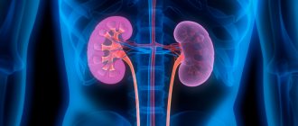
Renal hypoplasia in the fetus is a rare condition. With it, both kidneys are reduced, and the right one is stronger. In this case, the child is at risk of kidney failure. It will be necessary to regularly monitor the child’s health, take tests and register with a specialist. Unfortunately, with progressive pathology, the baby may die.
Ultrasound diagnostics of the fetal urinary system
The fetal bladder on ultrasound appears as a thin-walled formation in the lower parts of the body, having a round or pear-shaped shape. Its size increases throughout pregnancy. To assess the size, you need to take into account the possibility of emptying it, whether it is full or not. If there is no echo, you need to perform an ultrasound after 30-40 minutes. Congenital pathologies of the fetus can be diagnosed by screening ultrasound. The norms of indicators for a certain period are compared with those obtained during the survey. It feels no different from a regular ultrasound examination. Detection of abnormalities of the urinary system in later stages may affect the management of pregnancy.
Diagnostics
When the first symptoms of pathology appear, it is recommended to seek help from a doctor. The specialist will examine the patient and take a history. To confirm the preliminary diagnosis, endoscopy or radiological studies are recommended.
Thanks to endoscopy, it is possible to examine the organ from the inside. It is also used to examine the collecting system. To make a diagnosis, an ultrasound examination is necessary.
A CT scan is recommended for some patients. Magnetic resonance imaging is a fairly informative diagnostic method.